Original Scientific Article
Comparative anatomical studies on ductus venosus in fetuses of domestic ruminants
Pamela Bejdić
*
,
Amel Ćutuk
,
Amer Alić
,
Benjamin Čengić
,
Rizah Avdić
,
Faruk Tandir
,
Nedžad Hadžiomerović
,
Verica Mrvić
Received: 18 March 2020
Received in revised form: 07 October 2020
Accepted: 09 November 2020
Available Online First: 14 December 2020
Published on: 15 March 2021
Correspondence: Pamela Bejdić, pamela.bejdic@vfs.unsa.ba
Abstract
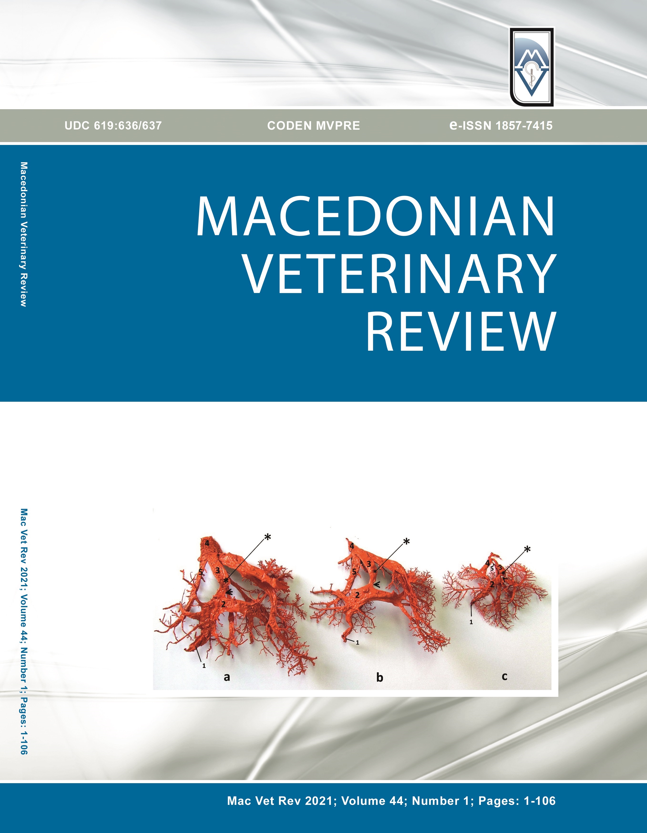
The study has aimed to investigate and determine the anatomical position, shape, size, and histological features of the ductus venosus, and its role as a shunt in the fetal circulatory system in domestic ruminants. The research was conducted on 19 bovine, 11 sheep and 5 goat fetuses, aborted at the late stage of pregnancy or deceased just after delivery. The general anatomy of the ductus venosus was investigated by in-situ dissection of the corrosive cast obtained by injection of 25% solution of Vinylite mass through the umbilical vein. For histological examination, the fetal tissue samples were stained with Hematoxylin and Eosin, Masson’s trichrome, Verhoeff-Van Gieson and Gomoriꞌs silver stain. The results showed that ruminant fetal ductus venosus is a curved, trumpet-shaped vessel, situated in the central part of the liver, above the porta hepatis. Its ventral part is constricted in the form of an isthmus, having a prominent lip-like thickening at the junction with the portal sinus. Histological examination showed the dominant presence of collagen and elastic fibers in its tunica media, with thin bands of smooth muscle fibers oriented in a longitudinal and circular direction indicating ability for vasoconstriction and vasodilatation.
Keywords: umbilicus, ductus venosus, ruminants, anatomy, morphology
References
- McGrady, T.A., Quinn, P.J., FitzPatrick, E.S., Ryan, M.T. (2006). Veterinary embryology. In: Cardiovascular system (pp. 105-130). Oxford, UK: Blackwell Publishing Ltd.
- Hyttel, P., Sinowatz, F., Vejlsted, M., Betteridge, K. (2010). Essentials of domestic animal embryology. In: Poul Hyttel (Ed.), Development of the blood cells, heart and vascular system (pp. 198-207). Edinburgh, London, New York, Oxford, Philadelphia, St. Louis, Sydney, Toronto: Elsevier Limited.
- Arpi, L.M.B. (2018). Histology. Histology of umbilical cord in mammals. [Internet]. (pp 47-58). IntechOpen. Open Access Peer-Reviewed Edited Volume. Provisional Chapter. [Available from: http://dx.doi.org/10.5772/intechopen.80766] https://doi.org/10.5772/intechopen.80766 PMid:31123483 PMCid:PMC6511591
- Shaker, N.A., El Gammal, S.M. (2018). Comparative anatomical studies on the fetal remnants in donkey (Asinus Equus) and camel (Camelus Dromedarius). IOSR-JAVS. 11(5): 12-21.
- Elgozouli, S.O., Osman, D.I. (2012). Histology of the constituents of the umbilical cord and ductus arteriosus and ductus venosus of the dromedary camel. JVA. 2(9): 449-456.
- Igbokwe, C.O., Muokwah, E.C., Ezeasor, D.N. (2015). Developmental changes in the morphology of some blood vessels in the foetuses of the red sokoto goat. J Anim Plant Sci. 25(3): 625-632.
- Botti, J.J., Edelstone, D.I., Caritis, S.N., Mueller-Heubach, E. (1982). Portal venous blood flow distribution to liver and ductus venosus in newborn lambs. Am J Obstet Gynecol. 144(3): 303-308. https://doi.org/10.1016/0002-9378(82)90583-X
- Edelstone, D.I., Rudolph, A.M., Heymann, M.A. (1978). Liver and ductus venosus blood flows in fetal lambs in utero. Circ Res. 42(3):426-433.
- Zink, J., Van Petten, G.R. (1980). Time course of closure of the ductus venosus in the newborn lamb. Pediatr Res. 14(1): 1-3. https://doi.org/10.1203/00006450-198001000-00001 PMid:7360516
- Coceani, F., Adeagbo, A.S., Cutz, E., Olley, P.M. (1984). Autonomic mechanisms in the ductus venosus of the lamb. Am J Physiol (Heart Circ Physiol). 247(1): H17-H24. https://doi.org/10.1152/ajpheart.1984.247.1.H17 PMid:6742210
- Rudolph, A.M. (1985). Distribution and regulation of blood flow in the fetal and neonatal lamb. Brief review. Circ Res. 57(6): 811-821. https://doi.org/10.1161/01.RES.57.6.811 PMid:3905044
- Rudolph, A.M. (1983). Hepatic and ductus venosus blood flows during fetal life. Hepatology 3(2): 254-258. https://doi.org/10.1002/hep.1840030220 PMid:6832717
- Kiserud, T., Eik-Nes, S.H., Blaas, H.G., Hellevik, L.R. (1991). Ultrasonographic velocimetry of the fetal ductus venosus. Lancet 338(8780): 1412-1414. https://doi.org/10.1016/0140-6736(91)92720-M
- Rudolph, C.D., Meyers, R.L., Paulick, R.P., Rudolph, A.M. (1991). Effects of ductus venosus obstruction on liver and regional blood flows in the fetal lamb. Pediatr Res. 29(4): 347-352. https://doi.org/10.1203/00006450-199104000-00004 PMid:1852527
- Schmidt, K.G., Silverman, N.H., Rudolph, A.M. (1996). Assessment of flow events at the ductus venosus - inferior vena cava junction and at the foramen ovale in fetal sheep by the use of multimodal ultrasound. Circulation 93(4): 826-833. https://doi.org/10.1161/01.CIR.93.4.826 PMid:8641013
- Kiserud, T. (1999). Hemodynamics of the ductus venosus. EJOG. 84(2): 139-147. https://doi.org/10.1016/S0301-2115(98)00323-6
- Kiserud, T., Acharya, G. (2004). The fetal circulation. Prenat Diagn. 24(13): 1049-1059. https://doi.org/10.1002/pd.1062 PMid:15614842
- Tchirikov, M., Schröder, H.J., Hecher, K. (2006). Ductus venosus shunting in the fetal venous circulation: Regulatory mechanisms, diagnostic methods and medical importance. Ultrasound Obstet Gynecol. 27(4): 452-461. https://doi.org/10.1002/uog.2747 PMid:16565980
- Kiserud, T. (1997). In a different vein: the ductus venosus could yield much valuable information. Ultrasound Obstet Gynecol. 9, 369-372. https://doi.org/10.1046/j.1469-0705.1997.09060369.x PMid:9239820
- Tchirikov, M., Schlabritz-Loutsevitch, N.E., Hubbard, G.B., Schröder, H.J., Nathanielsz, P.W. (2005). Structural evidence for mechanisms to redistribute hepatic and ductus venosus blood flows in nonhuman primate fetuses. AJOG. 192(4): 1146-1152.
- Adeagbo, A.S.O., Kelsey, L., Coceani, F. (2004). Endothelin-induced constriction of the ductus venosus in fetal sheep: Developmental aspects and possible interaction with vasodilatory prostaglandin. Br J Pharmacol. 142(4): 727-736. https://doi.org/10.1038/sj.bjp.0705849 PMid:15172962 PMCid:PMC1575056
- Bancroft, J.D., Layton, C. (2019). Connective and other mesenchymal tissues with their stains. In: Suvarna S.K., Bancroft, J.D. (Eds.), Bancroft's theory and practice of histological techniques (pp. 53–175). Printed in China: Elsevier Limited.
Copyright
© 2020 Bejdić P. This is an open-access article published under the terms of the Creative Commons Attribution License which permits unrestricted use, distribution, and reproduction in any medium, provided the original author and source are credited.
Conflict of Interest Statement
The authors have declared that no competing interests exist.
Citation Information
Macedonian Veterinary Review. Volume 44, Issue 1, Pages 29-36, e-ISSN 1857-7415, p-ISSN 1409-7621, DOI: 10.2478/macvetrev-2020-0034, 2021

 10.2478/macvetrev-2020-0034