Mac Vet Rev 2014; 37 (1): 83 - 88
 10.14432/j.macvetrev.2014.02.009
Received: 02 December 2013
Received in revised form: 05 February 2014
Accepted: 13 February 2014
Available Online First: 18 February 2014
Published on: 15 March 2014
Keywords: Harderian gland, lymphoid tissue, development
The Harderian gland in domestic birds lies behind the eyeball in the ventral and postero-medial part of the orbit (10, 22). Histologically, the gland was described as a compound tubulo-acinar organ covered by a thin connective tissue capsule (1, 8, 12, 17, 23). In domestic birds this gland is much larger than the lacrimal gland and represents a major source of tears (5, 6, 16). But, in addition to its exocrine role, the gland is also a secondary immune organ and represents an important part of the immune barrier CALT - Conjunctiva associated lymphoid tissue (6, 9, 13-16).
Different researchers have reported that in domestic birds the interstitium of the gland is colonized by a large number of lymphoid cells (2, 3, 4, 7, 11, 18). Among them, a large number of plasma cells were recorded in adult birds (2, 15, 19-23). Immunohistochemical investigations showed that these are IgA, IgM and IgG containing plasma cells (9, 13, 14). Recent findings indicate that in chicken, the lymphoid tissue of the gland is organized in two histologically different compartments, the head and the body (14).
However, relatively little information is available regarding to changes in distribution of lymphoid cells in the Harderian gland of laying hens during the different ages. Thus, the aim of this investigation was to determine the number of plasma cells and the distribution of lymphoid cells in the Harderian gland of laying hens, from the time of hatching to the period of 10 months of age. Through this investigation we will explain the process of glandular transformation into the secondary immune organ. This paper will provide valuable information for anatomist, immunologist and pathologist researchers.
The research was conducted on 110 healthy laying hens (Lohmann Brown strain). The morphological characteristics and development of the lymphoid tissue of the Harderian gland was determined by light microscope examination of glands taken from the chickens aged between 1, 2, 3, 5, 7, 20 and 40 days and later adult hens, aged between 4, 5, 7 and 10 months. Each age group consisted of 10 birds. All birds were hatched and later reared on the commercial hen farm Agreks, Donji Žabari.
From the day of hatching until 18 weeks (wk) of age, the birds were reared on the floor in deep litter system. At 18 wk of age hens were removed and placed into the standard laying cages (5 birds in one cage). The diets were provided as follows: commercial chick starter from 0 to 6 wk of age, grower from 16 to 21 wk, and layer´s mash from 21 wk until the end of the experiment. Water was available ad libitum for all birds. The photoperiod and ambient temperature were automatically controlled in accordance with recommendations for this strain. When birds reached certain age, ten of them (from each age group) were randomly collected and transported to the Department of Anatomy with Histology end Embryology, Veterinary Faculty in Sarajevo where they were decapitated in accordance with the law.
By careful dissection the Harderian gland was removed and fixed in 10% buffered formalin. The whole organ was embedded in paraffin blocks and cut into serial sections of 4 µm thickness. Tissue sections were stained with haematoxylin and eosin for general histological examination, periodic acid-shiff (PAS) for detection of carbohydrates and methyl green-pyronin for a detection of plasma cells (Sigma Aldrich). All samples were analysed under the light microscope Olympus UC 30 equipped with a digital camera. In the sections stained with methyl green pyronin technique, the number of plasma cells was counted in ten randomly chosen microscopic fields of standard size (55x55 µm). The average number of plasma cells was later tested by a statistical method of correlation (MS Excell) in relationship with birds ages. Correlation coefficients between studied traits were estimated at the level of significance P<0,05. Also, on all slides stained with periodic acid-shiff we examined the presence, shape and histochemical reaction of Russell bodies.
The Harderian gland is situated just behind the eyeball where it extends from the medial margin of the orbit to the place where n. opticus penetrates the eyeball. It is a compound tubulo-acinar gland covered by a thin connective tissue capsule from which the fine separates, that also divides the parenchyma of the gland into lobules of unequal size (Fig. 3). Blood vessels and nerve fibers pass through the capsule and its septa. Within each lobule it is easy to distinguish the epithelium of the acini and ducts, and the subepithelialy situated lymphoid tissue.
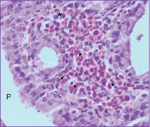
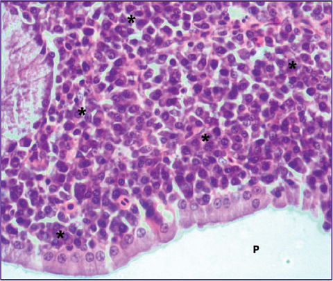
Figure 1. Large number of heterophils (→) and quite few lymphocytes and plasma cells (*) are present in the subepithelial region of the primary duct (P) of the Harderian gland in a 7-day-old chick. (H&E, 400X)
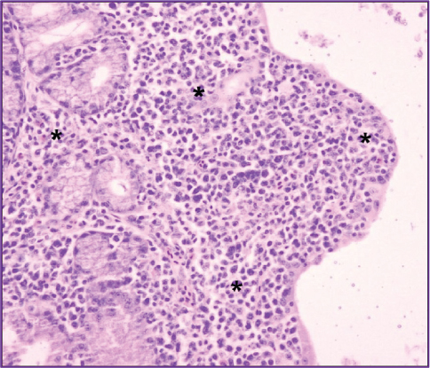
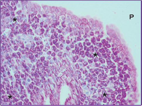
Figure 2. Interstitium of the gland in a 20-day-old chick is infiltrated by large number of lymphocytes (*). (H&E, 200X)
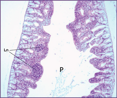
Figure 3. Two large lymphoid nodules (Ln) are visible in subepithelial region of the primary (P) duct in a 40-day-old chick. (H&E, 100X)
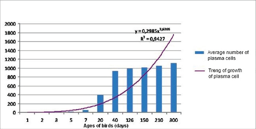
In birds of all ages the lymphoid tissue is colonised by three types of lymphoid cells: heterophils, lymphocytes and plasma cells. These cells are diffusely scattered and occupy mostly the central part of the lobules of the gland, between the primary and secondary ducts, while a far smaller number of them is situated at the periphery, between the acini of the gland. The number of lymphoid cells is directly dependent on the bird’s age. From the time of hatching to the period of 10 months of age (Fig. 7) there gradually comes a shift in the dominance of these three cell types.
Figure 4. Large number of mature plasma cells (*) infiltrates subepithelial region of the primary duct (P) in 7 month old laying hens. (H&E 400X)
Figure 5. Interstitium of the gland in 7 month old laying hens is colonized by large number of mature plasma cells (*) (Methyl green-Pyronin, 400X)
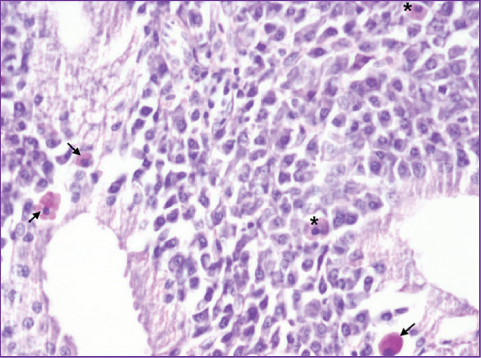
Figure 6. Plasma cells with small, rounded (*) and large, angular (→) Russell bodies in 7 month old laying hens. (Periodic acid-Schiff, 400X)
Figure 7. Increasing number of plasma cells from the time of hatching to the period of 10 months of age
In the first 7 days after hatching, the interstitium of the gland is colonized by large number of heterophil granulocytes (Fig. 1). Only a few lymphocytes and plasma cells are present among them. The first changes inside the lymphoid tissue come in the period between 7 and 20 days. In fact, in 20-day-old chickens the interstitium of the gland is infiltrated by a large number of lymphocytes (Fig. 2).
The number of heterophils in this age period is much reduced and there are also some mature plasma cells. Later, in 40-day-old chickens the typical lymphoid nodules (Fig. 3) are also present. Most of them are bounded by a thin connective tissue capsule and they have well defined outer mantle zone and inner germinal center. Lymphoid nodules are mostly situated in the subepithelial region of the primary ducts, through its entire length, from head to the body of the gland. Outside of them, the stroma contains many mature plasma cells, groups of lymphocytes, and some heterophils. The number of plasma cells continuously increases during the age periods. In hens of 4 months of age and all older birds, plasma cells are so numerous that they represent a dominant population (Figs. 4 and 5). In adult birds we noticed that plasma cells often represent 90% of cells in one microscopic field.
In all birds older than 3 days plasma cells with Russell bodies are also present. These cells always show a strong PAS positive reaction, so that they can easily be noticed (Fig. 6). The nucleus of plasma cells with Russell bodies is very small, pyknotic and situated eccentrically. The Russell bodies are different in size and shape. In some plasma cells they are very small and rounded, while in others they are quite large and have angular shape. Sporadically, in adult birds the group of lymphod cells is occupied with dense PAS positive material. The cells surrounded by this material are often affected with degenerative changes. Also, this material sometimes protrudes under and between the epithelial cells of glandular ducts.
The correlation coefficient r2=0,94 shows that the number of these cells significantly increases (p<0,05) from the time of hatching to the period of 10 months of age (Fig. 7). However, in the period between 4 and 10 months of age this is not a notable growth as it is the case in younger birds. Among numerous plasma cells small groups of lymphocytes and individual heterophils are visible, while the lymphoid nodules are no longer detectable.
Bang and Bang (2) first reported that in the first 7 days after hatching, the interstitium of the Harderian gland is colonised by heterophils, while later with aging, the number of plasma cells progressively increases. Whight et al. (22, 23) confirmed these findings and also reported that in adult birds the Harderian gland contains an exceptionally large number of plasma cells. Recently conducted investigations showed that these are IgA, IgM and IgG containing plasma cells whose number is variable, depending on bird’s strain, ages and health (9, 13, 14, 19-20).
Our findings have shown that from the time of hatching to the period of 10 months of age the lymphoid tissue is colonised by three types of cells: heterophils, lymphocytes and plasma cells. Although these cells have been previously recorded (2, 4, 18, 21-23), we noticed that their number is directly dependent on bird’s ages. In fact, we find that during the lifetime of the laying hens there gradually comes a shift in the dominance of these three cell types. This investigation showed that the gland develops in a similar way as other secondary immune organs.
However, the main morphological characteristic that distinguishes it from others secondary immune organs is the presence of a large number of plasma cells in hens of 4 months of ages and all older birds. This finding indicates that after sexual maturation the Harderian gland in laying hens is mainly responsible for humoral immunity. Additional studies are necessary to confirm this hypothesis. Also, in one of the first research it was assumed that the Harderian gland in chicken is the organ which contains the largest number of plasma cells, probably larger than any other (2). Our histological and statistical findings support this statement. In addition, our statistical results are in agreement with findings obtained by Pawar et al. (15) in White Leghorn and Wight et al. (22) in Shavers hens, indicating similarities between these strains.
Recently conducted research of the Oláh et al. (14) showed that lymphoid tissue of the Harderian gland in domestic fowl histologically can be divided into two major compartments, the head and the body. As previously stated, the head consists of follicle-associated epithelium and subepithelialy situated lymphoid cells and lymphoid nodules. The body is a significantly larger part and it contains a large number of plasma cells in different stages of maturation. However, we did not find these histological differences. Our research shows that lymphoid cells are diffusely scattered through the interstitium of the gland, equally in all its parts. Only in 40-days-old chickens we detected the typical lymphoid nodules. Also, opposite to Oláh et al. (14) we found lymphoid nodules in the subepithelial region of the primary duct, through its entire length, from head to the body of the gland. Lymphoid nodules appear just prior to the start of the tissue infiltration with plasma cells, while later they are no longer detectable. Among mature plasma cells we also noticed plasma cells with Russell bodies. These cells were previously recorded by Ohshima and Hiramatsu (13) and Rothwell et al. (17). However, in addition to the previous, we also noticed that Russell bodies come in two different sizes and shapes.
In conclusion, it could be stated that the Harderian gland in laying hens is the largest intraorbital excretory and secondary immune organ. In sexually mature hens, this gland contains an extremely large number of mature plasma cells.
© 2014 Bejdic P. This is an open-access article published under the terms of the Creative Commons Attribution License which permits unrestricted use, distribution, and reproduction in any medium, provided the original author and source are credited.
We are grateful to the Federal Ministry of Education and Science, Bosnia and Herzegovina which has provided financial support for this research. Also, we are indebted to our colleagues from the Agreks company who have provided all the material for this research.
The authors declared that they have no potential conflict of interest with respect to the authorship and/or publication of this article.
Macedonian Veterinary Review. Volume 37, Issue 1, Pages 83-88, p-ISSN 1409-7621, e-ISSN 1857-7415, DOI: 10.14432/j.macvetrev.2014.02.009, 2014