Mac Vet Rev 2014; 37 (1): 89 - 93
 10.14432/j.macvetrev.2014.02.010
Received: 17 December 2013
Received in revised form: 27 January 2014
Accepted: 05 February 2014
Available Online First: 17 February 2014
Published on: 15 March 2014
Keywords: ultrasonography, teat, cows
Pathological states resulting in disturbed milk release are the main indication for conducting udder ultrasonography. The primary causes for impaired milk ejection are teat damage and the milking technique (1, 4, 8, 11).
The first udder ultrasound in animals was performed by Caruolo and Mochrie (3), studying teats of lactating cows using an A-mode ultrasound apparatus and a 1 MHz probe. The first report for B-mode udder ultrasonography is that of Cartee et al. (2). The authors reported that teat canals, teat and gland cisterns, and the different teat wall layers could be clearly differentiated. Later, a number of researchers have explored the potential of ultrasound for diagnostics of physiological and pathological conditions of bovine udder (7, 9, 10, 19).
The ultrasound scans of teat structures allows for detailed measurement of teat canal length and width, as well as teat wall thickness (13, 21). Numerous researchers use the imaging technique to investigate the changes occurring in teat tissues of cows after milking (14, 17, 22). Gleeson et al. (13) studied the effect of milking on teat tissues in cows and demonstrated that ultrasonography could be used for evaluation of the effect of different milking systems on teat tissue response. Ultrasound measurements of teat length, teat diameter, cistern diameter and teat wall thickness performed by Gleeson et al. (12) were used to investigated the effect of milking cups, pulsation and vacuum level settings in lactating cows. Neijenhuis et al. (16) evaluated the recovery of bovine teats after milking through ultrasonography.
Literature data about teat structures size in lactating cows is inconsistent. Also, to the best of our knowledge, such studies are not available for cows from the Black-and-White breed in Bulgaria. That has been the incentive for the present experiment.
The purpose of the study was to determine the features and size of teat structures in Black-and-White cows through ultrasonography (in the pre and post-milking period).
Twelve clinically healthy Black-and-White cows, 3 years of age, weighing 350-450 kg, fed and housed uniformly, were included in the experiment. Feeding was based on a complete ration including maize silage, alfalfa haylage, concentrated feed, and vitamin mineral premix. The animals were reared in the cattle barn of the Experimental Farm of the Trakia University – Stara Zagora. All cows were primiparous, between the 3rd and 7th months of lactation. Animals were milked in the morning and in the evening by a milking machine (Milk-Rite, USA). The clinical udder examination included inspection, palpation and California Mastitis Test (6). When pathological changes in the udder were detected, the cows were excluded from the study.
Ultrasonography was performed on 48 teats using an ultrasound SonoScape A5v (SonoScape, China) with multifrequency linear transducer (5–12 MHz). The scans were performed via transcutaneous ultrasonography with vertically position of the probe and 12 MHz frequency. For a better contact between the probe and the teat skin, contact gel (Eco-ultra gel, Milano, Italy) was used.
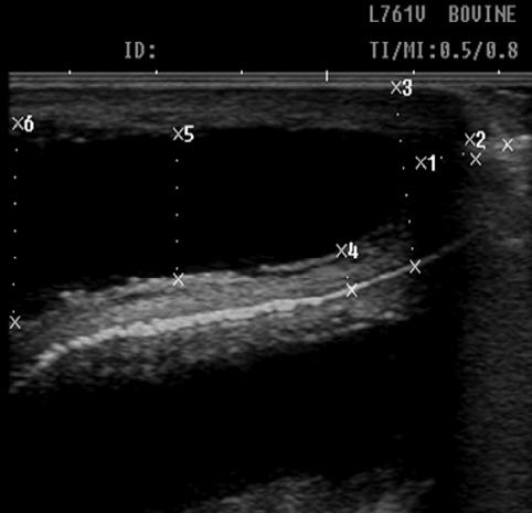
Scans were conducted 4 times - before milking, immediately after milking, 1 hour and 2 hours after milking. After detection of the possibility for teat structure visualization, the following parameters were measured: teat canal length and diameter, teat diameter in the area of the Furstenberg’s rosette teat wall thickness, teat cistern diameter in its middle part and teat cistern diameter in the teat base (Fig. 1).
Figure 1. Ultrasound measurements of studied parameters: 1 – teat canal length; 2 – teat canal diameter; 3 – teat diameter in the region of Furstenberg’s rosette; 4 – teat wall thickness; 5 – teat cistern diameter in its middle part; 6 - teat cistern diameter at teat base
The results were processed with statistical software (StatSoft, Statistica, Microsoft Corp. 1984-2000 Inc.) by means of ANOVA and non-parametric test of proportions, using the Student’s t-criterion. The differences were considered statistically significant at p<0.05.
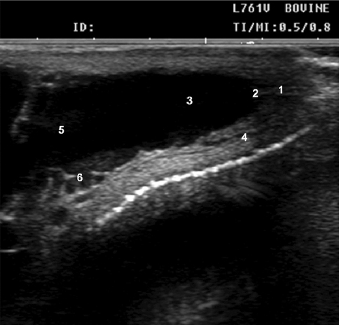
The teat canal was seen as a hyperechoic structure. The transition between the teat canal and the teat cistern, termed Furstenberg’s rosette, was hypoechoic. Three layers of the teat wall were clearly distinguished – outer hyperechoic (skin), hypoechoic (musculature) and inner hyperechoic (mucosa). The lumen of the teat cistern was anechoic when it was full of milk. At the boundary between the teat and the gland cisterns, anechoic blood vessels from the Furstenberg’s venous ring could be observed (Fig. 2).
Figure 2. Visualization of teat structures in cows: 1 – teat canal; 2 - Furstenberg’s rosette; 3 – teat cistern; 4 – teat wall; 5 – transition between teat and gland cisterns; 6 – Furstenberg’s venous ring
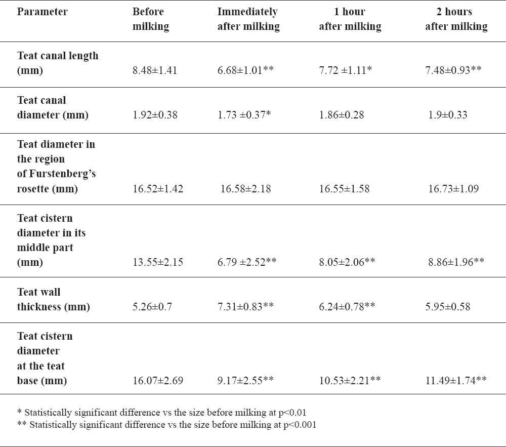
The average size of the studied parameters is presented in Table 1.
Table 1. Ultrasound measurements (Mean±SD) of teat structures in Black-and-White cows (n=48)
Before milking, the average teat canal length as measured by ultrasonography was 8.48±1.41 mm, and until the 2nd post-milking hour, decreased (7.48±0.93 mm) statistically significantly (p<0.001).
The teat canal diameter was 1.92±0.38 mm before milking, significantly lower (p<0.01) immediately after milking (1.73±0.37 mm). By the 2nd post-milking hour, its values were similar to baseline ones.
Teat diameter in the region of the Furstenberg’s rosette did not differ significantly between the individual measurements.
Teat cistern diameter (in its middle part and at the teat base) showed a clear trend towards reduction after milking vs premilking values (p<0.001).
Teat wall thickness was greater immediately after milking and by the 1st hour (p<0.001) compared to baseline size.
The numerous studies during recent years related to ultrasonographic measurement of teat structures in animals affirmed it as one of the main indications of the imaging technique. On the other side, contradictory literature data for the size of measured parameters depending on the breed, number of lactation, stage of lactation, udder health etc. support the necessity for such investigations.
Ultrasonographic characteristics of bovine teats are described in many publications (2, 8, 9, 18, 23), but not for the Black-and-white cows.
Franz et al. (9) observed the teat canal in cows using an 8.5 MHz linear probe as a central hyperechoic line, bounded by two parallel hypoechoic zones. A similar echogenicity pattern was observed by us with the 12 MHz linear probe. Cartee et al. (2), Şendağ and Dinç (20) and Franz et al. (8) affirm that by ultrasound, three different teat wall layers could be distinguished, whereas Couture and Mulon (5) differentiate 5 layers. According to our studies, three layers are clearly seen – outer hyperechoic, middle hypoechoic and inner hyperechoic.
Teat cistern lumen was anechoic as its wall filled with milk. Franz et al. (9) reported that when the cistern is empty, it is not visualized. On both sides of the boundary between teat and gland cisterns, we observed anechoic structures, which, according to Franz et al. (8) represent blood vessels from the Furstenberg’s venous ring.
With regard to the teat canal length, Klein et al. (15) observed statistically significant (p<0.001) breed differences – 15.7 mm in Brown Swiss cattle, 17.2 mm in Holstein-Friesian cattle and 18.3 mm in Simmental cattle. The authors suggest that teat length and diameter are positively correlated to udder health. Teat canal in cows with healthy udders were longer (17.4 mm) and narrower (1.8 mm), as compared with those with cows with inflamed udders (length 15.8 mm; width 2.1 mm).
In our studied Black-and-White cows, the average teat canal length before milking was 8.48±1.41 mm. Similar to ours is the data of Seker et al. (19) for teat canal length of first-lactation cows (0.91±0.04 cm) and cows between the 4th and 7th lactation months (0.95±0.04 cm).
Neijenhuis et al. (16) reported a statistically significant difference (p<0.05) in pre-milking teat canal length (11.2 mm), vs post-milking length (10.0 mm). Contrary to this result, our studies showed convincingly that teat canal length was statistically significantly lower (p<0.001) after milking. Moreover, even 2 hours after milking, teat canal length remained substantially lower (p<0.01). A similar relationship was observed for teat canal diameter. In our belief, this could be attributed to the effect of vacuum and mechanical influence of milking on teat tissues. This assumption is supported by the statistically significant increase (p<0.001) in teat wall thickness after milking (7.31±0.83 mm), in agreement with the reports of others (12, 13).
The teat cistern diameter in the middle and basal parts was considerably lower (p<0.001) after milking, which could be due to milk ejection. This finding is supported by the data of Neijenhuis et al. (16), Gleeson et al. (13) and Gleeson et al. (12). During the teat structure measurements, the only parameters that did not exhibit statistically significant differences before and after milking, was the teat diameter in the region of the Furstenberg’s rosette.
In conclusion, the analysis of results demonstrates that ultrasonography is a rapid and accurate technique for determination of features and size of teat structures. It allows for detection of alterations in teats of cows after milking – teat canal shortening, teat wall thickening and teat cistern diameter reduction.
© 2014 Fasulkov I. This is an open-access article published under the terms of the Creative Commons Attribution License which permits unrestricted use, distribution, and reproduction in any medium, provided the original author and source are credited.
The authors declared that they have no potential conflict of interest with respect to the authorship and/or publication of this article.
Macedonian Veterinary Review. Volume 37, Issue 1, Pages 89-93, p-ISSN 1409-7621, e-ISSN 1857-7415, DOI: 10.14432/j.macvetrev.2014.02.010, 2014