Abstract
This clinical review about the neurological examination in small animals describes the basics about the first steps of investigation when dealing with neurological patients. The knowledge of how to perform the neurological examination is important, however more important is how to correctly interpret these performed tests. A step-by-step approach is mandatory and examiners should master the order and the style of performing these tests. Neurological conditions can be sometimes very distressing for owners and for pets that might not be the most cooperating. The role of a veterinary surgeon, as a professional, is therefore to collect the most relevant history, to examine a patient in a professional manner and to give owners ad educated opinion about the further treatment and prognosis. However neurological examinations might look challenging for many. But it is only the clinical application of neuroanatomy and neurophysiology to an every-day situation for practicing veterinarians and it does not require any specific in-depth knowledge. This clinical review is aimed not only to provide the information on how to perform the neurological examination but it is also aimed to appeal on veterinarians to challenge their daily routine and to start practicing on neurologically normal patients. This is the best and only way to differentiate between the normal and abnormal in a real situation.
Keywords: neurological examination, dogs, cats, hands off examination, hands on examination
INTRODUCTION
Veterinary medicine has been developing very rapidly over the last decades and the owners of pets are more demanding for veterinary care. The veterinarians are therefore facing more challenging conditions in companion animals and are expected to make the correct diagnosis, give the correct prognosis to the owners and eventually to suggest the most appropriate treatment or recommendation. Neurological conditions are not more common than in the past but are more recognised and investigated because of this higher demand. The knowledge about the neurological condition in small animals is rapidly evolving and it is therefore expected that veterinarians keep up-to-date with this, generally considered “tough” field of veterinary medicine.
One would think that to make the correct diagnosis and to recommend the appropriate treatment for neurological conditions is not possible without sophisticated tools like Computed Tomography (CT) or Magnetic Resonance (MR). But this is not always true and to have a thorough knowledge about basic neurology is often necessary in order to make an educated judgment about the most likely diagnosis, severity of the condition and the prognosis.
The aims of the neurological examination is to answer the following questions:
-
Are we dealing with the primary neurological condition or not? So is this the neurological condition, or the condition affecting the function of the nervous system, or is this a completely different condition (orthopaedic, cardiovascular etc.)?
-
Can we localise the lesion within the nervous system?
-
What are the most common differential diagnoses?
-
How severe is the condition?
The first two questions are usually answered by the neurological examination. The third question is answered by combining the neurological examination, neurolocalisation and history. And the last question can help the clinician to advise owners about the prognosis and further diagnostic work-up (
1-
3).
History
It is very important to take a thorough history as this can give many clues in making the most likely differential diagnoses. It is important to know species, breed, sex and age of animals before taking the history. The signalment may influence the primary differential diagnosis. The veterinarian needs to carefully question owners about the main complaint. It is relatively easy to lose track from the important information and then to make the wrong judgement. The onset, evolution and course of the illness are most important for making the most likely differential diagnoses. The onset of the neurological signs should be defined as:
-
Acute (onset over minutes to hours)
-
Subacute (onset over days)
-
Chronic (onset over several days, weeks or months)
-
Episodic (the patient returns to normal between the episodes)
The evolution is described as progressive, static, improving or waxing and waning.
Physical examination
Complete physical examination needs to be performed before the neurological examination. It is of upmost importance to do so as many conditions that are not primarily neurological can be discovered here and the diagnostic work-up can take a different direction or can alter the final prognosis. An example would be a dog with paroxysmal episodes or suspected seizures in which a significant cardiac arrhythmia is found and the episodes might be a cardiac syncope. It is also important in the case with traumatic injuries, where the multiple injuries (e.g. diaphragmatic hernia, ruptured viscera, ruptured urinary bladder, multiple pelvis fractures etc.) can be discovered and this can alter the final prognosis and the primary goal for stabilising these patients in the emergency situation.
Neurological examination
The neurological examination should be performed in animals that are not sedated, have not received any analgesia or are recovering from seizures or general anaesthesia.
Hands off examination
This part of the neurological examination can be performed while collecting the history. The patient should be left to explore the examination room. The clinician can observe the awareness, behaviour, posture and gait in an undisturbed manner.
Consciousness, awareness, behaviour
State of consciousness is classified in order of severity as lethargy, depression, obtundation, stupor (semicoma) and coma. Generally if there is an altered state of consciousness then the lesion is affecting either diffusely both cerebral hemispheres or focally the ascending reticular activating system (ARAS) of the brainstem.
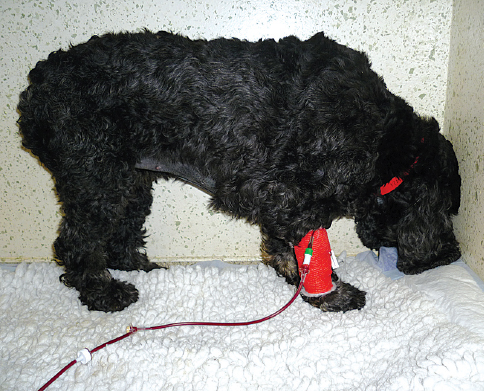
Changes in the patient’s level of awareness and behaviour include disorientation, delirium, aggression, compulsive walking, loss of learned behaviour (e.g. in-house urination, defecation etc.), vocalising and head pressing (
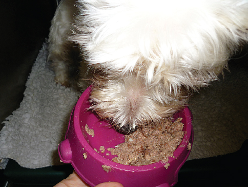
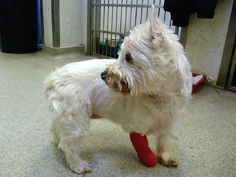
Fig. 1). Hemi-neglect or hemi-inattention syndrome is the abnormal behaviour in animals with forebrain lesions. The lesion in the forebrain is contralateral to the apparently “ignored” side by the animal (
Fig. 2).
Figure 1. Head pressing in a 4-year-old female neutered Cocker Spaniel with meningoencephalitis of unknown aetiology, immune-mediated hemolytic anaemia and immune-mediated thrombocytopenia. She was diagnosed with systemic lupus erythematosus.
Figure 2. Hemi-inattention syndrome in a 2-year-old female West Highland White Terrier with granulomatous meningoencephalitis. Please note that she is completely oblivious to food in the left side of the bowl. There must be a lesion affecting predominantly the right forebrain.
It should be remembered that extracranial diseases can influence the forebrain function, altering the behaviour and consciousness level (e.g. hepatic encephalopathy due to portosystemic shunt, hypoglycaemia, hypokalemia, hypernatremia etc.)
Posture and body position
Observation of the posture and body position at rest can reveal mild asymmetry and can also assess the balance at the stance. Common abnormalities are:
-
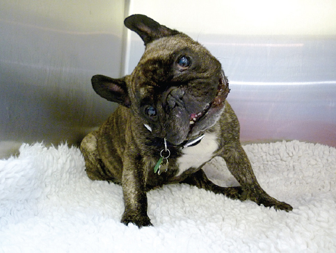
Head tilt - abnormal posture of the head when one ear is lower compared to the other one. A head tilt indicates a vestibular disorder (central or peripheral) (Fig. 3).
Figure 3. Right side head tilt and also ventro-lateral strabismus more appreciated in the left eye in a 2-year-old female neutered French Bulldog with granulomatous meningoencephalitis.
-
Head turn - characterised by the posture when the nose and often whole body (pleurothotonus) are turned to one side and ears are at the same median plane. This is most commonly associated with an ipsilateral forebrain lesion. (Fig. 4).
Figure 4. The same dog as in figure 2. Please note the head turn to the right side that confirms that the lesion is predominantly affecting the right forebrain.
-
Ventroflexion of the head - commonly associated with a neuromuscular disorder or spinal cord grey matter lesion.
-
Spinal curvature
-
Scolisosis (lateral deviation of the spine)
-
Lordosis (ventral curvature of the spine)
-
Kyphosis (dorsal curvature of the spine)
-
Torticollis (twisting of the neck)
-
Decerebrate rigidity - a posture when the patient is recumbent and has extension of all limbs and opithotonus (extension of the neck and head). The mental status is often stuporous or comatous and the lesion is commonly localised in the rostral brainstem.
-
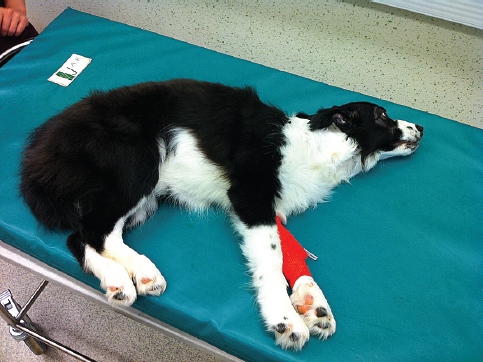
Decerebellate posture - a posture when the patient is recumbent, has extended thoracic limbs and opisthotonus but the pelvic limbs are usually flexed. The mental status is normal and the lesion is likely to occur in the cerebellum (Fig. 5).
Figure 5. Decerebellate rigidity in a 6-month-old female Border Collie with cystic lesion in the fourth ventricle.
-
Schiff-Scherrington posture - observed in animals with severe thoracic or cranial lumbar spinal cord trauma. The animal has extended thoracic limbs with the normal function but has paralysis of the pelvic limbs. This sign is present only in acute lesions and does not have any prognostic value.
Evaluation of gait
Abnormalities of the gait are one of the most common reasons to seek veterinary advice. It is therefore important to assess whether the animal is uncoordinated (ataxia), has an abnormality in the strength of voluntary movement (paresis) or is lame (neurological or orthopaedic).
Ataxia means uncoordinated gait. Ataxia can be a consequence of peripheral nerve or spinal cord dysfunction (general proprioceptive ataxia), vestibular system (vestibular ataxia) or cerebellum (cerebellar ataxia).
Paresis is defined as a loss of ability to support weight or to generate the gait. Plegia or paralysis refers to the complete loss of a voluntary movement, whereas paresis implies that the voluntary movements are still present. The animal can be tetraparetic (affected all four limbs), paraparetic (affected pelvic limbs), monoparetic (affected one limb) or hemiparetic (affected only one side of the body). There are clinical differences if the lesion is affecting the upper motor neuron (UMN) or lower motor neuron (LMN). An UMN lesion causes a delay in the onset of the protraction, which clinically looks like a stride being longer than usual. Whereas a LMN paresis causes difficulties in weight bearing which clinically looks like a short-strided and choppy gait with inability to support weight.
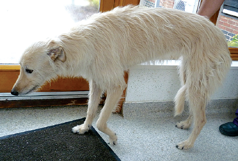
Lameness presents as a short stride of the lame limb and long stride in the contralateral limb. It is more commonly associated with orthopaedic diseases but diseases affecting the nerve root(s) can cause the same presentation (nerve root signature) (
Fig. 6).
Figure 6. Nerve root signature of the left thoracic limb in a 9-year-old female neutered Lurcher with C7 vertebral body tumour impinging left C8 nerve root.
Hands on examination
Cranial nerve examination
Olfactory Nerve – (CN I)
It is generally difficult to asses the smell in small animals and in a majority of cases it is the owners who complain about the loss of smell (anosmia) rather than the finding on neurological examination. Letting the animal to sniff something aromatic while blindfolded can test the smell. Irritating substances should not be used as this can stimulate CNV.
Optic Nerve (CN II)
This is not a peripheral nerve by strict definition but it is the extension of the brain. CNII is an important central visual pathway and it is afferent for menace response and pupillary light reflex (PLR).
The visual pathway has three important neurons(
1).
-
The bipolar cell in the retina is the first one in the row. This cell receives the information from neuroepithelial cells (i.e. rods and cones)
-
The ganglion cell in the retina is the second one. The axons of these cells form the optic nerve and the majority of them cross to the contralateral site in the optic chiasm (66% decussation in cats and 75% in dogs)(4).
-
The neuron body in the lateral geniculate body in the diencephalon is the last one in the row. The axons project to the visual cortex (mostly the occipital lobe – which is mostly contralateral to the stimulated retina).
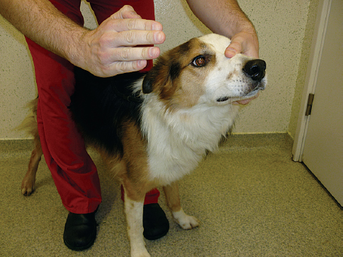
Menace response (
Fig. 7) is learned and cortically mediated blink produced by a threating gesture in front of the visual area of the patient. Puppies will not have this response prior to 10-12 weeks of age. The afferent part is the same as the visual pathway and the efferent is very complex but involves coordination of the visual cortex, motor cortex, facial nerve nucleus, cerebellum and facial nerve. The absent menace response can be a result of any of the parts involved in this pathway and does not always mean that the animal is blind.
Figure 7. The menace response is performed by making the threating gesture at the eye. The contralateral eye should be blinded. Care must be taken not to touch the eyelashes or to create air current as this stimulates the CN V and produces the palpebral or corneal reflex rather then genuine menace response.
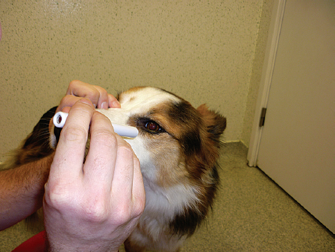
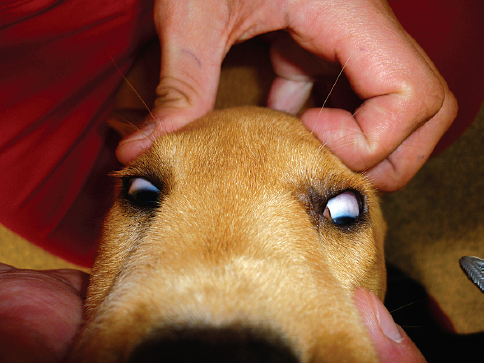
Pupillary light reflex (PLR) (
Fig. 8) has some parts in common with the afferent part of the visual pathway. The axons involved in the vision pathway reach the cortex via the lateral geniculate body, however axons involved in PLR have the third neuron in the pretecal nucleus. From there, most of the axons cross again to the parasympathetic nucleus of the oculomotor nerve (ipsilateral to the stimulated retina). The oculomotor nerve then constricts the pupil. This is a direct PLR. Consensual PLR (constriction of non stimulated eye) is explained by partial crossings along the PLR pathway (optic nerves in optic chiasm and axons from the pretecal nucleus in mesencephalon). PLR should be performed in all blind animals, as it shares only part of the pathway for vision.
Figure 8. The pupillary light reflex (PLR) is tested by shining a direct light into the eye. The normal response is the constriction of the ipsilateral pupil (direct PLR) and also contralateral pupil (consensual PLR).
Fundic examination should be also performed in all animals to visualise the optic nerve
Oculomotor Nerve (CN III)
This nerve innervates ipsilateral dorsal, ventral and medial recti muscles and ventral oblique muscle. It also innervates the levator palpebrae superioris muscle which is important for upper eyelid movement. And finally the oculomotor nerve plays an important role as an efferent arm of PLR. It controls the pupillary constriction by its parasympathetic component.
By observing the eyeball position and movement of the eyeball by testing for physiological nystagmus (see the vestibulocochlear nerve) this nerve can be easily assessed. Another observation needs to be done by assessing the normal position of the upper eyelid. PLR of course must be assessed and if non functional then thinking about the full PLR pathway needs to be remembered in order to localise the problem along this pathway.
An oculomotor nerve lesion results in ventrolateral strabismus and an inability to rotate the eye dorsally, ventrally and medially. It can also produce unresponsive mydriasis and narrowing of the palpebral fissure (ptosis of the upper eyelid). (
Fig. 9)
Figure 9. Ptosis of right upper eyelid and narrowing of the palpebral fissure in a 10-year-old male neutered Staffordshire Bull Terrier with right side CN III peripheral nerve sheath tumour.
Trochlear nerve (CN IV)
This is assessed by observing the position of the eyeball as well as by testing for physiological nystagmus. This nerve innervates contralateral dorsal oblique muscle. Dysfunction usually results in dorsolateral strabismus of the contralateral eye.
Trigeminal nerve (CN V)
The trigeminal nerve provides sensory innervation of the face as well as motor innervation of the masticatory muscles. It has three major branches:
-
Ophthalmic branch – innervates medial canthus of the eye, nasal septum, cornea and dorsum of the nose.
-
Maxillary branch – innervates lateral canthus, skin of cheeks, muzzle, palate and teeth of the upper jaw.
-
Mandibular branch – innervates mandibular area of the oral cavity.
The motor function is assessed by evaluating the symmetry and size of the masticatory muscles as well as by opening the jaw. The sensory function is assessed by corneal reflex which is done by touching the cornea with a sterile cotton bud. The palpebral reflex tests ophthalmic and maxillary branches (afferent arm of the reflex) by touching medial or lateral canthuses, respectively. A normal response for corneal and palpebral reflex is the blink of the tested eye that is mediated by the facial nerve (efferent arm of the reflex). Other tests that can assess the trigeminal nerve are nasal stimulation and pinching of the skin of the face that results in the ipsilateral blink or twitch of the facial muscles.
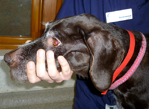
Unilateral dysfunction of the motor part results in unilateral masticatory muscle wastage, (
Fig. 10) whereas bilateral dysfunction results in the dropped jaw and inability to close the jaw voluntarily. Dysfunction of the sensory part results in facial hypoesthesia or anaesthesia and can also result in decreased tear production and neurotropic keratitis.
Figure 10. Unilateral wastage of the masseter and temporal muscles in an 8-year-old male neutered German Wire Haired Pointer with left side CN V peripheral nerve sheath tumour.
Abducent nerve (CN VI)
This nerve innervates the ipsilateral lateral rectus and retractor bulbi muscles. The assessment is therefore done by observation of the eye position, testing the physiological nystagmus and by corneal reflex (retracting of the eyeball). Dysfunction results in ipsilateral convergent strabismus, inability of the eye to cross the midline when testing physiological nystagmus and inability to retract the eyeball.
Facial nerve (CN VII)
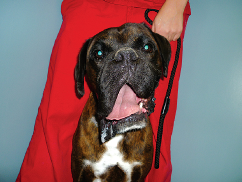
This nerve provides motor function to the muscles of the face and sensory function to the rostral two thirds of the tongue and palate. The facial nerve also carries the parasympathetic component that innervates the lacrimal glands, and mandibular and sublingual salivary glands. The motor function is assessed by observation of the symmetry of the face and spontaneous blink and movement of the nostrils. The facial nerve provides the efferent arm for palpebral reflex, corneal reflexes and menace response and can be assessed by performing these tests. The Schirmer tear test should be performed to assess the parasympathetic part of this nerve (
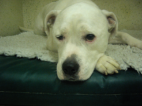
Fig. 11). Unilateral dysfunction produces the ipsilateral drooping of the face, inability to move the ear and nostril, widened palpebral fissure and absent blinking response (
Fig. 12). It can also produce keratoconjunctivitis sicca by inability to produce enough tears by loss of parasympathetic innervation to the lacrimal glands.
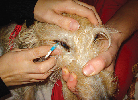
Figure 11. Schirmer tear test is an important part of neurological examination in dogs and cats.
Figure 12. Right side dropping of the lips and ear in a 4-year-old female neutered Boxer with right side idiopathic facial neuritis.
Vestibulocochlear nerve (CN VIII)
This nerve is involved in hearing and vestibular function. The vestibular system consists of the peripheral part (special proprioceptors in the inner ear and vestibular nerve) and central part (four nuclei in the brainstem and part of cerebellum). The hearing part involves the sensory receptors in the cochlea (inner ear). The input from the cochlea is then transmitted though the cochlear nerve, brainstem and midbrain to the contralateral auditory cortex, mainly in the temporal lobe.
Observation of the gait, body and head posture can give a lot of information about the vestibular function. Specifically physiological nystagmus can test the functional integrity of the vestibular system. This involves moving the head from side to side and up and down. A normal response is the involuntary “jerk” movement of both eyes to correct their position in relation to the position of the head.
To assess the hearing part is difficult but whistling or a handclap can be used to assess this.
Dysfunction of this nerve usually results in a head tilt (
Figure 3), falling to the side, leaning to the side, rolling, circling, pathological (abnormal) spontaneous or positional nystagmus, positional strabismus (
Fig. 13) or asymmetrical ataxia. The clinical signs can be a result of dysfunction of either the peripheral or central part. The presence of pathological nystagmus indicates vestibular dysfunction, however in some cases of bilateral vestibular dysfunction this might not be present. In this case wide based stance and symmetrical ataxia with inability to elicit physiological nystagmus is usually present.
Figure 13. Positional predominantly left side ventro-lateral strabismus in a 9-month-old male neutered Labrador Retriever with meningoencephalitis of unknown aetiology.
Glossopharyngeal nerve (CN IX) and Vagus nerve (CN X)
These two cranial nerves share the same motor and sensory nuclei in the brainstem. The glossopharyngeal nerve supplies the musculature of the pharynx and palate and it provides sensory innervation to the caudal third of the tongue and pharyngeal mucosa.
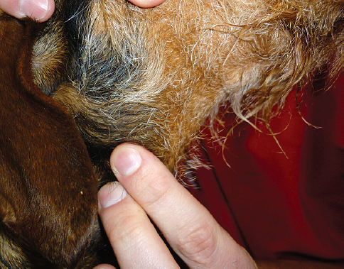
The parasympathetic part innervates parotid and zygomatic salivary glands. The Vagus nerve supplies musculature of the larynx, pharynx and oesophagus and it provides sensory innervation of larynx, pharynx and thoracic and abdominal viscera. The parasympathetic part of the nerve supplies all thoracic and abdominal viscera, except those of the pelvic region. The pharyngeal or gag reflex can assess the function of both nerves. Gently applying pressure to the thyroid cartilages provokes swallowing in a normal animal (
Fig. 14). Observing a patient while eating or drinking can also provide useful information about the function of both nerves.
Figure 14. Gag reflex can be elicited by applying gentle pressure on the thyroid cartilage and hyoid bone.
Dysfunction results in dysphagia, absent gag reflex, inspiratory dyspnea (due to laryngeal paralysis), voice change and regurgitation (due to megaoesophagus).
Accessory nerve (CN XI)
This nerve supplies motor innervation to the trapezius, sternocephalicus and brachiocephalicus muscles and so the dysfunction results in atrophy of these muscles and potential deviation of the neck. However isolated lesions of this nerve are rare.
Hypoglossal nerve (CN XII)
This nerve innervates the muscles of the tongue. Function of this nerve can be assessed by observing for symmetry of the tongue and movement of the tongue during the eating, or licking of food. Lesions of this nerve result in problems with prehension and mastication. Asymmetry of the tongue and fasciculation of the musculature of the tongue can also be seen.
Postural reactions
This part of the neurological examination is important in distinguishing neurological disorders from diseases of other body systems. Abnormality of this group of tests indicates a neurological lesion, however it does not localise the lesion within the nervous system. Proprioreceptors are specific receptors sensitive to detect the movements. They are located in joints, tendons and muscles (general proprioception) as well as in the inner ear (special proprioception). The responses have a complex pathway, but generally involve the afferent arc (proprioreceptors, peripheral sensory nerve, ascending spinal cord tracts and contralateral somatic sensory cortex) and the efferent arc (contralateral motor cortex, descending spinal cord tracts, peripheral motor nerve and the skeletal effector muscle).
Proprioceptive placing
This test is designed to evaluate the conscious awareness of limb position and movement in space. It is evaluated by flexing the patient’s paw so that the dorsal surface contacts the floor. It is important to support the patient with an arm under the abdomen if the patient is too weak (
Fig. 15). A normal response is immediate correction to the normal position. Another test involves putting the patient’s paw on a piece of paper and sliding the paper laterally. A normal patient will reposition its leg when the limb reaches an abnormal position. An abnormal reaction is delayed correction of the tested paw.
Figure 15. Proprioceptive placing is tested by placing the paw in the abnormal position, the patient’s body needs to be sufficiently supported with the other arm.
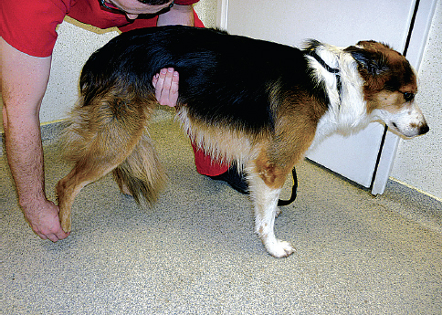
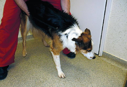
Hopping reaction
Hopping reaction is performed by holding the patient in a way so that the majority of its weight is placed on one limb (
Fig. 16). The animal is then moved laterally (do not move medially as this may cause an abnormal reaction even in a healthy animal) and the normal response is to “hop” and correct the centre of gravity when displaced laterally. An abnormal reaction is a delayed “hop”, however animals with severe orthopaedic disease will have some difficulties if the body weight is not supported sufficiently.
Figure 16. The hopping testing of the right thoracic limb. The majority of the weight is put on the tested limb and the animal is moved laterally.
Placing response
Visual and tactile placing are other complex postural reactions that can be used mainly if no other abnormalities are identified on previous postural tests. Visual placing also assesses also the vision whereas the tactile placing is tested with the eyes covered. The animal with covered eyes is lifted and then brought to the edge of the table so that the distal parts of the limbs touch the table. The animal should then place the limb onto the table. Visual placing allows the animal to see the table so it can reach and step before the limb touches the edge of the table. An abnormal reaction is absence or delay of this response.
Wheelbarrowing, Hemi-walking and Extensor postural thrusting are other complex postural reactions. I do not generally perform these tests unless I cannot find any abnormalities of the above-mentioned tests.
Spinal reflexes
Spinal reflexes evaluation needs to be done in conjunction with assessment of gait and postural reactions. The aim of the assessment of spinal reflexes is to narrow down the problem within the spinal cord segments or problems of the peripheral nervous system. The spinal cord segmental in small animals can be divided into four regions.
-
Cranial cervical (C1-C5)
-
Cervicothoracic (C6-T2)
-
Thoracolumbar (T3-L3)
-
Lumbosacral (L4-S3)
If the lesion that causes spastic tetraparesis is localised in the C1-C5 region then the spinal reflexes usually will be increased or intact, the lesions of C6-T2 that causes tetraparesis will usually produce increased or intact reflexes in pelvic limbs but decreased or absent in thoracic limbs. T3-L3 lesions that cause spastic paraparesis will usually cause increased or intact reflexes in pelvic limbs, whereas the lesion of L4-S3 that cause paraparesis will usually cause decreased to absent spinal reflexes of pelvic limbs. I need to stress that these reflexes must be evaluated in conjunction with evaluation of the gait and postural reactions. The spinal reflexes are decreased or absent if the lesion is affecting peripheral nerve or generally the peripheral nervous system. If the peripheral nervous system is affected then the animals will suffer flaccid tetraparesis.
The most reliable spinal reflexes of the limbs that are used in small animals are withdrawal reflexes of thoracic and pelvic limbs and the patellar reflex (
1,
4). There are other spinal reflexes (extensor carpi radialis reflex, biceps brachii and triceps reflex, cranialis tibialis and gastrocnemius reflex) that can be used but they are generally less reliable and I do not perform them routinely unless specifically indicated.
Withdrawal reflex in the pelvic limbs
This reflex evaluates the integrity of the L4-S2 spinal cord segment and sciatic and femoral nerves. In order to perform this test the digit of the paw needs to be pinched with the fingers (
Fig. 17). The normal response results in the flexion of the hip (femoral nerve), stifle and hock (sciatic nerve).
Figure 17. Withdrawal reflex tested on the right pelvic limb. Please note that all joints are sufficiently flexed after the toe was pinched.
Patellar reflex
This is a monosynaptic reflex that evaluates integrity of the L4-L6 spinal cord segment. The animal needs to be placed in to lateral recumbency with slight stifle flexion. The limb should be held in a neutral position with the examiner’s hand supporting the tested limb. The reflex hammer then hits the patellar tendon and extension of the limb should be observed (
Fig. 18).
Figure 18. Patellar reflex is tested by hitting the patellar tendon with the reflex hammer. The tested limb is supported by the other arm in a neutral position.
In some older dogs this reflex can be weak with no clinical significance. In some excited animals with no neurological dysfunction this reflex can appear hyperreflexive and this is again of no clinical significance.
Withdrawal reflex in the thoracic limbs
This reflex evaluates the integrity of the C6-T2 spinal cord segment and brachial plexus and peripheral nerves in the thoracic limb. Pinching of the digits needs to be performed and the flexion of all joints is considered to be a normal response.
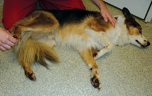
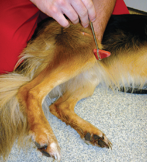
Perineal reflex
This reflex is often overlooked and an important part of the neurological examination. Stimulation of the perineum with the haemostat should result in the contraction of the anal sphincter and flexion of the tail (
Fig. 19). This reflex tests the integrity of the S1-Cd5 spinal cord segment and the pudendal nerve.
Figure 19. Perineal reflex should not be omitted during the neurological examination. It is done by gently pinching the perineal area.
Urinary bladder palpation
Palpation of the urinary bladder should not be omitted during the neurological examination. The innervation is very complex and involves normal sympathetic, parasympathetic and somatic nerve function. The normal function of the urinary bladder is beyond the scope of this review. Flaccid urinary bladder that is easily expressed is called lower motor neuron bladder and suggests an S1-S3 spinal cord segment lesion, whereas the full and turgid urinary bladder that is not easy to express and has overflow leakage of the urine indicates an upper motor neuron disorder. The abnormal function of the urinary bladder can as well be the result of dysfunction of the autonomic nervous system.
Sensory evaluation
Sensory part of the nervous system can be tested with postural reactions but also with the testing for pain perception (nociception). Assessment of the pain sensation requires a noxious stimulus and appropriate response of the animal.
Nociception testing
The sensory part of the peripheral nerve, the spinal cord and the brainstem must be intact to relay the nociception to the cerebral cortex. The nociception pathways are located deep in the white matter of the spinal cord and they also form multiple synapses along the length of the spinal cord. It is therefore an important test to do in the cases of spinal cord diseases because it reflects the severity of damage to the spinal cord. The noxious impulse (squeeze of the toe with the fingers or haemostat) is applied to the tested area and the animal must show a behavioural response (turning the head, trying to bite, vocalisation) to say that the nociception is intact (
Fig. 20). The most common mistake is to confuse the withdrawal reflex (flexion of the limb) with the conscious perception of the pain (behavioural response). Many animals that have lost nociception would still have intact withdrawal reflex and it is therefore important to distinguish these two.
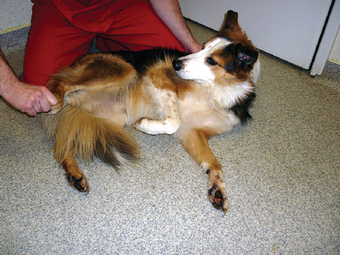
Figure 20. ‘Nociception is tested by applying the noxious stimulus to one of the digits in this patient. Please note that the dog seems to be uncomfortable and aware of the painful stimulus by looking and turning its head towards it. (Please compare it to the withdrawal reflex in
figure 17., when the patient is completely oblivious to the stimulus and only flexes the joints in the tested limb).
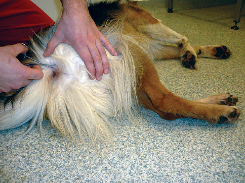
Cutaneous trunci reflex (panniculus)
This reflex is performed by pinching the skin of the dorso-lateral aspect of the body between T2 and L4-L5. The afferent arm tests the sensory nerves of the skin and the efferent motor part is emerging from the C8 nerve root and innervates the cutaneous trunci muscle on the side of the body. A normal reaction to the pinch of the skin is a twitch of the skin (bilaterally, but more prominent on the tested side) (
Fig. 21).
Figure 21. Cutaneous trunci reflex is tested by gentle pinching of the skin on both sides of the patient’s back with a hemostats.
If this reflex is lost completely along the whole spine then a lesion of C8 should be considered (brachial plexus), however in the absence of other neurological deficits this means very little as the absence can sometimes be observed in normal animals. With a presence of the spinal cord lesion there might be a cut off of this reflex in the certain area. In that case this reflex is sometimes absent caudal to the lesion but present cranial to this lesion. This is sometimes very useful to characterise better the exact localisation of the lesion in the T3-L3 region of the spinal cord.
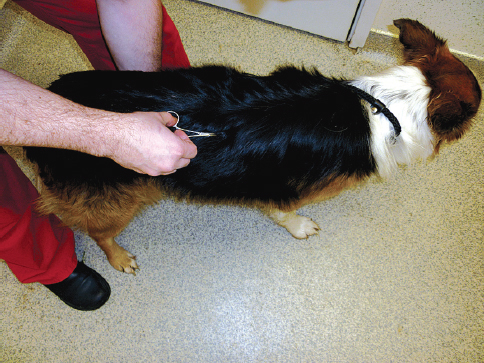
Palpation
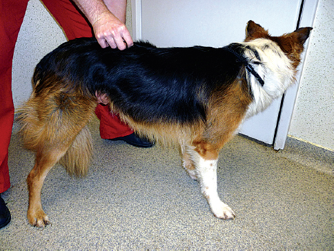
Palpation and manipulation is important to detect the muscle loss of the limbs and to detect the painful areas. It is important to palpate the head (assess the muscle mass, asymmetry, focus of pain or persistence of fontanels), spine (pressure to the spinous processes and then transverse processes can detect spinal hyperaesthesia) (
Fig. 22) and limbs (joint swellings, contractures of the muscles or loss of the muscle mass can be detected) to finalise the neurological examination.
Figure 22. Palpation of spine and potentially painful areas should be performed at the end of examination. Please note that while the spinous processes are palpated there is the other arm of the examiner under the abdomen. When a spinal hyperaesthesia is present patients sometimes respond by tensing up the abdominal musculature.
It is important to do this at the end of the examination as this can be painful for the animal and doing this earlier can discourage the animal to cooperate.

 10.14432/j.macvetrev.2014.02.011