Mac Vet Rev 2014; 37 (2): 129 - 134
 10.14432/j.macvetrev.2014.05.015
Received: 26 February 2014
Received in revised form: 02 April 2014
Accepted: 12 May 2014
Available Online First: 16 May 2014
Published on: 15 October 2014
Keywords: mammary tumours, bitches, cytology, histopathology
The mammary gland is a compound tubuloalveolar gland, divided into lobules by interlobular connective tissue. The mammary gland consists of parenchyma (alveoli), stroma (connective tissue), ducts, vessels and nerves. Bitches usually have five pairs of glands. The cranial two pairs are referred to as cranial and caudal thoracic mammary glands, the middle two pairs are referred to as cranial and caudal abdominal mammary glands, and the caudal pair is referred to as the inguinal mammary glands. After the skin, mammary gland is the second most common site for tumour development in dogs. Mammary tumours are the most frequent neoplasms in bitches. They represent approximately 42% of all tumours and 82% of those arising on the female reproductive organs (1, 2).
Benign mammary tumours appear earlier in life than malignant ones, and younger animals usually present with dysplasia or hyperplasia (3). Mammary gland tumours are more frequently found in bitches at the age of 5 years and older. Many studies have shown that dogs with malignant tumours were significantly older than dogs with benign tumours (4). Breed predisposition has been reported, but varies in different studies. Dachshunds, Cocker Spaniels, toy poodles, German Shepherds, mixed – breed dogs have been reported to have an increased incidence of mammary neoplasia (5). Mostly affected parts are caudal abdominal and inguinal mammary glands. Multiple tumours have been seen in 50% to almost 70% of dogs with mammary tumors (2, 4, 6). In case of multiple tumours, different tumour types may be present within one animal. According to some authors 50% of the tumours are malignant, the majority of malignant mammary tumors in dogs are carcinomas and <5% are sarcomas (7).
Group of authors have concluded that malignant tumours are significantly larger than benign tumours. Also, evidence of histological progression towards malignancy has been seen with increasing tumour size. These findings suggest that mammary tumours can progress from benign to malignant (4).
The action of ovarian hormones (estrogens and progesterone) on mammary gland tissue during different stages of development is a risk factor associated with the development of mammary tumors. A preventive effect of spaying on the development of mammary tumors has been reported. Early ovariectomy in dogs and cats has a protective effect against both benign and malignant mammary tumours. (8) The risk of developing a mammary tumour increases as the number of estrous cycles increases. The risk of developing mammary gland tumours is 0.05% if the bitch is spayed prior to the first estrous cycle. An increased incidence of tumour development also has been observed in dogs that received injectable progestins for the prevention of estrus (9).
It has been reported that most of the dogs are clinically healthy when they are initially presented for clinical examination (2). If metastases are present, dogs may show nonspecific symptoms such as fatigue, lethargy, weight loss, dyspnoea, cough, lymphoedema or lameness. The extent and location of the metastases determines the occurrence and severity of the clinical signs (2, 6). Initial assessment of canine mammary tumour is based on basic patient data (age, age at ovariohysterectomy, history of clinical signs duration, reproductive cycles, lactation, synthetic progestagens therapy), general condition and physical examination (2, 6, 7). Surgery remains the basic treatment for dogs with most common type of mammary gland tumours (benign mixed tumor, adenoma, and adenocarcinoma). The adjuvant therapies (radiation therapy and chemotherapy) are used for inoperable tumours and in inflammatory carcinoma. The survival time after the mastectomy and the possibility of metastases have been reported as main clinical complications (10).
The aim of this retrospective study was to present clinical cases of canine mammary tumours recorded at the University Veterinary Hospital in Skopje in the period of two years, to evaluate the type of mammary tumours in the patients; to analyze the relationship between tumor incidence and the age of the patients, heat cycles and sterilization, as well as the survival period after surgery.
During a period of two years, canine patients have been submitted to medical and surgical treatment after diagnosing various types of tumours at the University Veterinary Hospital at the Faculty of veterinary medicine in Skopje. Ten patients were diagnosed with mammary tumours.
Breed, age, location of the affected mammary glands, tumour size (maximal diameter of the tumour), period (in days) between the first detection by the owners and the time the patient was brought for treatment, reproductive history (spaying status, age at spaying, whelping and history of false pregnancy), application of exogenous hormones and type of surgery (radical mastectomy) were obtained from review of medical records. Patients were thoroughly examined (assessment of general condition, temperature, pulse, respiration, palpation of the lymph nodes, CBC and serum biochemical profile (ALT, AST, AP, creatinine, urea).
After the initial examination and the lab results, fine needle aspiration (FNA) was performed using a 22-G needle on each mammary mass, in order to make the initial determination of the mass. At least 4 samples from different areas were obtained from each mass and at least 1 slide was prepared in a standard procedure for each aspirate. The slides were air-dried and stained with Diff Quick staining (Merck, Darmstrad, Germany) and evaluated under a light microscope (Nikon; 40 X magnification).
In addition, chest radiographs were taken before the planned surgical treatment. Surgical removal of the masses was treatment of choice (complete mastectomy with or without ovariohisterectomy). The owners were offered an adjuvant therapy (chemotherapy) however, none of them accepted the procedure due to the side effects of chemotherapy as well as the high price of chemotherapeutic medications. Histopathological examination of the excised tumours was performed at the Department of Pathology at the Faculty of Veterinary Medicine in Skopje. Following surgical excision, tissue samples were fixed in 10% formaldehyde, embedded in paraffin, cut into 4 µm sections, and stained with hematoxylin and eosin. Histological evaluation was performed according to the WHO classification scheme for canine mammary tumours. Canine mammary tumours are histologically classified into four categories: malignant tumours, benign tumours, unclassified tumours, and mammary hyperplasia/dysplasia (11).
The majority of the patients (6 out of 10) with diagnosed mammary tumours were at the age of 12 – 13 years. The youngest patient with mammary tumour was 6 years old. From a reproductive point of view, five of the patients with malignant mammary tumours have never been whelped and never treated with any exogenous hormones; one was treated with synthetic progesterone before every cycle, two were with unknown history. The patients were of different breed (Doberman Pincher, French Poodle, Miniature Schnauzer, Dalmatian, Cocker Spaniel, Irish Setter, Belgian Sheepdog and two of mixed breeds).
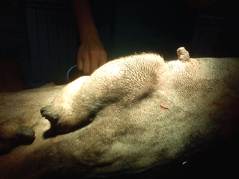
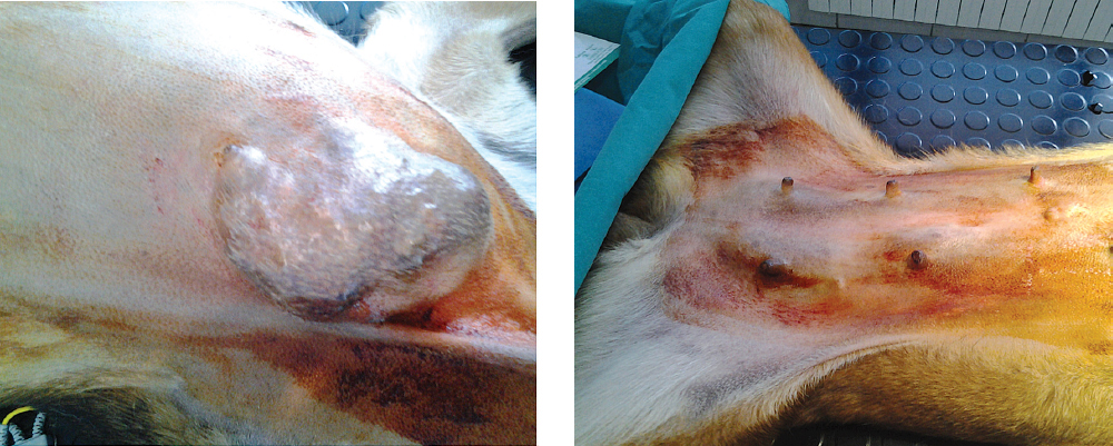
Multiple nodular appearance of the tumour was found in 6 patients (Figure 1, 2), while 4 patients had solitary tumour mass.
Figure 1. Mammary tumour of the cranial and caudal abdominal mammary gland- left side (6 year old Doberman Pinscher)
Figure 2. Mammary tumour on the cranial thoracal mammary gland (right) with metastases on the ingvinal mammary gland (9 year old Belgian Sheepdog)
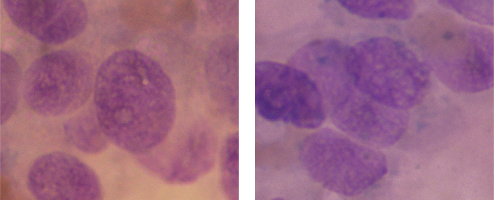
In nine patients, with malignant tumors (90%), one was diagnosed as adenoma. Cytological examination of the samples (n=5) revealed lobular adenocarcinoma (Figure 3). Pathohistological examination of the mammary tumor samples (n=5) revealed tubuloalveolar adenocarcinoma (n=4) and ductal adenocarcinoma (n=1). Chest x-ray imaging was recommended for all patients with adenocarcinomas, but only 5 made it. In 3 out of 5 of the patients with diagnosed adenocarcinoma lung metastases were noted by the X-ray imaging. Dimensions of the malignant mammary tumours were between 5 and 10 cm.
Figure 3. FNA sample from canine mammary tumour (Diff Quick, 100x)
The only treatment was surgical removal of the tumours (simple or radical mastectomy). The owners of seven patients agreed to additional ovariohysterectomy. Radical mastectomy was performed in patients that had multiple mammary tumours. Three patients died between 20 days and 2 months after the surgery. They were presented in the Hospital with sings of dyspnea due to lung metastases, anorexia, vomiting, decreased body temperature and lethargy. The rest of the treated patients (n= 7) were monitored on a monthly basis. Six months after the treatment, these patients remained in good body condition, without any signs of health disorders. They are recommended and visit the hospital on regular check-ups every 6 months.
In this study, older dogs (12-13 years), were predominantly affected by mammary tumours. Similarly, Murphy (9) reported predominance of mammary tumours in bitches at the age of 10 – 11 year, and seldom before the age of 2 years (9). Other research group reported the highest incidence of mammary tumours in patients between the age of 6 and 10 years (up to 63%), 32% of incidence in patients older than 10 years and only 5% in patients younger than 5 years. (12)
Ovarian steroids stimulate growth of normal mammary tissue under physiological conditions in bitches. Their proliferative effect on the epithelium may create conditions for neoplastic proliferation (13, 14). This occurs during every cycle and renders the bitch more susceptible to carcinogenesis. Oestrogens promote ductal growth, whereas progestins are able to induce a lobuloalveolar development of the mammary glands with hyperplasia of secretory and myoepithelial cells (5). Bitches that are spayed before the first oestrus have a 0.5% risk; animals spayed before the second oestrus have an 8% risk, and those spayed after the second oestrus and before two and a half years of age have a 26% risk of developing mammary tumours compared with the intact ones (15). It had been reported that exogenous administration of progesterone derivates, which are used to prevent oestrus in dogs, may increase the incidence of canine mammary tumours. Synthetic progestins (such as medroxyprogesterone acetate) induce the same effect on the mammary gland as endogenous progesterone. One clinical study on dogs treated with medroxyprogesterone acetate for oestrus prevention reported a greater risk of developing mammary tumours, the majority being malignant. (16)
In our study as well as in other studies (17, 18) dogs were admitted in the clinic for examination and treatment, roughly a year after the first detection of the mass. In our study 6 patients (66%) were with multiple mammary tumours. Similarly, other authors reported 50% to almost 70% of multiple tumours in dogs with diagnosed mammary tumours (2, 4, 6, 19). The majority of malignant tumours of the mammary gland in dogs are carcinomas (7), in our study carcinoma was diagnosed in 89% of the patients. Other study has shown that dogs with malignant tumours were significantly older than dogs with benign tumours, and malignant tumors were significantly larger than benign tumours (4). Five mammary tumours were examined at the Department of histopathology and were diagnosed as carcinomas (adenocarcinoma), which is the most common type of canine mammary tumour. (20)
The main treatment of canine mammary tumours is surgery, except for inoperable highly metastatic disease (6, 7). Surgical excision allows histopathological diagnosis and can be curative if the margins are clean and if the cancer has not spread (21). Dogs with benign tumours and approximately 50% of dogs with malignant mammary tumours will successfully recover if treated on time and depending on size of the tumour (22). The other 50% of the patients with malignant mammary tumours already has metastases at the time of surgery, which will eventually lead to death of the patient (6). The incidence of malignant mammary tumours in our study was 43.5%, similar to Moe (2001) who reported an incidence of 53.3% (23). Certain authors advice more radical surgery (24) because their study showed new tumour development in 58% of the patients in the ipsilateral mammary tissue after local removal of a solitary canine mammary tumour. In our study, radical mastectomy was done in 6 (66%) patients that had multiple mammary tumours. Literature shows contradicting results regarding the benefit of ovariectomy at the time of the excision of the mammary tumours (25). Only 2 patients in our study, with large tumor masses, were submitted to ovariohysterectomy and until present no signs of distant metastasis were detected.
Mammary tumours are common tumors and often present in older intact female patients. Late diagnosis is one of the major problems that results in lethal outcome due to lung metastases. Dogs with canine mammary tumours need a full diagnostic work-up, and every mammary tumour should be excised and submitted for histopathological examination. Since the ovarian steroids play an important role in the aetiology, the most effective prevention of mammary tumours is elective ovariectomy of the bitch at an early age.
© 2014 Atanaskova Petrov E. This is an open-access article published under the terms of the Creative Commons Attribution License which permits unrestricted use, distribution, and reproduction in any medium, provided the original author and source are credited.
The authors declared that they have no potential conflict of interest with respect to the authorship and/or publication of this article.
Macedonian Veterinary Review. Volume 37, Issue 2, Pages 129-134, p-ISSN 1409-7621, e-ISSN 1857-7415, DOI: 10.14432/j.macvetrev.2014.05.015, 2014