

INTRODUCTION
One of the most common respiratory diseases in horses is recurrent airway obstruction (RAO). The term derived from human medicine Chronic Obstructive Pulmonary Disease (COPD), has not been used since the late 1990s. This disease is encountered in horses older than 7 years, that are kept in stables, standing on straw bedding and fed poor-quality hay (1). So far, no breed predisposition to the disease has been found; however, genetic background should be taken into account, as more frequent cases of the disease have been described in certain breeding lines.
PATHOGENESIS
The development of the disease involves two types of allergic reactions: type I in an initial stage and type III in a later stage (2). Airway inflammation in horses predisposed to RAO is caused by inhalation of organic pollutants contained in dust, the largest source of which are hay and bedding. Endotoxins, dust mites and fungal spores contained in the dust, such as Feaniarecti virgula, Aspergillus fumigatus, Thermoactinomyces vulgaris, on entering the airways activate inflammatory cytokine production and neutrophil influx into the lumen of the bronchi (3). When an animal is led from the pasture to the stable and exposed to allergens, inflammatory changes occur within 6-8 hours. As inflammation develops, lower respiratory tract stricture increases. Inflammatory mediators are responsible for bronchoconstriction that causes both smooth muscle contraction acting on their cholinergic endings, as well as a large amount of mucus to accumulate in the bronchi as a result of goblet cells hyperplasia (Fig. 1). The mucus is denser and less transparent than that produced under physiological conditions. This makes it difficult to remove secretions and leads to prolonged retention in the airway lumen. Reconstruction of the bronchial walls involves both acute (intercellular edema) and chronic (smooth muscle hypertrophy, peribronchial fibrosis) changes (4). Changes in the airway wall are due to chronic processes and the lower effectiveness of the therapy and often make it impossible to return the animal to its normal condition.
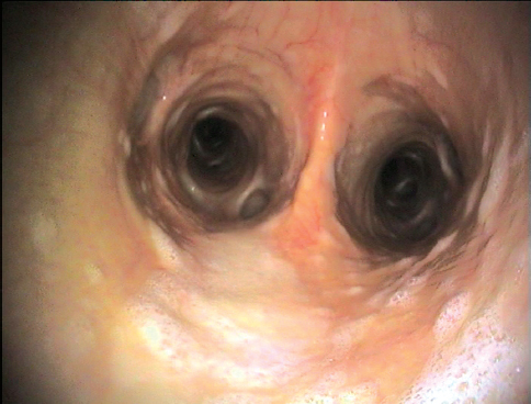
Figure 1. The bronchoconstriction with large amount of mucus accumulated in the bronchi as a result of goblet cells hyperplasia
Although the condition has an allergic background, eosinophils do not play a significant role in the pathogenesis of RAO.
CLINICAL SYMPTOMS
The initial stage of RAO is difficult to diagnose. Non-specific symptoms in the form of single coughing attacks may suggest a number of other diseases. At a later stage, the frequency of the dry cough, often paroxysmal in nature, increases.
RAO exacerbation is manifested by an increase in the respiratory rate, a frequent cough, nostril dilatation, mucosal nasal discharge and exercise intolerance (Fig. 2). Severe symptoms usually occur during exercise or are associated with an increase in the amount of dust in the air (e.g. while cleaning the stable or feeding hay). In horses suffering from chronic disease, a two-phase process of exhaling supported by the abdominal muscles is observed. This results in hypertrophy of the external abdominal oblique muscle, which manifests itself as a so-called “heave line” (Fig. 3).
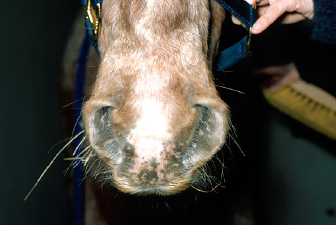
Figure 2. Nostril dilatation and mucosal nasal discharge in RAO
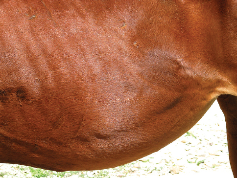
Figure 3. Hypertrophy of the external abdominal oblique muscle which manifests itself as a so-called “heave line”
In advanced untreated cases, the intake of food by the horse can be reduced, which leads to weight loss and devastation of the animal’s organism. At the same time, a pale coloration of the mucous membranes or, in extreme cases, cyanosis may appear. In the area of the foreskin or mammary gland congestive edema is observed, and exhalation is accompanied by a pushing of the anus. Each time the animal is used for work it causes fatigue disproportionate to the effort.
In a horse at rest, auscultation of the lungs in the early stages of the disease will not reveal any pathological changes. During the effort test, wheezing and rattling coming from the peripheral lung can be heard.
DIAGNOSTICS
The diagnosis of advanced RAO involves mainly a history and characteristic clinical signs. In the early stage of the disease additional tests are required. A blood test is helpful in excluding pneumonia and other infectious diseases, because in the course of RAO blood parameters do not deviate from the reference values (5).
A useful examination showing the state of the cellular infiltration of the lung is bronchoalveolar lavage (BAL). In cytological preparations made from the fluid, the largest group of cells are non-degenerated neutrophils (15-85% of all cells), whereas in healthy horses there are no more than 10% (6). There are also a number of compact formations of bronchial mucus (Curschmann’s spirals). Neutrophil count positively correlates with the severity of lesions and the course of the disease.
Radiological examination (X-ray) of the lung is helpful in a differential diagnosis. It is used to rule out other diseases of the lower respiratory tract. Lesions imaged using an X-ray are not pathognomonic for RAO, while on a radiograph only fibrosis of the lung parenchyma can be seen. By using a bronchoscope, tracheal wall congestion, tracheal bifurcation (bifurcatio tracheae) and residual mucous can be confirmed (1, 7).
An examination evaluating the severity of lesions and their susceptibility to therapy is the determination of the acid-base balance of arterial blood. The most decisive parameters are the partial pressure of oxygen (PaO2) and carbon dioxide (PaCO2). PaO2 falls a lot below 80 mmHg in animals with the initial form of the disease and up to 50 mmHg in horses with advanced RAO. PaCO2usually does not deviate from the reference value or is slightly elevated (5).
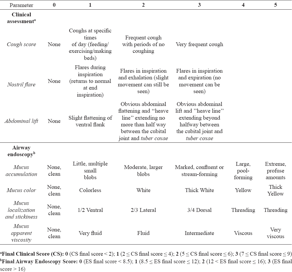
For RAO staging, the author correlates clinical (CS) and endoscopic (ES) scores in horses with RAO (Table 1).
Table 1. Modified clinical staging of RAO in horses according to Tilley et al. (17)
TREATMENT
Modification of the environment
The best method of treatment of recurrent airway obstruction in horses is a modification of the environment, which leads to a decrease in air aeroallergens. Pharmacological therapy of RAO includes treatment with glucocorticoids and bronchodilators. This procedure should involve both horses with advanced disease, as well as animals with a slight exacerbation of clinical symptoms. Pharmacological treatment without environmental modification is not effective (8).
The best solution is year-round maintenance of the animal in pasture. This is possible in almost every climate, because horses can withstand temperatures down to -30ºC without any health consequences. Only during frosts should they be provided with a winter shelter and fed high quality food. Remission of clinical symptoms is noticed after about 3-4 weeks from the time of moving the horse from the stable to the pasture (9,10). Repeated contact with the stable environment should then be avoided, because even a few minutes’ of exposure to the contaminants contained therein may cause a recurrence of the disease. If the therapy is unsuccessful, it should be carefully checked whether horses are being maintained in accordance with the recommendations. It often happens that the horses are brought into the stables at night or during feeding.
If for some reasons the owner is not able to provide an animal with such conditions, the treatment should be carried out within the stable. Since the feed is one of the main sources of dust, special attention should be paid to its quality. If it is not possible to provide adequate quality hay, it should be soaked before use in water and put in a hay net. Merely sprinkling it is not enough, because the water quickly evaporates from the surface and does not penetrate into the deeper layers (11). A good, but expensive solution is keeping the hay in silos or feeding a complete pelleted hay. However, in the former case the horse owner needs to use the appropriate silos due to the high sensitivity of horses to botulism.
To improve the conditions of maintenance, it is necessary to change the type of bedding. Keeping the horse on straw, which is most commonly used, significantly increases the amount of dust in the stable. Sawdust, peat, paper or cardboard can be used as straw replacements. This increases the cost of maintenance; however, it brings good results. The factor that has a major impact on air quality in the stable is ventilation. Five air changes per hour in the stable are sufficient to significantly reduce the amount of dust in the air (12).
Modifying the environment must involve the entire stable to be effective. This is the only way that, in cooperation with pharmacological therapy, respiratory system functions may improve sufficiently to allow the horse to live normally.
Pharmacological therapy
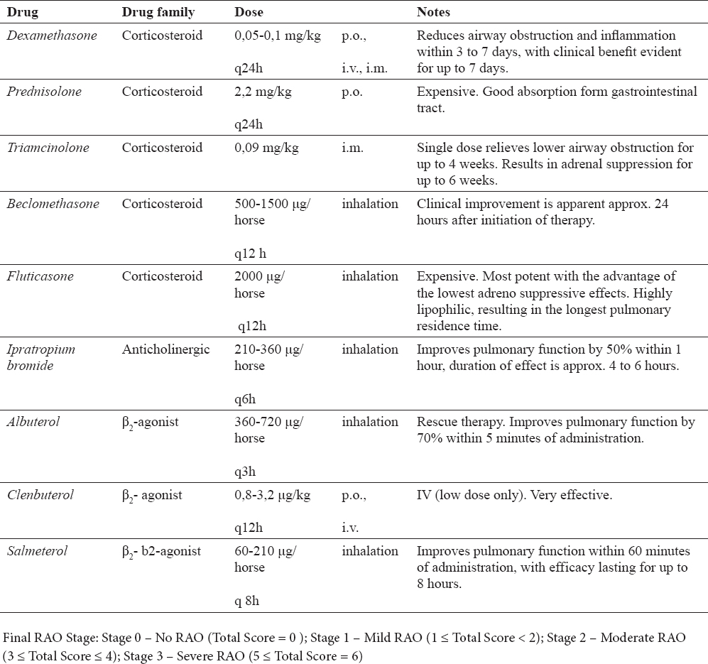
RAO treatment involves the simultaneous use of corticosteroids and bronchodilators (Table 2). Steroids reduce airway inflammation and bronchodilators improve bronchial airflow. Bronchodilators are not recommended for use as the sole therapy, because they do not exert any anti-inflammatory activity. Due to the synergistic effect of these drugs and the possibility of side effects, one should not exceed the recommended dose (13).
Table 2. Drugs commonly used in RAO treatment
Non-steroidal anti-inflammatory drugs (NSAIDs) and antihistamines are not applicable in the treatment of RAO.
Corticosteroids may be administered systemically or inhaled. The first solution is cheaper and simpler to be implemented, but long-term use entails the risk of iatrogenic Cushing’s syndrome. Systemic drugs are generally recommended for horses with severe respiratory ailments, since in these cases inhalants have limited access to the airways. In cases with mild symptoms, it is recommended to use corticosteroids inhaled using a special inhalation masks (Equine AeroMask or Equine Haler). Drugs used via inhalation act directly on the lower respiratory tract, which means that their doses are not high. Such treatment is very effective and usually there are no side effects, but this increases the cost of treatment (14,15).
Short-acting, inhaled bronchodilators are used in severe, life-threatening cases. Properly applied, they improve breathing as soon as 5 minutes after administration and their effect is maintained for about one hour. Long-acting bronchodilators are used for the prevention of exercise-induced bronchospasm or that from contact with a dusty stable environment (16). It should be noted, however, that the rules and regulations used in the majority of equestrian competitions and shows do not allow the use of these drugs before taking part in competition.
CONCLUSION
The prognosis for RAO is only good if the disease is diagnosed early, the appropriate treatment is introduced and the owner can provide the animal with an environment devoid of sensitizing allergens. On starting treatment, one should also take into account the high cost and long-term nature of the therapy and the possibility of the recurrence of the disease in the case of horse exposure to an adverse environment. Adequate conditions will enable the normal use of the animal for many years.


