Mac Vet Rev 2014; 37 (2): 165 - 170
 10.14432/j.macvetrev.2014.08.021
Received: 02 June 2014
Received in revised form: 31 July 2014
Accepted: 10 August 2014
Available Online First: 14 August 2014
Published on: 15 October 2014
Keywords: pig ovary, selenopyran, glutathione peroxidase (GPx)
Selenium is an essential and important trace element for reproductive biology and health due to its high antioxidative properties. The evidence of that is its wide distribution between tissues in the organism with varying density of storage: 30% in the liver, 30% in muscle, 15% in the kidney, 10% in the plasma, and the remaining 15% throughout other organs (1). Concentrations of free selenium are greatest in the renal cortex and the pituitary gland, followed by the thyroid gland, adrenals, testes, ovaries, liver, spleen, and cerebral cortex (2, 3).
For the achievement of the optimal performances, the diets for the reproductive swine female require additional feed additives, including selenium. Selenium is included as a main active part in the selenoproteins, which are coded of 25 genes in mammalians and exert multiple actions on the endocrine, immune and inflammatory functions (4). The most important for reproduction and pregnancy are the six antioxidant glutathione peroxidases (GPxs), which play a pivotal role in reducing hydrogen peroxide (H2O2) and lipid peroxides to harmless products (water and alcohols) (5, 6). As antioxidants, the GPxs help maintain membrane integrity, limit the propagation of oxidative damage to lipids, lipoproteins, and deoxyribonucleic acid (DNA) (5).
Data regarding selenium content related to the GPxs activity and female fertility are limited. The investigations in humans had shown that the unexplained infertility and low success rate in in vitro fertilization procedures are related to the decreased follicular selenium concentrations and lower GPxs activity (7, 8). In animals in vivo effects of selenium on ovaries were observed in beef cattle (9) and sheep (10), where parenteral administration of Se maximized fertilization rates of oocytes, but no mechanism of selenium action was discussed.
Little information is available about the effect of dietary supplemented selenium on the antioxidative status of pig ovaries. Recently there has been an increased interest to replace the inorganic selenium in the diet with organic. The selenium-containing compound designed to supplement is expected to have the following properties: hypotoxicity and biological availability of selenium in the composition. In this way, the organic selenium is more preferable, than sodium selenite. A number of research has reported increases of selenium serum levels and its positive effect on reproductive properties in sows fed by Se from an organic (Se-content yeast) versus inorganic (sodium selenite) source (11, 12, 13). The main disadvantage of this organic source is the difficulty to do the precise dosages of the selenium, because the yeast accumulation of the selenium from medium depends on different factors and shows wide variability.
The present work aimed to investigate the effect of the organic selenium compound-selenopyran on the state of antioxidative defense in ovary of growing pigs.
18 gilts of Danube white breed, ages between 120 - 228 days, were randomly divided into two groups, spread in 4 pig pens. The animals received equal basal diets without selenium additives. The animals were fed a basal diet with the following nutrition composition: 15.5% crude protein, 0.86 % lysine, and 12.56 MJ (metabolic energy) for the live weight 30-60 kg and 13.40% crude protein, 0.72% lysine and 12.83MJ for live weight 60-100 kg. The content of Se in basal diet was 0.15 mg per kg of forage.
The experimental gilts (n=9) were injected intramuscularly with oil solution of selenopyran (9-phenyl-symmetrical octahydroselenoxanthene) once per month with dose 0.1 mg Se/kg live weight. This organic source of selenium has a content of 24% of Se and has a lot of advantages. Besides the possibility for precise dosage of the compound, the toxity of it is lower than sodium selenite (LD50=1600 mg/kg against LD50=3.25 mg/kg). With regards to its chemical structure, selenopyran plays a role in the selenium storage and liberates the selenium slowly according the needs of the organisms (14). After slaughtering, one ovary from each animal was used for the histological analysis. The second ovary was used for the estimation of the selenium content in ovarian tissue by the atomic absorption spectroscopy method in the State Laboratory for Food Control. The atomic absorption spectroscopy method by apparatus SpectrAA 220Z „Varian” was applied for measuring the quantity of selenium in the ovary. Se was analyzed by standard addition, range 0-20 μg/L-1.
The blood for the investigation was obtained from sinus ophtalmicus. The analytical method used in this study for detection of Se in serum was a modified Classen and Bode method (15). It is a chemical technique based on complexing selenium with diaminobenzidine. The GPx activity in ovary homogenates using the colorimetric assay kit (BioVision, cat. no. K762-100) was measured, where the generated glutathione (GSSG) is reduced to GSH with consumption of NADPH. The decrease of NADPH (measured at 340 nm) is proportional to GPx activity. The assay was used to measure all of the glutathione dependent peroxidases with a detection sensitivity of ~ 0.5 mU/ml of GPx in samples.
For the preparation of histological sections, specimens were fixed in 4% paraformaldehyde (PFA) for 48 h. All samples were embedded in paraffin and sectioned at an approximate thickness of 5 µm. Some sections (10 per ovary) were stained with H&E for histological observation. The expression of γ-glutamyl transpeptidase (GGT) in ovaries was estimated by immunochistochemical method using antirat-GGT polyclonal antibody (16). To detect the primary antibody, sections were treated with streptavidin/biotin complex, using a Vectastain Elite ABC kit (Vector Laboratories, Burlingame, CA, USA) according to the manufacturer’s instructions and visualized with diaminobenzidine. The sections were counterstained with hematoxylin.
The statistical processing of the data was done by the STATISTIC computer programme (Stat Soft Inc., Ver.10.0). The one-way and regression analysis were done. Significance of mean differences was estimated by Student‘s t-test.
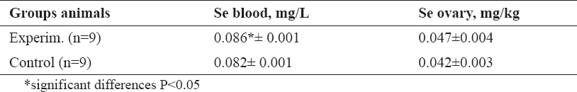
The Se content in blood and ovary is given in Table 1. Data shows that the selenopyran treatment leads to increase of the selenium level in blood (P< 0.05).
Table 1. Selenium content in blood and ovaries of pigs
Selenium content in ovarian tissues was higher in the experimental group compared to the control, but not significantly (P > 0.05).
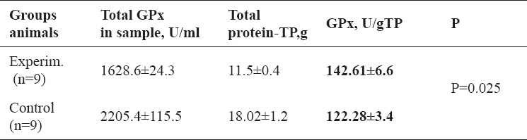
The significant increase (P < 0.05) of the activity of GPx per g total protein in the ovaries of experimental group was observed (Table 2).
Table 2. Glutation peroxidase activity (GPx) in the ovary of growing gilts
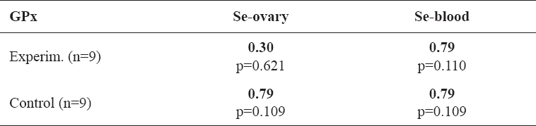
There were no significant correlations between the selenium content in blood and ovary and activity of GPx in ovaries (Table 3). Despite that the absolute values of the correlative coefficients (r = 0.79), reflecting the relationship between activity of GPx in ovaries and the selenium content in blood and ovary, were high in the control group. The same value of r was found for the relation of GPx activity and selenium content in blood in the experimental animals (Table 3). It seems that the activity of GPx in ovary is more dependent on the selenium content in the blood than in the ovary.
Table 3. Correlative relationship between activity of GPx in ovarian tissue and selenium content in blood and ovaries of growing gilts
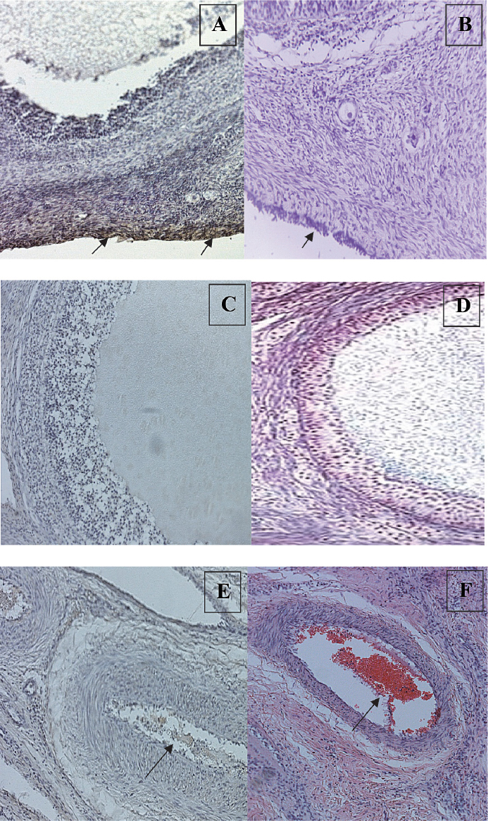
Our analysis of GGT expression in the pig ovary has shown that GGT is not expressed in the oocytes, germinal epithelium or most stroma in control and experimental animals, but it presents in the ovarian cortex cells (Fig. 1, A-B) follicular fluid (Fig. 1, C-D) and in the erythrocytes of ovarian blood vessels (Fig. 1, E-F) in treated gilts.
Figure 1. Localization of γ-glutamyl transpeptidase (GGT) in ovaries of growing pigs treated with selenopyran: A-immunochistochemical reaction in ovarian cortex cells; B-histological stained on ovarian cortex cells; C-immunochistochemical reaction in follicular fluid; D-histological stained on follicular fluid; E-immunochistochemical reaction on ovarian blood vessel; F-histological stained on ovarian blood vessel
The most important metabolic role of selenium in mammalian species is its function in the active site of the selenoenzyme glutathion peroxidase and this enzyme, together with superoxide dismutase and catalase, protects cells against damage caused by free radicals and hydro- or lipoperoxides (17).
In the present study, the results show significant increase of the selenium level in blood (P<0.05) as well as higher activity of GPx (P<0.05) in the ovaries of the experimental group. Harrison and Conrad (1984), reported increased content of Se in whole blood, plasma and ovary (18). Whole blood Se was chosen in the present experiment as an indicator of circulating Se. It possibly reflects better the animal Se status, because more than 50% of the overall Se in circulation is presented in erythrocytes, mainly bound to hemoglobin (19). The studies in poultry (20) and goats (21), failed to show any effect of Se treatment on the activity of blood GPx. In our experiments we did not investigate the GPx activity in blood, but we observed the increase the GPx activity in ovaries.
Fortier et al. (2012), reported that the GPx in yellow bodies (CL) from ovaries of treated with Se pigs was greater that in the control group. Concerning the other main characteristics of CL including weight, diameter, total number or Se content, authors didn‘t find the treatment effect (22).
The important sign of the enhancement of the antioxidative defense is the activation of the glutathione transport that is the main constitutional part of GPx enzyme. The enzyme related to this process is γ-glutamyl transpeptidase (GGT). We found GGT expression in the ovarian cortex cells, follicular fluid and in the erythrocytes of ovarian blood vessels in treated gilts, which seems as an evidence of active transport of glutathione from blood to ovary tissue.
Enhancement of selenium content in blood (significant), in ovary (non-signi icant) and the GPx activity in ovaries as well as the evidence of activation of glutathione transport to ovary (by γ-GGT expression) has shown that the organic selenium compound - selenopyran promotes the GPx dependent antioxidative defense in the ovary of growing gilts.
© 2014 Abadjieva D. This is an open-access article published under the terms of the Creative Commons Attribution License which permits unrestricted use, distribution, and reproduction in any medium, provided the original author and source are credited.
The investigations were supported by the projects № DKOF7RP02/17 under the auspices of the Ministry of Education and Science of Bulgaria. This work was supported by the grant № BG051PO001-3.3.06-0059, financed by the European Social Fund and Operational Programme Human Resources Development (2007 – 2013) and co-financed by the Bulgarian Ministry of Education and Science.
The authors declared that they have no potential conflict of interest with respect to the authorship and/or publication of this article.
Macedonian Veterinary Review. Volume 37, Issue 2, Pages 165-170, p-ISSN 1409-7621, e-ISSN 1857-7415, DOI: 10.14432/j.macvetrev.2014.08.021, 2014