Mac Vet Rev 2015; 38 (1): 73 - 78
 10.14432/j.macvetrev.2014.11.033
Received: 21 July 2014
Received in revised form: 14 November 2014
Accepted: 21 November 2014
Available Online First: 26 November 2014
Published on: 15 March 2015
Keywords: histopathology, immunohistochemistry, porcine circovirus type 2, post-weaning multisystemic wasting syndrome
The circovirosis presents one of the most significant virus diseases in pig production in the world. The etiological agent of this disease is a small, nonenveloped, spherical virus with a single stranded DNA genome which belongs to the Circoviridae family (1, 4, 5).
The virus was first isolated on a PK-15 cell culture and was considered non-pathogenic (25). Later, a virus was isolated from pigs with PMWS with a different genotype than the virus present on the PK-15 cell culture (11, 19). The virus present in the pigs with PMWS was marked as Porcine circoviruse type 2 (PCV2), and the virus present on the PK15 cell culture was marked as Porcine circovirus type 1 (PCV1) (17, 19).
PCV2 is the causal agent of several pig diseases which are collectively known as Porcine circovirus diseases - PCVD in Europe, or porcine circovirus-associated diseases (PCVAD) in North America (9). Most significant among them is the Post-weaning multisystemic wasting syndrome (PMWS); and the other diseases in this group are: Porcine dermatitis and nephropathy syndrome (PDNS); porcine respiratory disease complex (PRDC); proliferative and necrotizing pneumonia (PNP); enteritis; as well as some reproductive diseases (4, 8, 9, 12, 23).
It is noted that the PCVD, especially the PMWS, have the greatest impact on pig production. In the European Union alone it is estimated that the annual loss caused by the PCVD is more than 600 million Euros. PMWS affects pigs between the age of 7 and 20 weeks, mainly older pigs and in the early fattening period, but the diseases has also been described in pigs between the age of 1 and 6 months (1, 10).
The most characteristic damages of the PMWS develop in the immunological system of the pigs expressed by depletion of the lymphocytes in the lymphoid tissues, changes of the cell subpopulation in the peripheral blood and change in the cytokines expression in the diseased pigs (1, 3, 7).
The diagnosis for the porcine circovirosis is based on three criteria. The first criterion is the clinical picture which is consisted of loss of body weight, skin pallor, respiratory disorders and occasionally jaundice (14, 15, 21). The second criteria is the finding of characteristic histopathological lesions in the tissues, and the third criteria is the finding of PCV2 (antigen and/or nucleic acid) in the microscopic lesions (6, 7, 22).
Lately the PCR method gets more attention in the diagnosis of the porcine circovirosis (2, 13, 16, 20, 24). This method is based on the isolation of DNA (from viruses, bacteria, other microorganisms etc.) from the tissue samples and its amplification for easier demonstration of the existence of the causal agent in the examined sample. There are several types of the PCR method out of which the most used are the quantitative (qPCR) and the nested (nPCR). The advantage of the PCR method compared to the other previously mentioned is the short time necessary for establishing the diagnosis and quantification of the amount of the virus present in the examined material.
The “golden standard” in the PCV2 diagnosis is the method that includes finding of tissue changes and in the same time detection of the PCV2, which makes the in situ hybridization and immunohistochemical method the most often used method (18, 22).
In this article, we have reviewed the investigation of four farms from different regions in the Republic of Macedonia, where a clinical diagnosis for PMWS has been previously set. All farms were farrow to finishing units (the common way of breeding in Macedonia). The first farm had 400 sows in the herd and a total of 8000 finishing pigs produced per year. The second farm has produced 20000 finishing pigs per year and had 1000 sows. The third and fourth farm have produced 14000 finishing pigs per year per farm and had 650 sows in each of the farms. The examination was performed post mortem on thirty pigs from these farms, age from two to five months old, which had the most severe symptoms of the disease. The materials were taken not later than twelve hours after the animals’ death. A complete necropsy was performed on all of the pigs and samples were collected from the following tissues: lymph nodes (mesenteric, superficial inguinal, mediastinal and submandibular), lungs, liver, spleen, kidneys, tonsils, jejunum, ileum and colon. The samples for histopathology and immunohistochemistry were fixed in a 10% formalin solution. Samples from all the previously listed tissues were frozen at -80°C for further PCR investigation.
Tissue samples from the lymph nodes (mesenteric, superficial inguinal, mediastinal and submandibular), lungs, liver, spleen, kidneys, tonsils, jejunum, ileum and colon were collected at the necropsy and were fixated in 10% formalin, dehydrated, embedded in paraffin wax, sectioned at 3-4 μm and stained with haematoxylin and eosin (HE). F217 2C6-H9-A2 monoclonal antibodies and En Vision Kit (Dako ChemMate, Denmark) were used for immunostaining of the tissue sections which allowed us to assess low, moderate or large amounts of PCV2 antigen.
The most important clinical symptoms observed before death were: wasting, weight loss, decreased rate of weight gain, lymph node enlargement, respiratory distress, dyspnea, skin pallor and occasionally icterus were diagnosed.
In some of the pigs the presence of red papules and macula was noted, as well as hemorrhagic and necrotizing skin lesions. In the necropsy, the most evident finding was the enlarged lymph nodes (especially the inguinal, mediasinal and the mesenteric lymph nodes). The lungs had the following findings: cranio ventral lung consolidation in ten pigs, lack of pulmonary collapse in almost all of the pigs, interstitial pneumonia and lung edema. The liver was atrophic and discolored; white spots on the kidneys and catarrhal to hemorrhagic enteritis in twenty pigs were predominantly found.
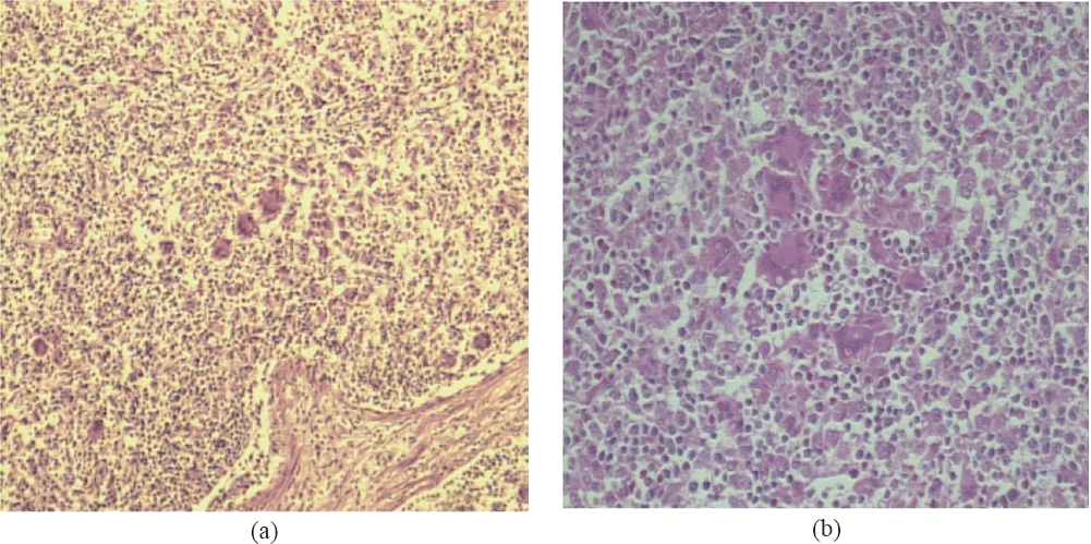
The microscopic lesions in the lymphatic organs (the lymph nodes, tonsils, spleen and the Payer’s patches) in all of the pigs were mainly expressed in the form of lymphocyte depletion and necrosis in the cortex and the paracortex of the lymph nodes, as well as the presence of giant cells in the same areas (Fig. 1).
Figure 1 Giant (syncytial) cells in the lymph node of the infected pigs. H.E.x10 (a) and x 20 (b)
In the lymph follicles and the parafollicular areas lymphocyte depletion and infiltration with large histiocytic cells was found. Large multinucleated cells were found in the lymph nodes, Payer’s patches and lamina propria of the intestinal villi.
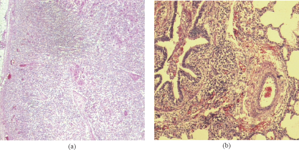
In the lungs, a multifocal lymphohistiocytic to granoulomatous interstitial pneumonia with the presence of histiocytes and multinucleated giant cells in the interalveolar walls was found (Fig. 2). Five of the pigs had necrotizing pneumonia.
Figure 2 Lymphoid depletion in the tonsil (a) and lymphohistiocytic interstitial pneumonia in the lungs (b). H.E.x10 (a) and x 20 (b)
Changes in the liver consisted of lymphohistiocytic infiltration, disorganization of the hepatic lobules, and in few samples, a perilobular fibrosis.
Lymphohistiocytic infiltration was also found in the kidneys, the intestines and also in most of the other tissues. The finding in the kidneys consisted of tubulointerstitial lymphoplasmatic and granulomatous nephritis.
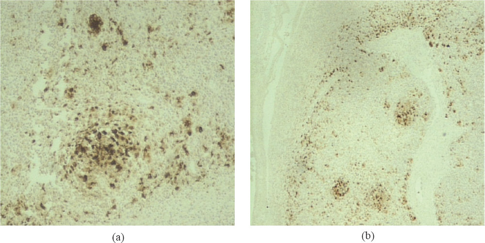
The immunohistochemical method revealed the presence of the PCV2 antigen in most of the examined pigs. The presence of the antigen is found in all the lymphoid tissues (specifically in the cells’ cytoplasm), while in the non lymphoid tissues it was less present. The presence of the antigen in the tissues shows positive correlation with the presence of the histopathological lesions in the same tissues. The antigen is mostly present in the necrotic areas of the lymph follicles and less present in the giant cells and the mononucleated phagocytes of the lymph follicles (Fig. 3).
Figure 3 Abundant presence of porcine circovirus antigen in nuclei of lymphocytes and histiocytes of tonsil. IHC x 20 (a) and x10 (b)
In the lungs, the PCV2 antigen is found in the large cells in the hyperplastic bronchial lymph tissue (BALT), in the epithelial alveolar cells, interstitial mononucleated cells, as well as the inflammatory exudates of the bronchi, bronchiole and alveoli.
In the liver, the antigen is mostly present in the centroacinar areas, especially in the hepatocytes, the epithelial cells of the bile ducts and in the Kupffer cells.
In the kidneys, the PCV2 antigen is widely present not only in the inflammatory cells, but also in the epithelial cells, in the interstitial inflammatory infiltrate and in the tubular cells.
This article described the histopathological and immunohistochemical findings and also made a comparison of these findings in thirty pigs from four farms in the Republic of Macedonia. All the pigs have PMWS symptoms. From the presented histopathological and immunohistochemical diagnoses it is evident that there is a close correlation between the finding of the PCV2 antigen, and the degree of the tissue lesions. The examinations included the parenchymatous organs of the animals and changes which are characteristic for the porcine circovirosis were found. Most significantly affected are the lymphoid tissues expressed by lymphocyte depletion, histiocytic infiltration and presence of a giant multinucleated cells. The findings suggest immunosuppression.
The use of the immunohistochemical method helped to determine the presence and the distribution of the PCV2 antigen in the tissues. The previously presented histopathological and immunohistochemical findings can confirm that the virus has a tropism towards the lymphoid tissue as it has been previously concluded by the other articles (1, 4, 7). It has been confirmed that both methods give satisfying results in the diagnosis of the porcine circovirosis. The “golden standard” in the diagnosis of porcine circovirosis consists of three parts: clinical symptoms (weight lose, pallor, respiratory distress, jaundice), histopathological findings (lymphocyte depletion and the presence of multinucleated giant cells) and confirmation of the presence of the virus DNA in the tissues using immunohistochemistry and in-situ hybridization.
In recent time, the PCR (polymerase chain reaction) method is being widely used. This method consists of only several steps which makes the time necessary for diagnosis significantly shorter (with the conventional method, about 7-78 hours, and with the real time quantitative PCR method, only about 45 minutes). Most of the studies recommend the real time quantitative PCR (qPCR) method. However, there are differences in opinion on what amount (threshold) of the virus in the serum should be present in the diagnosis of PCV2: 106.21, 106.91, 107 and 107.43 viral copies/ml. It is important that the sensitivity and the specificity obtained with the qPCR tests (used separately or combined with serology) are not enough to replace the histopathology plus the detection of PCV2 in the tissues (8).
According to the proposal of some authors the diagnosing of a herd should be based on two elements: 1) significant increase in the mortality of post weaning pigs followed by clinical signs compatible with PCV2 compared to the histological data for the herd, and 2) individual diagnosing of PMWS in at least one in three to five necropsies of pigs combined with the mentioned increase in the mortality. At the same time, other reasons for increased mortality should be excluded.
The results gained in this examination showed that the histopathological and the immunohistochemical method combined with the clinical diagnostics are sufficient to confirm or deny the presence of PCV2 infection in the pig population.
In future investigation, in order to improve diagnostic accuracy, beside the histopathological and the immunohistochemical method, the PCR method has to be introduced in the diagnosis of the PCV2, as well as comparison of the results between these methods.
© 2015 Gjurovski I. This is an open-access article published under the terms of the Creative Commons Attribution License which permits unrestricted use, distribution, and reproduction in any medium, provided the original author and source are credited.
The authors declared that they have no potential conflict of interest with respect to the authorship and/or publication of this article.
Macedonian Veterinary Review. Volume 38, Issue 1, Pages 73-78, p-ISSN 1409-7621, e-ISSN 1857-7415, DOI: 10.14432/j.macvetrev.2014.11.033, 2015