Mac Vet Rev 2015; 38 (2): 159 - 166
 10.14432/j.macvetrev.2015.04.042
Received: 20 January 2015
Received in revised form: 27 March 2015
Accepted: 31 March 2015
Available Online First: 16 April 2015
Published on: 15 October 2015
Keywords: ketosis, hematological parameters, μ-hydroxybutyrate, dairy cows
Metabolic disorders in ruminants are most frequently found in the transition period from late pregnancy to early lactation, as the animal’s body suffers substantial changes. During the pregnancy, the metabolism of cows is adapted for the developing embryo (1), while during the early lactation the metabolism follows the increased production of milk.
Ketosis occurs most commonly after calving during the 2nd lactation (3-7 week postpartum) (2, 3) and considerably less afterwards (4). This period corresponds to an inadequate energy intake and increasing milk secretion, determining the occurrence of a negative energy balance (5).
Subclinical ketosis (SCK) is a pathological condition associated with an increased level of ketone bodies in the organism without symptoms for clinical ketosis (6). Health and economic consequences of subclinical ketosis are reduced milk yield (7, 31), reproductive disorders (8), low insemination index (9), prolonged service period, clinical and subclinical mastitis (10), abomasal dislocation (11), and/or clinical ketosis (12).
The periparturient period with metabolic and hormonal changes, stress factors has a significant impact on the health of dairy cows and decreases the resistance to various infections (13). The period of negative energy balance is of critical for appearance of clinical and subclinical ketosis. The increased blood concentrations of non-esterified fatty acids (NEFA) or β-hydroxybutyrate (BHBA) correlate positively with disturbances in dairy cows’ health, reproduction and milk yield during the postpartum period (12, 14).
Marked hyperketonaemia is manifested clinically with reduced appetite, rapid weight loss and reduced milk yield. Sometimes, the animals exhibit nervous signs as pica, biting and licking unusual object, blindness (15). The faeces is usually hard, dry and scanty. Sometimes very high blood BHBA concentrations, clear clinical signs of ketosis could be absent (16).
Determination of the Body Condition Score (BCS) provides available information for the body reserves, for the determination of how dairy cows are prepared for the period of negative energy balance, full of stress and inappropriate diet (17). It can be used as an indicator for potential health problems in dairy cows, sheep and goats. The changes in BCS suggest the presence of inadequate energy supply and occurrence of postpartum metabolic disorders (18). Literature data about BCS before calving shows wide variety: ≤ 3.0 (19); 3.25 (20) and 3.00-3.50 (5). It is demonstrated that cows with BCS before calving which is > 3.5 has 2.5 times higher risk of developing type II ketosis (21).
Some hematological and biochemical parameters are indicators of physiological, nutritional, metabolic and clinical status of production animals, as an important part of health and welfare management (22). Several authors report that blood BHBA concentrations are a basic parameter for proper evaluation of ketosis, where BHBA is more stable than other ketone bodies (acetone and acetoacetate) (16, 23).
For diagnostic SCK in lactating cows, mainly 3 values of blood BHBA are noticed in the literature: more than 1.0 mmol/l (24, 25), more than 1.2 mmol/l 7, 26) and more than 1.4 mmol/l (12, 16, 27). If the blood BHBA is more than 1-1.4 mmol/l, there is 3 times greater risk for dislocation of the abomasum and/or development of ketosis (12, 16, 27). In our previous studies of goats with subclinical ketosis (28), blood BHBA concentrations between 0.8 and 1.9 mmol/l were established between the 10th and the 30th day of lactation. This data is comparable with type I subclinical ketosis in dairy cows. Subclinical ketosis and type I clinical ketosis (CK) occur usually between the postpartum days 14–21 and 5–50, respectively (16). Type II clinical ketosis is generally present during the first 14 days after calving, but could be encountered in cows up to the 30th day of lactation.
Blood BHBA values for clinical ketosis are: over 2.0 mmol/l (29), over 2.6 mmol/l (30) and over 3.0 mmol/l (2). Blood BHBA concentrations >3.0 mmol/l were established only in 20% of ketotic cows (14).
Considering the widespread occurrence of bovine ketosis at a global scale (31) and economic losses incurred by farmers, the early and accurate diagnosis is essential for prevention of this disease. On the other hand, it is known that circulating ketone bodies (acetone, acetoacetate and BHBA) have negative influence on all organs and physiological processes of the animal body. Alterations in blood biochemistry can be used for early diagnosis of metabolic diseases and taking preventive measures for herd health management and against economic losses in dairy production.
The aim of this study was to investigate changes in the clinical and hematological parameters in high-yielding cows suffering from subclinical and clinical ketosis.
Studies were performed in dairy farms in the Republic of Bulgaria and Republic of Serbia between February and September 2014.
A total of 47 Holstein cows from 1st to 4th lactation were included in the study (n=29 from R. Bulgaria and n=18 from R. Serbia). Dairy cows were fed rations corresponding to the physiological state (lactation) of the studied groups. All cows were up to 45 days in milk (DIM). In this study, the blood BHBA threshold value for subclinical ketosis was set to ≥ 1.2 mmol/l, and for clinical ketosis – to ≥2.6 mmol/l. All cows were submitted to physical examination, body condition score evaluation and analysis of blood β-hydroxybutyrate concentrations.
Cows were divided into three groups:
- first group (n=24) with blood BHBA level <1.2 mmol/l – control cows;
- second group with blood BHBA between 1.2-2.6 mmol/l (n=15) – cows with subclinical ketosis;
- third group with blood BHBA >2.6 mmol/l (n=8) – cows with clinical ketosis.
All cows were submitted to examination of the rectal body temperature, heart rate, respiratory and rumen contraction rates, and visible mucous inspection using routine clinical diagnostic procedures.
Body condition scores were evaluated using a 5-point scale (1.0-5.0, at intervals of 0.25). The cows were scored visually by two investigators (32).
Blood samples were collected through puncture of the coccygeal vein using sterile 21G needles and vacutainers either anticoagulated with K2 EDTA - 3 ml or with gel and clot activator - 6 ml. (Biomed, Bulgaria). Samples were obtained in the morning before feeding.
Blood BHBA concentrations were determined immediately using a portable Xpress-I system (Nova Biomedical, UK). Samples for CBC analysis were transported and stored at 4°C. Analysis was performed within 2 hours after sampling. The following parameters were determined: Red Blood Cell (RBC, 1012/l), Hemoglobin (HGB, g/l), Hematocrit (HCT, %), Mean Corpuscular Volume (MCV, fl), Mean Corpuscular Hemoglobin (MCH, pg), Mean Corpuscular Hemoglobin Concentration (MCHC, g/l), White Blood Cell (WBC, 109/l), Lymphocytes (LYM, 109/l), Monocytes (MON, 109/l), Granulocytes (GRA, 109/l), Red Blood Distribution Width (RDW, %), Red Blood Cell Distribution Width Absolute (RDWa, fl), Platelets (PLT, 109/l) and Mean Platelet Volume (MPV, fl). Hematological investigations were analyzed on an automated analyser Exigo EOS Vet (Boule Medical AB, Sweden).
Statistical analysis was done with Statistica 6.0 (Windows) software, StatSoft, Inc. (USA, 1993) and ANOVA test. Results were presented as mean (x) ± standard deviation (SD). The level of statistically significance was p < 0.05.
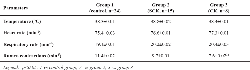
Values of physical examination of cows with SCK showed no statistically significant changes vs. control group. Cows with CK, had a reduced average rumen contraction rate (p<0.05): 7.6±0.02 vs control values (11.4±0.02). Data from physical examinations (rectal temperature, heart rate, respiratory rate, rumen contraction rate) are presented in Table 1.
Table 1. Clinical parameters of cows from the control group (Group 1), with subclinical ketosis (SCK) (Group 2) and clinical ketosis (CK) (Group 3)
There were no changes in both groups of ketotic cows where inspection was performed of the color of visible mucosae, swelling, discharges and coat.
Reduced rumen contraction rate (hypotonia) was recorded, cows with clinical ketosis also exhibited anorexia and body weight loss.
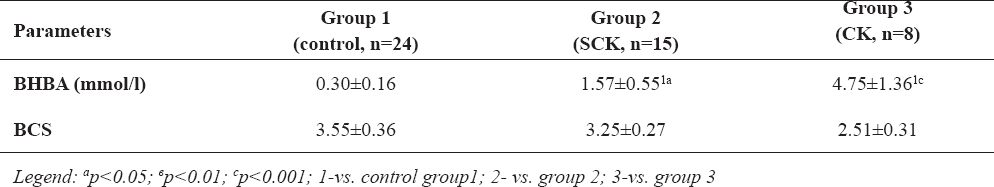
The results of blood β-hydroxybutyrate concentrations and BCS in all cows included in experiment are presented in Table 2. Cows from group 2 (SCK) and group 3 (CK) BHBA levels were higher –1.57±0.55 mmol/l (p<0.05) and 4.75±1.36 mmol/l (p<0.001) respectively, compared to cows from the control group - 0.30±0.16 mmol/l.
Table 2. Average blood concentrations of β-hydroxybutyrate (BHBA) and body condition scores (BCS) in cows from the control group (Group 1), with subclinical ketosis (SCK) (Group 2) and clinical ketosis (CK) (Group 3)
Average BCS in control animals was 3.55±0.36, in SCK cows – 3.25±0.27 and in cows with clinical ketosis (group 3) – 2.51±0.31.
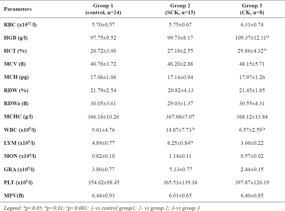
Data reflecting changes in hematological parameters of both study groups are presented in Table 3. Cows with CK had higher HGB and HCT values - 109.37±12.11g/l and 29.86±4.32% respectively, vs. healthy cows (97.75±9.52 g/l and 26.72±3.00%; p<0.05). Total WBC values in cows with CK (group 3) were lower – 6.57±2.59 (109/l) (p<0.05) as compared with controls: 9.61±4.76 (109/l). Unexpectedly, higher WBC values were detected in cows with subclinical ketosis - 14.87±7.73 (109/l) (p<0.05), than in the control cows – 9.61±4.76 (109/l). In cows with SCK higher values were established of lymphocytes - 8.25±0.84 (109/l) (p<0.05) vs. control group - 4.89±0.77 (109/l).
Table 3. Hematological parameters in cows from the control group (Group 1), with subclinical ketosis (SCK) (Group 2) and clinical ketosis (CK) (Group 3)
The other hematology parameters (RBC, MCH, MCHC, MCV, RDW, RDWa, MON, GRA, PLT and MPV) were not significantly different than values in control animals.
This research confirmed that blood BHBA concentrations in cows with SCK is higher than 1.2 mmol/l and in cows with CK is higher than 2.6 mmol/l, which are set as limited values for subclinical and clinical ketosis, respectively. Blood BHBA is an indicator of inappropriate oxidation of non-esterified fatty acids in the liver (33). It is used as early marker for detection of ketosis in ruminants (16, 23). Some researchers (24, 25) set a limit value >1.0 mmol/l, others (12, 16, 27) over 1.4 mmol/l, while in the present study value over 1.2 mmol/l is chosen as a limit of exhibited clinical sings (7, 26). Reduced blood glucose level and low insulin secretion are triggers for enhanced mobilization of lipids from the adipose tissue and deposition of triglycerides in the liver parenchyma and stimulation of ketogenesis (34).
Low values of BCS in cows with clinical ketosis correlated negatively with higher blood BHBA concentrations. Loss of appetite is a further reason for high blood ketone bodies concentrations. The weight loss until the 30th day of lactation has a considerable impact on the risk for development of ketosis, dislocation of the abomasum, milk yield reduction, disturbance of the reproductive performance and early embryonic death (35). Other authors (36) found that there was no relationship between the weight loss during the lactation and the incidence of metabolic diseases. Excessive fat deposition in the dry period correlates with increased occurrence of clinical ketosis in cows after calving (37). Literature and these results confirmed that the BCS is an important data for nutrition management of dairy herd.
The results from the complete blood count showed increased hemoglobin and hematocrit values only in cows with clinical ketosis. We suggest that these changes were due to higher erythrocyte counts in cows from group 3, although the differences were not statistically significant. Most authors (38) suggest that clinical and subclinical ketosis is not accompanied by changes in RBC, hemoglobin and hematocrit. There is literature data (39) for lower hemoglobin level and RBC counts in ketotic cows, as in these cases the erythropaenia was accompanied by anisocytosis and poikilocytosis.
Statistically significant increase of WBC is reported in this research in cows with SCK. It could be assumed that higher leukocyte counts were related to the wide spread of bovine enzootic leucosis in Bulgaria (40), and to the fact that 2 of cows (13.3%) presented signs of metritis. The leukocytosis in cows during the postpartum period is most commonly attributed to acute or chronic inflammations (mastitis, endometritis, metritis etc.) (22).
Reduction in WBC counts was shown in cows from group 3 (with clinical ketosis). Results in this investigation are in agreement with other reports (41, 42, 43). Lower WBC counts are reported in cows with enhanced catabolism in the periparturient period and increased blood BHBA levels (44). High levels of ketone bodies inhibit the cell proliferation in bone tissue (42), the in vitro chemotaxis of leukocytes (43) and respiratory activity of polymorphonuclear leukocytes (41).
In this study, we found statistically significant changes in lymphocyte counts (lymphocytosis) in cows suffering from SCK. This is consistent with the increased neutrophil and lymphocyte counts reported in lactating cows in consequence to the enhanced lipomobilisation, ketogenesis and hypoglycemia (45). On the other hand, high neutrophil and lymphocyte counts could result from stress and accompanying levels of glucocorticoids (cortisol) (46). The distinct immunosuppressive effects of high ketone bodies concentrations (BHBA and acetoacetate) (47) and oestrogens (48) are also acknowledged.
Clinical and subclinical ketosis among dairy cows in the first 45 days after delivery is quite prevalent. In practice, early detection is useful for treatment and assurance of good milk yields. The blood parameters, especially BCS and blood BHBA are excellent parameters for the nutritional status and health of dairy cows and could be utilized as markers for timely detection of metabolic disorders in cattle. Identification of problems at the herd is a signal for correction of the diet, preventive supplementation with rapid sources of energy etc., in order to reduce economic losses. Changes in hematology have a limited diagnostic value in clinical and subclinical ketosis, but our results provoke interest for further studies to clarify their meaning.
Copyright
© 2015 Marutsova V. This is an Open Access article distributed under the terms of the Creative Commons Attribution-Non Commercial License (http://creativecommons.org), which permits unrestricted non-commercial use, distribution, and reproduction in any medium provided the original work is properly cited.
Conflict of Interest Statement
The authors declared that they have no potential conflict of interest with respect to the authorship and/or publication of this article.
Citation Information
Macedonian Veterinary Review. Volume 38, Issue 2, Pages 159-166, p-ISSN 1409-7621, e-ISSN 1857-7415, DOI: 10.14432/j.macvetrev.2015.04.042, 2015