Mac Vet Rev 2015; 38 (2): 203 - 208
 10.14432/j.macvetrev.2015.07.050
Received: 29 April 2015
Received in revised form: 02 July 2015
Accepted: 13 July 2015
Available Online First: 27 July 2015
Published on: 15 October 2015
Keywords: infertility, sperm abnormalities, spermatogenesis, Trypanosoma congolense, Yankasa rams
Sheep is a major source of animal protein in Nigeria (21), playing an important role in the livelihood of most Nigerians (26). Their distribution is majorly affected by socio-economic and environmental factors, such as availability of feeds, animal traction, marketing systems, cultural preferences and disease (8), which is a major constraint to livestock production in Nigeria (15). Trypanosomosis is an economic and zoonotic disease caused by protozoa of the genus Trypanosoma (20). It affects the cardiovascular, nervous, respiratory, digestive and reproductive systems of the body (12).
In the male reproductive tract, pathological disorders attributed to trypanosomosis include testicular degeneration, scrotal inflammation, penile protrusion, prepucial inflammation, testicular odema, epididymitis and abnormal spermatogenesis (1, 33, 34, 35). In the female, there is abortion, irregular oestrus cycle, cystic degeneration of the ovary, follicular cyst, flaccidity of the uterine horn, decreased conception rate, low birth-weights and neonatal death (10, 13, 16, 28). In addition, pregnant animals infected by trypanosomes may die before or after parturition (4, 6).
Trypanosoma congolense has a wide host range (11, 19). It is transmitted biologically (17) although mechanical (12) as well as congenital transmissions (14) have been reported. Infection of males with T. congolense causes severe testicular degeneration, penile protrusion, haemorrhage, prepucial inflammation, decreased testosterone levels, increased cortisol concentration, depressed pituitary and adrenocortical functions in sheep, cattle and pig (18, 31, 35). Although Adeyemo et al. (3) studied the pathogenesis of T. congolense and T. bruceiinfections on West African Dwarf ram, while Sekoni (33) the effect of T. vivax on sperm morphology in Yankasa rams, there is no study on the effect of T. congolense on reproduction in Yankasa rams to the best of our knowledge. Aspects of the study involving genital lesions, reaction time, and semen characteristics have been described elsewhere (23, 24). This paper therefore reports on the effect of T. congolense on the incidence of sperm morphology of Yankasa rams.
The study was carried out at the experimental animal house, Faculty of Veterinary Medicine, Ahmadu Bello University Zaria, Nigeria. Nine mature healthy Yankasa rams from an initial flock of sixteen rams purchased from local markets around the study facility were used. Their age was 24 – 30 months old and they were fed on legume hay (harawa), ground nut, maize offal, concentrate (100gm/head/day) multi-mineral nutrient block and fresh pasture. Water was also provided ad libitum throughout the experiment. The animals were acclimatized for 4 months in fly and tick proof pens. Trypanosoma congolense used for this study was obtained from the Nigerian Institute for Trypanosomiasis Research (NITR) Vom, Nigeria. This trypanosome was initially isolated from cattle but inoculated into mice and maintained by continuous passage until use. The study was approved by the ethical board of the Faculty of Veterinary Medicine, Ahmadu Bello University, Zaria and adequate measures were taken to minimize pain or discomfort.
The rams were divided into 2 groups of six infected and three uninfected control. They infected group of six (6) animals were inoculated with 1 x 10⁶ Trypanosoma congolense through the jugular vein. All the rams were closely monitored for clinical signs suggestive of trypanosomosis. Semen was collected weekly from each ram, for seven weeks using electro-ejaculator and evaluated according to the methods of Chemineau and Cagnie (9). Sperm morphological abnormalities were estimated by dilution of semen sample with buffered formal saline and by staining with eosin-nigrosin stain then counting at least 500 sperms per slide as described by Sekoni et al. (29). Data obtained were analyzed using unpaired student t-test on SAS computer package. Values of P<0.001 were considered statistically significant.
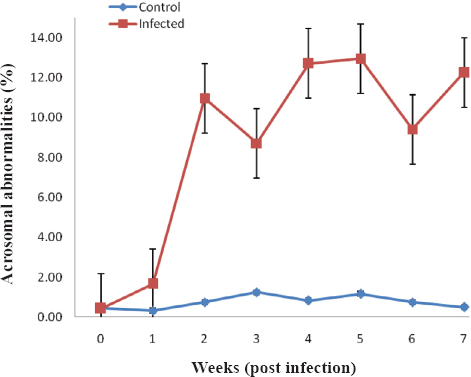
The parasites were detected in the infected Yankasa rams within 7-11 days post infection (pi). There was a steady increase in mean acrosomal abnormalities of the infected Yankasa rams from 0.43 % to between 1.66 % and 12.25 % pi. These values were significantly increased (P<0.001) from week 1 pi compared to the control group, that ranged from 0.33 % - 1.25 % pi (Fig. 1).
Figure 1 Mean percentage acrosomal abnormalities of Yankasa rams experimentally infected with T. congolense
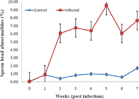
The mean head abnormalities rose from 0.04 % pre-infection to a pi value of 0.88 % - 9.55 % for the infected group, which were significantly (P<0.001) increased compared to the control group (0.67 % - 1.00 %) from week 2 pi (Fig. 2).
Figure 2 Mean percentage sperm head abnormalities of Yankasa rams experimentally infected with T. congolense
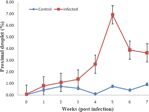
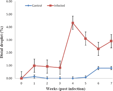
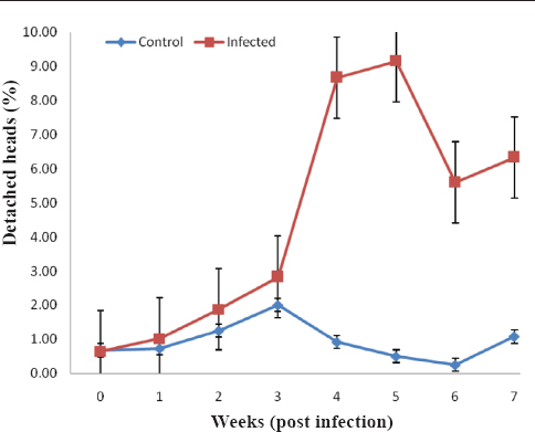
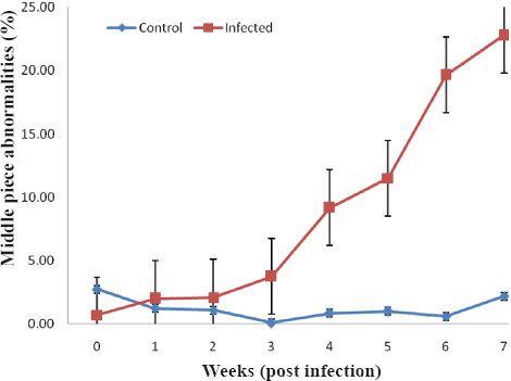
The mean proximal droplet values of infected Yankasa rams (0.33 % - 6.15 %) were significantly increased (P<0.001) compared to the control (0.08 % - 0.92 %) from week 4 pi till the end of the study (Fig. 3). There was also significant increase (P<0.001) in the mean distal droplet of the infected group (0.83 % - 4.33 %) compared to the control group (0.00 % - 0.42 %) from week 1 pi till the end of the study (Fig. 4). The mean percentage of detached sperm heads of control and infected rams is presented in Figure 5. There was a significant (P<0.001) increase in the mean detached heads of the infected rams (1.02 % - 9.15 %) compared to the control (0.25 % - 2.00 %) from week 2 pi till the end of the study. The middle piece abnormalities of the infected rams (1.98 % - 22.79 %) were significantly (P<0.05) increased compared to rams in the control group (0.08 % - 3.63 %), from week 3 pi till the end of the study (Fig. 6).
Figure 3 Mean percentage proximal droplet of Yankasa rams experimentally infected with T. congolense
Figure 4 Mean percentage distal droplet of Yankasa rams experimentally infected with T. congolense
Figure 5 Mean percentage detached heads of Yankasa rams experimentally infected with T. congolense
Figure 6 Mean percentage middle piece abnormalities of Yankasa rams experimentally infected with T. congolense
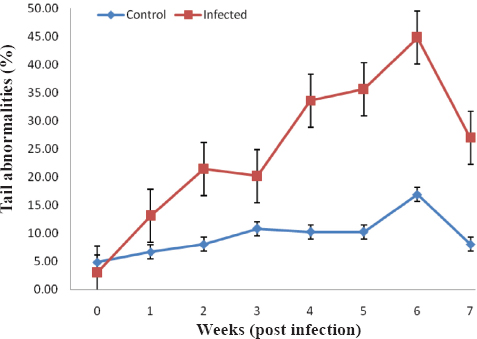
Tail abnormalities also significantly (P<0.05) increased, but from week 1 pi till the end of the study in the infected rams (13. 99 % - 44.75 %) compared to rams in the control group (6.72 % - 16.72 %) (Fig. 7). Sperm tail abnormalities were more prominent in infected rams than abnormalities associated with the sperm head.
Figure 7 Mean percentage tail abnormalities of Yankasa rams experimentally infected with T. congolense
The infection of Yankasa rams with T. congolense showed some clinical signs (fluctuating, pyrexia, ruffled hair coat, dullness, weight loss and pallor of the mucous membrane) that have been published in our previous investigations (24).
In this study we demonstrated the adverse effect of T. congolense infection on the sperm morphology of Yankasa rams. The morphological abnormalities seen were acrosome abnormalities, sperm head abnormalities, proximal droplet, distal droplet, middle piece abnormalities, detached head and tail abnormalities. Sperm morphology is one of the factors determining semen quality besides sperm motility and concentration (27). The occurrence of sperm morphological abnormalities in the semen of animals is associated with infertility and sterility (30). From the first week of infection, substantial acrosomal, tail and distal droplets defects were observed. This was capable of compromising acrosomal reaction and sperm motility and by extension the fertility of infected animals. Other morphological abnormalities were seen in the second (sperm head and detached head abnormalities); third (middle piece) and fourth (proximal droplet) weeks post infection. However, signs of infertility in the infected rams would have been evident as early as the first week and will continue to increase till the end of the study. This progressive increase in abnormalities for the 7 weeks study period, which is also the duration for a spermatogenic cycle in rams, suggest that T. congolense infection in Yankasa rams may not just affect the spermatogenesis in the testicles alone, but also the maturation process at the tail of the epididymis. This is supported by the lesions observed in the epididymis and the testis in our earlier study (23). The sperm morphological abnormalities seen in this study are also similar to those reported by Sekoni (33) in T. vivax infected Yankasa rams where abnormalities were seen from the second week post infection. However, in this study, abnormalities were seen from the first week post infection. The variation in trypanosome specie maybe responsible for this, since its infectivity and pathogenesis depends on the specie and strain of the trypanosome (22). This may suggest that T. congolense in more virulent than T. vivax in Yankasa rams. Previous studies in bulls have also supported this fact (32, 34). Osaer et al. (25) observed minor morphological abnormalities following infection of Djallonke rams with T. congolense. They also observed return of sperm morphology to their pre-infection state as the infection progressed in this breed of sheep. This is contrary to the report in this study, which may have been influenced by breed difference. The Djallonke sheep is a West Africa Dwarf sheep found in Gambia and are generally known to be trypanotolerant surviving in tsetse infested areas (7). In contrast, the Yankasa breed used in this study are highly susceptible to trypanosomosis (2, 5, 33).
The T. congolense used in this study is pathogenic to Yankasa rams with substantial percentage of sperm morphological abnormalities. The outcome of this on Yankasa rams in field situation may increase the incidence of infertility which is detrimental to sheep production.
Copyright
© 2015 Okubanjo O. O. This is an Open Access article distributed under the terms of the Creative Commons Attribution-Non Commercial License (http://creativecommons.org), which permits unrestricted non-commercial use, distribution, and reproduction in any medium provided the original work is properly cited.
Conflict of Interest Statement
The authors declared that they have no potential conflict of interest with respect to the authorship and/or publication of this article.
Citation Information
Macedonian Veterinary Review. Volume 38, Issue 2, Pages 203-208, p-ISSN 1409-7621, e-ISSN 1857-7415, DOI: 10.14432/j.macvetrev.2015.07.050, 2015