Mac Vet Rev 2016; 39 (2): 159 - 166
 10.1515/macvetrev-2016-0080
Received: 25 November 2015
Received in revised form: 20 January 2016
Accepted: 25 March 2016
Available Online First: 06 April 2016
Published on: 15 October 2016
Keywords: river barbel, genotoxic effects, erythrocyte abnormalities, pollution
Since the first description of the morphological nuclear alterations in fish erythrocytes (1), a number of papers published during the past decades reported the appearance of erythrocyte nuclear abnormalities in fish (2, 3). These modifications of the shape and dimension of the nucleus lead to genetic instability, cell dysfunction and/or cell death (4, 5). Although these changes can occur naturally, it is proposed that pollutants can cause a manifold increase in incidence of nuclear anomalies. One of the methods of studying genotoxic effects of different mutagens on the living organisms is the detection of frequency of micronuclei (MN) which is often used in studies of teleost fish (6). Other most commonly noted nuclear abnormalities caused by genotoxins are vacuolated nucleus (VN), binucleus (BiN), notched nucleus (NotchN) and blebbed nucleus (BlebN) (2, 3).
Fish are shown to be excellent subjects for detection of potential mutagenic and/or carcinogenic effects of contaminants in water, because they can metabolize, concentrate and store different pollutants. This makes them a good model organism for experimenting with potential toxins that can influence humans, as well (7). In general, changes in fish will reflect the changes in the environment because of their sensitivity to low concentrations of genotoxins (8). In addition, as top predators, fish will accumulate even higher amounts of genotoxins when feeding on other organisms that have consumed the pollutant, thereby being an indicator of the shape of whole ecosystem (9).
The river Bregalnica is situated in the eastern part of the Republic of Macedonia and represents the main water resource for all water-related activities (from irrigation to drinking water supply) in that region. Recent studies have revealed intensive negative anthropogenic pressure on the river, resulting in high water pollution (10, 11). Uncontrolled discharge of polluted waters into the river, directly or indirectly, threatens the ecosystem health. Examination of the exposed wildlife is necessary for the early detection of environmental damage and prediction about the potential human health risks.
Considering the increasing pollution trend of the river Bregalnica, especially in its middle and lower stretches, the aim of this study was to examine whether pollutants that are released in this river can generate genotoxic changes such as micronuclei and erythrocyte nuclear abnormalities in local barbel (Barbus peloponnesius, Valenciennes, 1842) population.
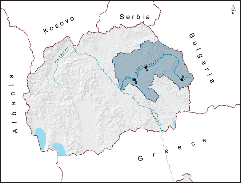
Fish were collected from three localities along the Bregalnica River (Figure 1), during the period from October 2007 to September 2008. The locations were selected based on the presence of different pollution sources determined in previous studies on the river Bregalnica (11, 12, 13). For a referent – control site (Site R), a locality near the town of Berovo was chosen (22°51’27,9’’E, 41°41’59,8’’N), because of an absence of urban, industrial and agricultural influences. The second locality (Site A) was near the village of Teranci in the middle course of the River Bregalnica (22°20’58,5’’E,41°51’45,4’’N). Site A has an influx of pollutants from agricultural activities, untreated communal wastewaters from the town of Kočani, and metal pollution from the nearby active zink and lead mine (Zletovo) via its tributary – the River Zletovska (13). In this locality, toxic substances enter the River Bregalnica directly through surface runoffs and indirectly through the River Zletovska. The third chosen locality (Site B) was near the town of Štip (22°10’27,0’’E, 41°43’55,0’’N), where contamination by untreated communal and chemically burdened wastewater from the textile industry was noted (12).
Figure 1. Map of Republic of Macedonia. The studied region in Eastern Macedonia is indicated. Letters correspond to the sample sites along the course of Bregalnica River: R – Control Site; A - Site A; B - Site B
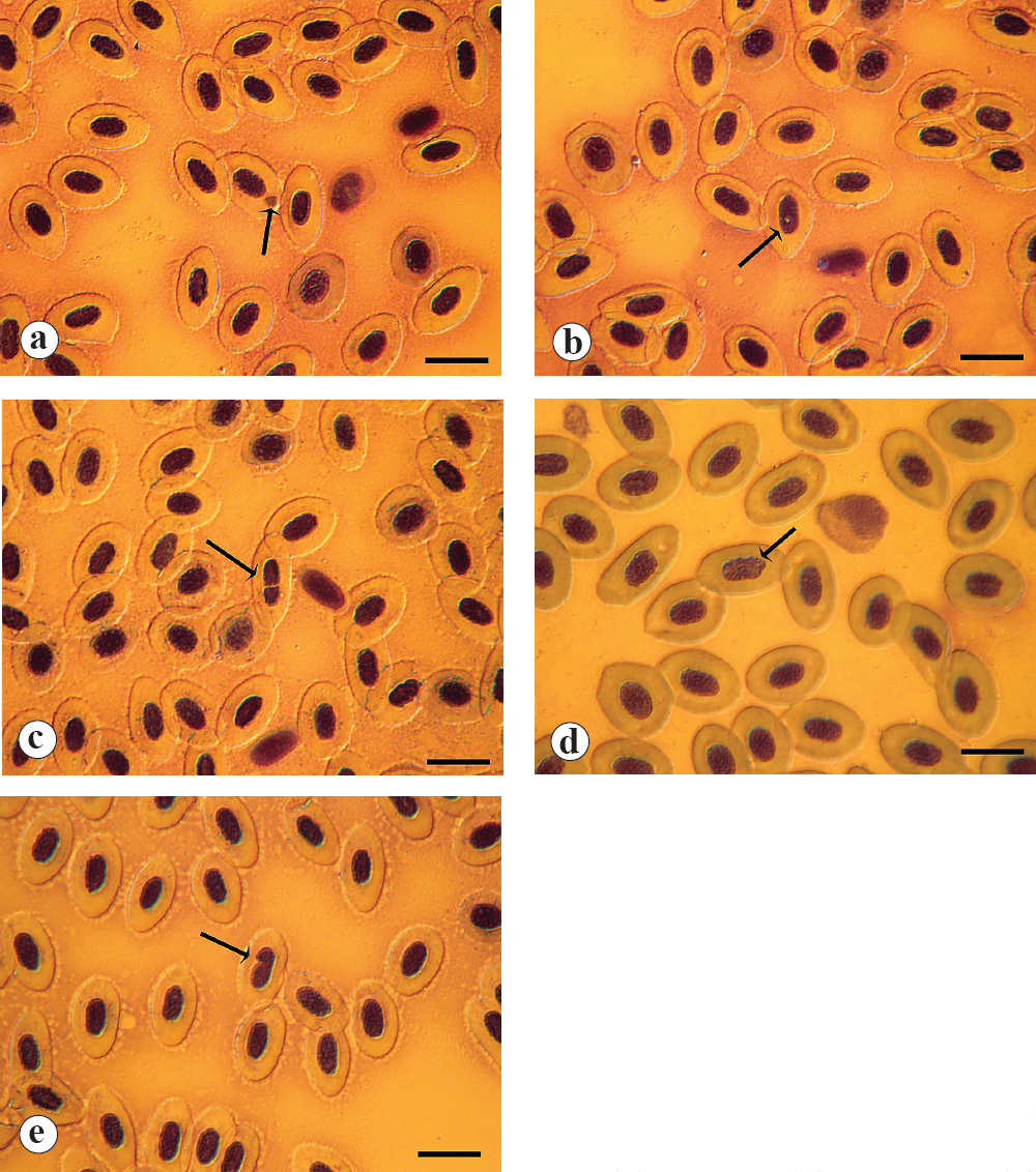
From each of localities, thirty barbel were collected with electro-fishing, covering a distance of nearly 1000 m downstream from the chosen points. Blood samples were taken from the caudal vein with heparinized 2.5ml sterile plastic syringes and needles from 0.45 mm x 16 mm (27G x 5/8 inches) to 0.8 mm x 40 mm (21G x 1.5 inches). Promptly after the collection, blood smears were made (5 slides per fish), air-dried and fixed 10 minutes in methanol. The blood smears were afterward stained with May-Grünwald/Giemsa (MGG) method, modified according to Strunjak-Perovic et al. (14). On each slide, areas with uniform spread in monolayer without overlapping cells were targeted. From each slide 20 fields were analyzed (total - 100 fields per fish) at a magnification of 1000x, chosen by systematic random sampling method. Total number of analysed erythrocytes per fish varied from 8000-10000, depending on the density of cells distribution on the smear. Analyses consisted of detection and count of barbel’s erythrocyte MN, VN, BiN, NotchN and BlebN classified according to Carrasco et al. (1). Frequency of each nuclear anomaly was calculated relative to the total count of erythrocytes for each fish. All results were presented in per mill.
The obtained results are presented as mean values accompanied with coefficient of variation (CV = SD / mean). Software STATISTICA 7.0 for Windows was used for the statistical analysis. One-way ANOVA, followed by post-hoc Newman-Keuls test were used for testing the effect of locality on the measuring parameters. Results were considered significant for probabilities greater than 95% (p < 0.05).
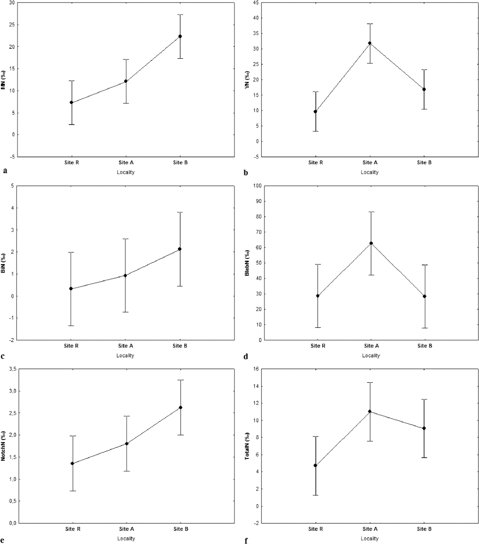
The frequency of MN (Table 1, Fig. 2a) showed lowest value in the control site (Site R), compared to the other two sites, while this parameter was significantly greater in Site B in comparison with the Sites R and A (Fig. 3a).
Table 1. Frequency of barbel micronuclei* (MN), vacuolated nuclei (VN), binuclei (BiN), blebbed nuclei (BlebN) and notched nuclei (NotchN), expressed as permiles from the Site R, Site A and Site B
Figure 2. Light micrograph of the blood smear of a barbel shows micronuclei (2a), vacuolated nuclei (2b), binuclei (2c), blebbed nuclei (2d) and notched nuclei (2e)
Figure 3. Changes in the frequency of the a) micronuclei – MN; b) vacuolated nuclei – VN; c) binuclei – BiN; d) blebbed nuclei – BlebN; e) notched nuclei - NotchN and f) total nuclear deformations – TotalN sampled from the selected sites. The data are expressed as mean, and vertical bars denote a 0.95 confidence interval
Values with different lowercase superscript letters in the same column represent significant differences (p<0.05) between sampling sites, according to the ANOVA followed by post-hoc Newman-Keuls test.
The highest and significant frequency values for the VN and BlebN were recorded in Site A (Table 1, Fig. 2b and 2d), compared to the other two sites (Fig. 3b; 3d). Concerning the BlebN, almost equal frequencies were recorded in Sites R and B. On the other hand, the frequency of NotchN (Fig. 2e) revealed significantly higher values in fish collected in Site B (Table 1), compared to the other two locations (Fig. 3e).
The only parameter that did not show significant difference between investigated localities was BiN (Fig. 2c, Fig. 3c), in spite of the three-fold increased frequency of fluctuation in Site A (0.93 ‰) and six-fold increase in Site B (2.12 ‰), compared to the control Site R (0.33 ‰) (Table 1).
The analysis of all nuclear abnormalities (TotalN) showed the lowest value in Site R and the highest value in Site A, which was significant compared to values in Sites R and B. (Fig. 3f).
In general, the Coefficient of Variation showed extremely high variability for all examined parameters. Minimal biological variation was noticed for MN in Site A (0,40) and maximal for BiN in Site B (2,01) (Table 1).
River Bregalnica has been intensively monitored in the past decade, and some researches revealed high water pollution, especially in Site A, where a complex mixture of pollutants derived from household wastewater, heavy metals from the nearby Zletovo mine and pesticides and fertilizers from agricultural activities admixes (12, 15). The results of this study showed the highest TotalN and a two- and three-fold higher VN and BlebN frequencies in fish from Site A, probably caused by the mixture of pollutants. Namely, literature data showed that synergistic effect of pollutants is more deleterious than their isolated influence (16).
Compared to the Site R, although MN was not the highest at this point (Site A), we have recorded extremely higher frequencies of VN and BlebN, which are considered as predecessor for MN (17, 18). Although the etiology of VN is not known, some authors suggest that it is a result of aneuploidy leading to micronuclei (18). Verstraeten et al. (19) described donut-shaped nuclei induced by genotoxins formed due to de-condensation of chromatin in late mitosis. In their study (19), the described donut-shaped nuclei exhibited defects in karyokinesis, aneuploidy and often multinucleation. These defects are in accordance with the proposed association of VN, as aneuploidies that lead to MN by Anbumani and Mohankumar (18). Although there is no clear support yet, we presume that both VN and the donut-shaped nuclei might have similar mechanism of formation. If we presume that VN is analogous to the donut-shaped nuclei described by Verstraeten et al. (19), the results from our study, i.e. the high frequency of VN would lead to a conclusion of the presence of genotoxic substances into the water of Site A. However, this cannot be confirmed without further investigation.
BlebN whose frequencies are also extremely high in Site A is described as a mechanism of the cell to cope with excess chromatin that will become MN and then expelled from the cell as double minute - DM (17). Acute exposure causes significant damage that could induce apoptosis as opposed to chronic exposure that leaves a time to adapt and repair damage (18). It is possible that a prolonged exposure of barbel to pollution in this site leads to activation of certain mechanisms, e.g., a nuclear budding for removing excess chromatin, which could explain the extremely high frequency of BlebN.
Although we already noticed insignificantly higher MN frequencies in Site A compared with Site R, we should take into account that MN circulation frequencies caused by different pollutants are dose, time and species dependent, reaching different peak-time, while after a prolonged exposure their frequencies decline (20, 21). Thus, in experiments with different concentrations of cadmium as genotoxin, the frequency of blood erythrocytes MN in Nile tilapia (Oreochromis niloticus) appears to have a peak at the fourth day and then decrease gradually, starting from the sixth day from the treatment (21). Experiments with higher concentrations of elemental or metallic mercury showed significant increase in MN frequencies in peripheral erythrocytes of Cyprinus carpio, which was higher after 31 days of exposure, followed by slight stabilization and gradual decrease (20).
Nepomuceno and Spanó (22) showed that exposure of Cyprinus carpio to different concentrations of pesticides induced MN formation in the blood erythrocytes in the first 24h, except for the highest concentration where highest MN frequencies were observed at 48h. According to these authors, this is a result of cell death at high concentration in the first 24h, which masked the appearance of MN. However, decrease of the MN frequencies after prolonged time might be a defensive mechanism/adaptation that stabilizes MN and nuclear abnormalities frequency due to removal of the damaged cells (20). Moreover, increased erythropoiesis can dilute or mask the number of MN and cell cycle and maturation time should be taken into account (23). In that line, insignificantly higher frequency of MN recorded in Site A compared with Site R, suggests that in this site genotoxic influence is prolonged. Prolonged exposure may allow decreasing of MN frequency by the removal of dying out or defective cells from circulation.
Highest frequency of MN, BiN and NotchN were observed in Site B. The frequency of MN was significantly higher compared with other two sites, while the frequency of NotchN was significantly higher compared with the Site R. Although BiN frequency in fish from the Site B was twice bigger than frequency of this parameter in Site R, those differences were not significant probably due to big inter-individual variation observed for this nuclear abnormality. In Site B organic pollution and many detergent surfactants from the household wastewater, as well as textile industry wastewater are floweing into the river without pretreatment (11, 12). Urban wastewater is a complex mixture that is responsible for multiple effects in fish including genotoxic effects (24). Field based investigations with native fish populations (25) and in situ cage exposure (26) in river sectors after big cities, where the water quality deteriorates, revealed significantly increased MN frequencies. Only one specific stressor does not influence formation of MN and other nuclear abnormalities; it is rather multifactorial (14). In this vein, organic contaminants in the sediments have stronger genotoxic potency than heavy metals, while the presence of both heavy metals and organic pollutants can decrease the overall genotoxic effects (27). Although textile industry wastewater could be a source of copper, chromium and nickel (28), metal toxic effects on barbel in the Site B is less expected than in the Site A. This is a result of the reduced bioavailability of metals which in this type of wastewater appear in form of complexes (28). Thus, we can assume that organic pollutants may have stronger genotoxic influence resulting with higher MN frequency in Site B.
Although the MN and other nuclear abnormalities revealed different pattern of variation along the river flow, the lowest values for all measured parameters were observed in the Site R. In general, significantly higher values for erythrocyte deformations, including MN noted in the middle and lower part of the river showed pollution burden of this water ecosystem. The results of this study suggest that all toxic materials that finally end into the river display genotoxic effect on aquatic organisms. Frequencies of MN and nuclear abnormalities may vary, depending on type of pollutants, their combination and time of exposure. The presence of MN and nuclear abnormalities should be taken as an early warning sign for the current status of this ecosystem concerning genotoxicity. The pollution pressure on the ecosystem by genotoxins should be controlled, in order to avoid reduction of reproductive potential and decrease in size of barbel population. This study provides a scientific basis for future investigations regarding the genotoxicity in the River Bregalnica ecosystem.
1. Carrasco K, Tilbury K.L, Myers M.S, Assessment of the piscine micronucleus test as an in situ biological indicator of chemical contaminant effectsCan J Fish Aquat Sci 1990; 47: 2123-2136. http://dx.doi.org/10.1139/f90-237
2. Ayllón F, Garcia-Vazquez E, Micronuclei and other nuclear lesions as genotoxicity indicators in rainbow trout Oncorhynchus mykissEcotoxicol Environ Saf 2001; 49: 221-225. http://dx.doi.org/10.1006/eesa.2001.2065 PMid: 11440474
3. Çavaş T, Ergene-Gözükara S, Induction of micronuclei and nuclear abnormalities in Oreochromis niloticus following exposure to petroleum refinery and chromium processing plant effluentsAquat Toxicol 2005; 74: 264-271. http://dx.doi.org/10.1016/j.aquatox.2005.06.001 PMid: 16023743
4. Fenech M, Cytokinesis-block micronucleus assay evolves into a “cytome” assay of chromosomal instability, mitotic dysfunction and cell deathMutat Res 2006; 600: 1-258-66. http://dx.doi.org/10.1016/j.mrfmmm.2006.05.028 PMid: 16822529
5. Fulda S, Gorman A.M, Hori O, Samali A, Cellular Stress Responses: Cell Survival and Cell DeathInt J Cell Biol 2010 2010; 214074- http://dx.doi.org/10.1155/2010/214074
6. Al-Sabti K, Metcalfe C.D, Fish micronuclei for assessing genotoxicity in waterMutat Res 1995; 343: 121-135. http://dx.doi.org/10.1016/0165-1218(95)90078-0
7. Ali F, El-Shehawi A.M, Seehy M.A, Micronucleus test in fish genome: A sensitive monitor for aquatic pollutionAfr J Biotechnol 2008; 7: 5606-612.
8. Furnus G.N.A, Caffetti J.D, García E.M, Benítez M.F, Pastori M.C, Fenocchio A.S, Baseline micronuclei and nuclear abnormalities frequencies in native fishes from the ParanáRiver (Argentina)Braz J Biol 2014; 74: 1217-221. http://dx.doi.org/10.1590/1519-6984.13712
9. van der Oost R, Fish bioaccumulation and biomarkers in environmental risk assessment: a reviewEnviron Toxicol and Phar 2003; 13: 57-149.
http://dx.doi.org/10.1016/S1382-6689(02)00126-6
10. Balabanova B, Stafilov T, Šajn R, Bačeva K, Spatial distribution and characterization of some toxic metals and lithogenic elements in topsoil and subsoil from copper mine environsInt J Environ Prot 2013; 3: 91-9.
11. Dimitrovska O, Markoski B, Apostolovska Toshevska B, Milevski I, Gorin S, Surface water pollution of major rivers in the Republic of MacedoniaProcedia Environ Sci 2012; 14: 32-40. http://dx.doi.org/10.1016/j.proenv.2012.03.004
12. Spasovski O, Heavy and Toxic Metals and Nutrients in Separate Places in the River Bregalnica (Eastern Macedonia)Annual of the university of mining and geology “St. Ivan Rilski” 2013; 54: 2118-120.
13. Ramani S, Dragun Z, Kapetanović D, Kostov V, Jordanova M, Erk M, Hajrulai-Musliu Z, Surface water characterization of three rivers in the Pb/Zn mining region of north-eastern MacedoniaArch Environ Contam Toxicol 2014; 66: 4514-528. http://dx.doi.org/10.1007/s00244-014-0012-z PMid: 24626792
14. Strunjak-Perovic I, Topic Popovic N, Coz-Rakovac R, Jadan M, Nuclear abnormalities of marine fish erythrocytesJ Fish Biol 2009; 74: 2239-2249.
http://dx.doi.org/10.1111/j.1095-8649.2009.02232.x PMid: 20735550
15. Popov S.I, Stafilov T, Šajn R, Tănăselia C, Bačeva K, Applying of factor analyses for determination of trace elements distribution in water from river Vardar and its tributaries, Macedonia/GreeceSci World J 2014 2014; 809253- http://dx.doi.org/10.1155/2014/809253
16. de Castilhos Ghisi N, Siundararajan R.P, Relationship between biomarkers and pesticide exposure in fishes: A reviewPesticides–advances in chemical and botanical pesticides 2012; InTech Open Access Publisher; http://dx.doi.org/10.5772/48604
17. Shimizu N, Itoh N, Utiyama H, Wahl M.G, Selective entrapment of extrachromosomally amplified DNA by nuclear budding and micronucleation during S phaseJ Cell Biol 1998; 140: 61307-1320. http://dx.doi.org/10.1083/jcb.140.6.1307
18. Anbumani S, Mohankumar M.N, Gamma radiation induced micronuclei and erythrocyte cellular abnormalities in the fish Catla catlaAquat Toxicol 2012; 122–123: 125-132. http://dx.doi.org/10.1016/j.aquatox.2012.06.001 PMid: 22771702
19. Verstraeten V.L, Peckham L.A, Olive M, Capell B.C, Collins F.S, Nabel E.G, Young S.G, Fong L.G, Lammerding J, Protein farnesylation inhibitors cause donut-shaped cell nuclei attributable to a centrosome separation defectProc Natl Acad Sci U S A 2011; 108: 124997-5002. http://dx.doi.org/10.1073/pnas.1019532108 PMid: 21383178 PMCid: PMC3064351
20. Nepomuceno J, Ferrari I, Spanó M.A, Centeno A, Detection of micronuclei in peripheral erythrocytes of Cyprinus carpio exposed to metallic mercuryEnviron Mol Mutagen 1997; 30: 3293-297. http://dx.doi.org/10.1002/(SICI)1098-2280(1997)30:3<293::AID-EM7>3.0.aCO;2-M
21. Özkan F, Gündüz S.G, Berköz M, Özlüer Hunt A, Induction of micronuclei and other nuclear abnormalities inperipheral erythrocytes of Nile tilapia, Oreochromis niloticus, following exposure to sublethal cadmium dosesTurk J Zool 2011; 35: 4585-592.
22. Nepomuceno J.C, Spanó M.A, Induction of micronuclei in peripheral erythrocytes of Cyprinus carpio fish by methyl parathionRev Int Contam Ambie 1995; 11: 19-12.
23. Udroiu I, The micronucleus test in piscine erythrocytesAquat Toxicol 2006; 79: 201-204. http://dx.doi.org/10.1016/j.aquatox.2006.06.013 PMid: 16846653
24. Bolognesi C, Hayashi M, Micronucleus assay in aquatic animalsMutagenesis 2011; 26: 1205-213. http://dx.doi.org/10.1093/mutage/geq073 PMid: 21164204
25. Minissi S, Ciccotti E, Rizzoni M, Micronucleus test in erythrocytes of Barbus plebejus (Teleostei, Pisces) from two natural environments: a bioassay for the in situ detection of mutagens in freshwaterMutat Res 1996; 367: 245-251. http://dx.doi.org/10.1016/S0165-1218(96)90084-1
26. Klobučar G.I.V, Štambuk A, Pavlica M, SertićPerić M, KutuzovićHackenberger B, Hylland K, Genotoxicity monitoring of freshwater environments using caged carp (Cyprinus carpio)Ecotoxicology 2010; 19: 77-84. http://dx.doi.org/10.1007/s10646-009-0390-6 PMid: 19626438
27. Costa P.M, Lobo J, Caeiro S, Martins M, Ferreira A.M, Caetano M, Vale C, DelValls Á, Costa M.H, Genotoxic damage in Solea senegalensis exposed to sediments from the Sado Estuary (Portugal): Effects of metallic and organic contaminantsMutat Res 2008; 654: 29-37. http://dx.doi.org/10.1016/j.mrgentox.2008.04.007 PMid: 18534898
28. European Integrated Pollution Prevention and Control Bureau (EIPPCB). Reference Document on Best Available Techniques for the Textile Industry 2003; European commission. BREF; 77-85.
© 2016 Ivanova L. This is an open-access article publishedunder the terms of the Creative Commons Attribution License whichpermits unrestricted use, distribution, and reproduction in any medium,provided the original author and source are credited.
The authors declared that they have no potential conflict of interest with respect to the authorship and/or publication of this article.
Macedonian Veterinary Review. Volume 39, Issue 2, Pages 159-166, p-ISSN 1409-7621, e-ISSN 1857-7415, DOI: 10.1515/macvetrev-2016-0080, 2016