Mac Vet Rev 2018; 41 (1): 33 - 37
 10.1515/macvetrev-2017-0028
Received: 09 May 2017
Received in revised form: 30 August 2017
Accepted: 14 September 2017
Available Online First: 23 November 2017
Published on: 15 March 2018
Keywords: tuna loins, Katsuwonus pelamis, canned tuna, Staphylococcus aureus, histamine
Tuna is a salt water fish belonging to the tribe Thunnini, a part of the Scombridae family. Normally the name tuna encompasses more than 50 species; of which 5 (yellowfin Thunnus albacares, bigeye Thunnus obesus, bluefin Thunnus thynnus, albacore Thunnus alalunga, and skipjack Katsuwonus pelamis) are important to feed a global supply chain in which the European Union represents the primary market (10). Widely consumed worldwide as fresh or canned (2), tuna is caught in all the major oceans (15). After harvest it is normally eviscerated, steam cooked (pre-cooking) and chilled to facilitate the cutting and the separation (cleaning) of the bones and skin (13). The light meat, obtained from the cleaning step, can be directly canned or frozen in plastic films vacuum packed (loins) and then shipped worldwide to producers, where it is thawed and canned.
Considering that tuna fish, like other scombroid fish, is commonly associated with cases and outbreaks of histamine intoxication, the pre-cooking step is important for reducing bacterial contamination, in particular Gram-negative bacteria responsible for histamine production. Additionally, since humans represent the first reservoir of Staphylococcus aureus (8), handling and manipulation during the “cleaning” step may contaminate tuna. Furthermore, frozen storage can be carried on for a long time before the loins are used by the industry, whereas Staphylococcus aureus has been demonstrated to persist in frozen tuna loins at -20°C for 4 weeks and to rapidly increase population up to 3 Log cfu/g when thawed (17). Enterotoxin, a single thermostable chain protein, normally produced when Staphylococcus aureus reaches 5-6 Log cfu/g (14), is able to resist the canning process therefore this hazard has to be considered in risk assessment.
The aim of this work is to assess the microbiological quality and safety of precooked tuna loins imported by a canned tuna producer in the Calabria region (Italy) from two different fishing areas, focusing on Staphylococcus aureus enterotoxin and histamine.
Fifty samples of frozen precooked loins of skipjack tuna (Katsuwonus pelamis), were obtained from a canning plant. 38 were from fishing area n°71 and 12 from fishing area n°87 of the Food and Agriculture Organization of the United Nations (FAO). Two hundred grams of tuna meat were aseptically sampled after thawing overnight (12-14 hours) in refrigerator at 7°C, and the samples were prepared within 1 h. Samples preparation for microbiological analysis was made according to UNI EN ISO 6887-3:2004. On every sample the following analyses were carried out. Total bacterial count (TBC) was evaluated according to UNI EN ISO 4833-1:2013 using Plate Count Agar (Oxoid, Hampshire, UK) as culture medium incubating plates for 24h at 30°C. Coliforms and Enterobacteriaceae were enumerated on Violet Red Bile Agar (Oxoid, Hampshire, UK), according to ISO 4832-2006 and ISO 21528–2:2004.
Enumeration of beta-glucuronidase-positive Escherichia coli was performed according to UNI EN ISO 16649–1:2003; suspect isolates were confirmed by negative oxidase reaction (Oxoid, Hampshire, UK) and in positive indole reaction (Oxoid, Hampshire, UK).
Detection of Listeria monocytogenes was assessed according to UNI EN ISO 11290-1:2005; Listeria monocytogenes like-colonies were tested by Vitek2 System in accordance to the manufacturer’s instructions (bioMérieux SA, Marcy-l’Etoile, France).
Detection of Salmonella was done in accordance with ISO 6579:09.2006; colonies were biochemically tested by using Vitek2 System in accordance with the manufacturer’s instructions (bioMérieux SA, Marcy-l’Etoile, France).
Enumeration of Staphylococcus aureus (UNI EN ISO 6888 – 1:2004) was carried out after dilution in peptone water (Oxoid, Hampshire, UK) and using Baird-Parker Medium (Oxoid, Hampshire, UK) for culturing. The identification of suspect colonies was performed by the detection of coagulase (Denka Seiken, Tokyo, Japan) and confirmation using ViTek (bioMérieux SA, Marcy-l’Etoile, France).
Vibrio parahaemolyticus and Vibrio cholera were researched following ISO/TS 21872-1:2007; biochemical identification was carried out using Vitek2 (bioMérieux SA, Marcy-l’Etoile, France).
pH measurement was performed as reported by Kyrana (12) using a 5:1, water:fish homogenate and a glass electrode at 20°C (PCE instruments UK).
Staphylococcus aureus enterotoxin presence was assessed using miniVidas® (BioMèrieux, France) according to manufacturers instruction.
Histamine levels were evaluated using a colorimetric assay method with a commercial rapid histamine test kit (RIDA ® QUICK Histamine from R-Biopharm AG, Darmstadt, Germany) in 50 g of fish muscle according to the manufacturer’s instruction.
All bacterial counts were analysed in triplicate; results are the means of three determinations and expressed as Log values for statistical analysis. To asses if the samples coming from the two fishing areas were different a t test was performed. Furthermore, the correlation between pH and microbiological parameters was analysed by linear regression. The statistical software GraphPad Prism version 7.00 for Windows (GraphPad Software, La Jolla California USA, (www.graphpad.com) was used.
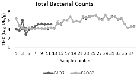
The total bacterial count ranged from 2,94 to 6,04 Log cfu/g for samples from FAO fishing area 71 and from 4,08 to 7,26 for samples from FAO fishing area 87. Results are showed in Fig 1.
Figure 1. Total bacterial count (TBC) of microorganisms in tuna loins from two different FAO fishing areas
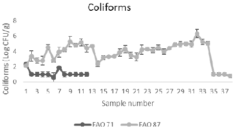
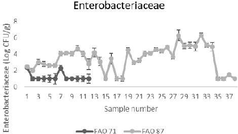
Results for coliforms, Escherichia coli and Enterobacteriaceae are reported in Fig. 2, 3 and 4 respectively.
Figure 2. Coliforms in tuna loins from two different FAO fishing areas
Figure 3. Enterobacteriaceae in tuna loins from two different FAO fishing areas
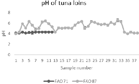
Figure 4. pH in tuna loins from two different FAO fishing areas
Total bacterial count, Coliforms, E. coli and Enterobacteriaceae were statistically different between the fishing areas (p<0,05).
A positive correlation was evidenced between pH and TBC (r = 0,3831 p<0,05), between pH and coliforms (r = 0,7834 p<0,05), pH and E. coli (r = 0,6962 p<0,05) and between pH and Enterobacteriaceae (r = 0,7249; p<0,05).
Staphylococcus aureus was evidenced in 45 samples below 2 Log cfu/g and in 5 samples, from FAO fishing area 71, exceeding 3 Log cfu/g. A total of 7 staphylococcal species were also isolated from 9 samples; the most frequent species was S. warneri (3 isolates), followed by S. saprophyticus (2 isolates), S. epidermidis (1 isolate), S. hominis (1 isolate), S. intermedius (1 isolate), S. vitulinus and S. sciuri (1 isolate). Staphylococcus aureus enterotoxin was not evidenced in any of the samples tested.
Listeria monocytogenes, Salmonella spp., Vibrio parahaemolyticus and Vibrio cholera were not detected in any of the samples tested.
Histamine levels ranged between 0 to 5 mg/kg; no differences were evidenced between harvesting areas.
The results of total bacterial count, Enterobacteriaceae and coliforms indicated a high contamination rate of tuna loins. In previous studies Wu and Su (17) reported that loins, chunk and flake of albacore and skipjack, analysed after thawing, had a total bacterial count ranging from 3,46 to 3,99 Log cfu/g. pH values, in tuna from the FAO 71 fishing area, are in agreement with previous studies (17) where a range from 5,60 to 5,61 was reported; on the other hand samples from FAO 87 fishing area had a statistically lower pH. The difference in the bacteriological and pH values between fishing areas can be due to capture and post-capture practices used in the different FAO areas or to a temperature abuse before freezing (6).
The presence of Staphylococcus aureus in all the samples tested can be due to the manual handling of pre-cooked tuna loins prior to canning. Although contamination reached 3 log cfu/g in 5 samples, enterotoxin production has been demonstrated at 27°C, within 8 h in albacore and within 8 to 10 h in skipjack (11). Additionally Staphylococcus warneri, S. intermedius and S. sciuri has been demonstrated to produce enterotoxin (1, 4) and enterotoxins genes were evidenced in S. saprophyticus and S. hominis as reported in previous studies (5, 16). Even though European regulation did not lay down limits for staphylococci in fishery products, but only for shelled and shucked products of cooked crustaceans and molluscan shellfish, Staphylococcus spp., presence has to be taken into account when risk analysis of tuna product is carried out, since temperature abuse can allow bacteria reaching 5-6 log cfu/g value normally indicated as threshold for enterotoxin production (3, 9).
Commission Regulation (2073/2005/EC) on microbiological criteria for foodstuff and its amendments (Regulation 1441/2007/EC and Regulation 365/2010/EC) lays down food safety criteria for histamine in fishery products for fish species (Scombridae, Clupeidae, Engraulidae, Coryfenidae, Pomatomidae, Scombresosidae) associated with a high amount of histidine between 100 mg/kg (m) and 200 mg/kg (M) and for fishery products which have undergone enzyme maturation treatment in brine, manufactured from fish species associated with a high amount of histidine between 200 mg/kg (m) and 400 mg/kg (M). In addition, Regulation 853/2004/EC lays down specific hygiene rules for food of animal origin, providing a possibility to set freshness criteria and limits for fishery products with regards to histamine and place the responsibility on food business operators to ensure that limits with regard to histamine are not exceeded in the context of health standards for these products. Histamine fish poisoning still represents the main hazard for canned tuna since no available method of preparation, including freezing, canning and smoking, is able to destroy the causative toxin (7). Our results, far from the EU limits, showed the safety of the analysed tuna loins.
For both histamine and enterotoxin, attention has to be paid to cold chain maintenance especially after thawing and before canning.
Tuna loins analysed in this paper have to be considered safe; the pre-cook step, normally used in processing of tuna loins, was successful in reducing bacterial load, destroying pathogens and halting gram negative growth responsible for histamine production. The main concerns are represented by histamine and staphylococci presence: temperature used in the canning process can destroy all vegetative cells, but enterotoxin and histamine are able to resist canning temperature. Minimizing the time between thawing and canning can reduce risk to an acceptable level.
© 2017 Casalinuovo F. This is an open-access article published under the terms of the Creative Commons Attribution License which permits unrestricted use, distribution, and reproduction in any medium, provided the original author and source are credited.
Special thanks to Dr Elonora Sarno for all advices and support.
The authors declared that they have no potential conflict of interest with respect to the authorship and/or publication of this article.
Macedonian Veterinary Review. Volume 41, Issue 1, Pages 33-37, p-ISSN 1409-7621, e-ISSN 1857-7415, DOI: 10.1515/macvetrev-2017-0028, 2018