Mac Vet Rev 2018; 41 (1): 47 - 53
 10.1515/macvetrev-2017-0030
Received: 13 October 2016
Received in revised form: 17 November 2017
Accepted: 23 November 2017
Available Online First: 09 December 2017
Published on: 15 March 2018
Keywords: Mycobacterium avium subsp. hominissuis, granuloma, myofibroblast, TGF-β1, TGF-β1RI
Mycobacterium avium, belonging to the Mycobacterium avium complex (MAC), is divided into the subspecies avium, paratuberculosis and silvaticum (1). More recently, M. avium subsp. aviumhas been further divided into M. avium subsp. avium and M. avium subsp. hominissuis (2). M. avium subsp. hominissuis (MAH) is a ubiquitous microorganism which lives both in water and soil and causes mycobacteriosis of pigs and immunocompromised human population (3, 4, 5, 6). The hallmark of MAH infections in pigs are granulomatous lesions localized on gastrointestinal and mandibular lymph nodes (7), but generalized disease is less common and can affect liver, spleen or kidneys. Nevertheless, the morphological changes are not always visible during meat inspection in slaughterhouses (8-12).
Granuloma is a unique form of tissue reaction on presence of agents which are able to provoke a chronic inflammation (10), but development and sustainability of granulomas are controlled by interferon γ (INF-γ), tumor necrosis factor α (TNF-α) and transforming growth factor β1 (TGF-β1) (10, 11). Although cytokine mediated immunosuppressive effect is well known in different infection diseases of pigs, TGF-β1 represents an important molecule with distinctive imunomodulating features (12, 13). A connection between transforming growth factor - (TGF-β1) and TH-17 cells has been also investigated and confirmed (14). The cells demarcating granulomas expressing alpha-smooth muscle actin and vimentin, suggest that these cells are myofibroblasts. It is believed that TGF-β could affect the induction of myofibroblast proliferation. The aim of this study is to point out the importance of myofibroblasts in the formation and sustainability of granulomas during natural infection of pigs with M. avium subsp. hominissuis.
Examinations have been performed on the samples of gastrointestinal lymph nodes (Lnn. jejunales, Lnn. ileocolici and Lnn. colici) of 100 pigs (Norwegian Landrace breed), 5-10 months old and all positive on intradermal comparative tests for bovine and avian tuberculin (Bovitubal and Avitubal, Bioveta, Czech Republic). The pigs imported in Serbia originating from Lithuania were kept in quarantine. Immediately after the euthanasia using T-61 (Merck animal health Intervet, Holland) in accordance with Serbian national animal welfare regulation, autopsy and sampling of gastrointestinal lymph nodes were performed. The tissue samples for microscopic and molecular examinations were fixed 48 hours in a 10% buffered formalin. After standard tissue processing and paraffin embedding, 4-5 µm thick sections were stained with hematoxylin-eosin (HE) and selected for immunohistochemistry. Formalin fixed and paraffin embedded tissue sections (FFPE) of gastrointestinal lymph nodes of 10 healthy pigs were used as negative control.
Molecular examination on lymph nodes has been performed according to the Norwegian Veterinary Institute protocol described in a previos study (15).
Briefly, one hundred and fifteen FFPE lymph nodes from 100 pigs were sent to the Norwegian Veterinary Institute, and real-time PCRs for detection of DNA sequences of M. avium and MTC were performed. For extraction of DNA, Nucleon HT (Tepnel Life Sciences, Manchester, UK) was applied, following the protocol provided by the manufacturer. From mycobacterial isolates and non-fixed tissue samples, DNA was extracted by Nuclisens® easyMag® (BioMerieux, Inc., Durham, NC, USA) following the manufacturer’s instructions. Three singleplex real-time PCRs were developed, for IS1245 and IS6110 specific for M. avium and MTC respectively, and for the porcine P-globin gene. Amplification of the latter sequence was used as a positive control for successful DNA extraction from the samples. Primers and 6FAM labelled TaqMan MGB probes were designed using the program Primer 3.0 (http://frodo.wi.mit.edu/primer3/) (Table 1). Real-time PCR was performed using Stratagene Mx3005P (Stratagene, La Jolla, CA, USA). Reaction mixtures had a total volume of 20 |Π, consisting of 2 |Π template DNA at a concentration of 10-15 ng/|Π, 10 |Π PerfeC Ta® qPCR FastMix® (Quanta Biosciences, Gaithersburg, MD, USA), and primers and probes at a final concentration of 400 and 150 nM, respectively.
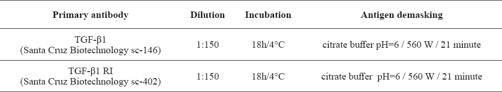
Table 1. Primary antibodies used in IHC (LSAB2) procedure
DNA extracted from FFPE lymph nodes from animals clinically infected with M. avium subsp. hominissuis or M. bovis, verified by culture prior to fixation, were used for optimization of PCR conditions. DNA extracted from M. avium ATCC 25291, M. bovis BCG (Danish strain 1331) and from a porcine spleen, were used as positive controls for IS1245, IS6110 and P-globin. Ultrapure Milli-Q water was used for adjustment of volume and concentrations and as a template substitute in negative controls.
The real-time PCR reactions were initiated with denaturation at 95 °C for 10 min, followed by 45 runs of the following thermal cycle: 95 °C for 3 sec and 60 °C for 30 sec. All samples and controls were run as single measurements. The results were analyzed using Stratagene MxPro 4.10 software (Stratagene), applying the automatic calculation of the threshold fluorescence. Ct values above 40 were regarded as negative as recommended by the manufacturer (Stratagene).
According to microscopical features, granulomas were classified into three groups: 1. Granulomas without necrosis, 2. Granulomas with an initial necrosis and 3. Granulomas with distinctive necrosis and calcification.
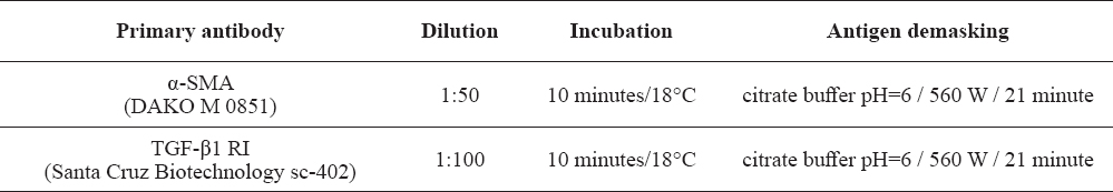
Immunohistochemical examinations were carried out on formalin fixed and paraffin embedded tissue sections. Streptavidin-biotin imunohistochemical method (DAKO, LSAB2) and the double IHC staining method for simultaneous detecting of two antigens (αSMA/ TGF-β1RI) in the same tissue section (DAKO Envision GI2 Doublestain) were used. The description of primary antibodies dilution, incubation and antigen retrieval are given in Table 1 and Table 2. Briefly, in LSAB2 endogenous peroxidase was blocked in 3% H2O2 at 18°C, 15 minutes followed by antigen retrieval in citrate buffer pH=6, 560W, 21 min. For vizualization of positive reaction, 3’ diaminobenzidine tetrachloride (DAB+), was used as a chromogene. Counterstaining was performed in Mayer hematoxylin and the slides were mounted in a water mounting medium, Glycergel (DAKO, C 563). The tissue sections without primary antibodies were used as negative control, while the tissue sections already positive to examined antigens served as positive control. For simultaneous visualization of two different antigens on the same tissue section “DAKO Envision GI2 Doublestain” system (K5361) was used. Microsscopic examinations were performed using microscope Olympus BX51.
Table 2. Primary antibodies used in the method of double IHC staining (DAKO Envision GI2 Doublestain)
The Norwegian Veterinary Institute received 115 FFPE lymph nodes from the Serbian pigs in order to confirm the presence of MAC. Twenty-three samples were excluded from real-time PCR analysis. Only one sample was included from each pig. Additionally, samples with uncertain sample ID or poor DNA quality measured by negative result on real-time PCR for the porcine P-globin gene were excluded. Of the 92 remaining samples analysed for the presence of IS1245 by real-time PCR, 51 (55.4%) were regarded positive. Ten (10.9%) of the 92 samples analysed by IS6110real-time PCR were considered positive, all but three were however simultaneously positive in IS1245 PCR. The PCR products of the samples positive for IS6110 were additionally sequenced and compared to published sequences by NCBI BLAST (http://blast.ncbi.nlm.nih.gov/Blast.cgi) to confirm their identity. Of the pigs with neither microscopic nor macroscopic lesions, only one of eight samples examined was positive for IS1245 by PCR, and none were positive for IS6110. In samples from 38 of the tuberculin reactors no IS element could be detected.
The microscopic examination of all PCR positive lymph node samples for IS1245 (51) stained by the routine Hematoxylin-eosin (HE) method showed the presence of granulomatous lymphadenitis.
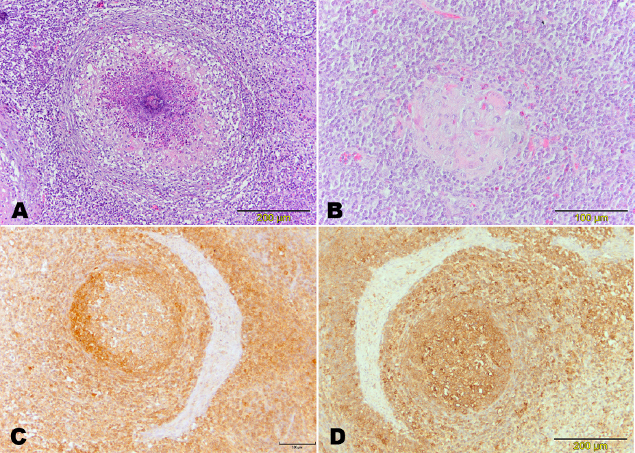
Positive cytoplasmic reaction for transforming growth factor TGF-β1 was detected predominantly on macrophages in granulomas, as well as on some lymphocytes. Expression of TGF-β1 was of lower intensity in granulomas with prominent fibrosis, necrosis and calcification. The high expression of TGF-β1 was present in macrophages in the early stages of granuloma development, while the very necrotic center of a granuloma was always negative to TGF-β1. (Fig. 1C).
Figure 1. Microscopic and immunohistochemical finding in a gastrointestinal lymph node of pigs infected with Mycobacterium avium subsp hominissuis. Macrophages and lymphocytes around the area of caseous necrosis in a granuloma, HE (A); Numerous giant cells in a granuloma, HE (B); Expression of TGF-β1RI, LSAB2 (C); Expression of TGF-β1, LSAB2 (D)
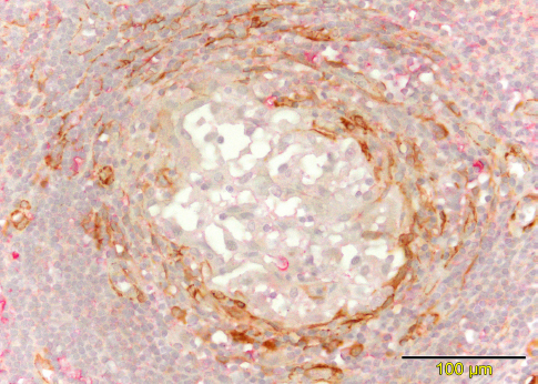
Receptor TGF-β1 RI for TGF-β1 molecule was exclusively expressed on myofibroblasts. Besides this signalling molecule, in the tissue samples stained by double immunohistochemical staining many of myofibroblasts simultaneously expressed smooth muscle actin αSMA and TGF-β1RI. Reaction to αSMA was homogenous and cytoplasmic. The αSMA expression was commonly detected in the cytoplasm of myofibroblasts at granuloma peripheries (Fig. 2).
Figure 2. Immunochistochemical finding in a gastrointestinal lymph node of pigs infected with Mycobacterium avium subsp hominissuis. Expression of αSMA/ TGF-β1RI, double immunohisochemical staining, DAB/PR. (αSMA - brown chromogen, TGF- β1RI - pink chromogen)
Granulomatous inflammation is a basic morphological characteristic of immune response to infection caused by mycobacteria. Granulomas are formed as immune response to infection of mycobacterium and they represent lesions where epithelioid cells, giant cells, fibroblasts and lymphocytes of various immunophenotypes are located usually around a necrotic and/or calcified center (16, 17). However, all immunophenotypic characteristics of effector cells in granulomas important for pathogenesis of swine mycobacteriosis are not well described. The ratio of myofibroblasts, epithelioid cells and occurrence of necrosis is controlled by various factors, but attention is paid mainly to TGF-β1 (18). It is believed that cytokines and growth factors which cause necrosis also lead to myofibroblast stimulation (TGF-β) supporting that that myofibroblasts have a significant role in formation of granulomas caused by mycobacterium (19). When activated, myofibroblasts express adhesive molecules for lymphocytes, mast cells and neutrophil granulocytes. They also participate in formation of tissue granulomas and play a significant role in inflammatory reactions, having in mind that they produce important inflammatory mediators (18). It is assumed that TGF-β1 induces proliferation of myofibroblasts in granulomas, which changes interpretation of its role considering that this factor has been also ascribed an inhibitory role in activation of macrophages in granulomatous lesions in tuberculosis. Some authors showed that myofibroblasts have a particularly important role in formation of granulomas caused by MAC in pigs (18, 20). They also emphasize the connection between the number of myofibroblasts and expression of TGF-β1 (21). Our previous results indicate that TGF-β1 induces proliferation of myofibroblasts. However current investigation reveals the important role of expression of TGF-β1 RI receptors in the development of granulomatous lymphadenitis in pigs infected by M. avium subsp. hominissuis.
Similary, in humans the number of myofibroblasts increases with the maturity of tuberculous granuloma, while these cells were found in low numbers in an early granulomatous lesion formed predominantly from epithelioid cells (18). Finally, immunoreactivity of TGF-β1 decreases with the progression fibrosis in granuloma (18). TGF-β1 is also considered responsible for reduced sensitivity to INF-γ in human infections caused by MAC and experimental infection with M. bovis in cattle (11, 18, 21). Immunohistochemistry of lung granuloma reveals the location of TGF-β primarily in giant cells of Langerhans type, and to a lesser extent in other macrophages (18). Using the immunohistochemical method, our study showed that the principal source of TGF-β1 molecules in granulomas of pigs infected with MAC are macrophages. Additionally, the cells demarcating granulomas expressing alpha-smooth muscle actin and vimentin, suggest that these cells are myofibroblasts.
Similarity between immunopathogenesis of infections by MAC in pigs and humans is pointed out by Hybiya et al. (17) showing the same pattern of MAC granuloma development in pigs and immuno-compromised humans both infected with M. avium.
Our results underline the role of myofibroblasts in morphogenesis of granulomatous lymphadenitis of pigs naturally infected with M. avium subsp. hominissuis. The double immunohistochemical staining showed that myofibroblasts which express TGF-β1RI and αSMA play a key role in the morphogenesis of granulomatous lymphadenitis in pigs infected with Mycobacterium avium subsp. hominissuis. Infections of pigs with M. avium subsp. hominisuis have characteristics similar to infections with MAC in humans and therefore could be used also for studying pathogenesis of micobacterial infections in humans.
©2017 Polaček V. This is an open-access article published under the terms of the Creative Commons Attribution License which permits unrestricted use, distribution, and reproduction in any medium, provided the original author and source are credited.
We thank Tone Bjordal Johansen and Angelika Agdeinstain from the Norwegian Veterinary Institute for their help with real time PCR diagnostics and Danka Vukasinovic for helping translate the article. The research was partly conducted within the project of the Ministry of Education and Science of the Republic of Serbia, under the registration number TR31071, TR31084 and III46002.
The authors declared that they have no potential conflict of interest with respect to the authorship and/or publication of this article.
Macedonian Veterinary Review. Volume 41, Issue 1, Pages 47-53, p-ISSN 1409-7621, e-ISSN 1857-7415, DOI: 10.1515/macvetrev-2017-0030, 2018