Mac Vet Rev 2018; 41 (1): 95 - 98
 10.1515/macvetrev-2017-0032
Received: 05 June 2017
Received in revised form: 24 July 2017
Accepted: 14 September 2017
Available Online First: 14 December 2017
Published on: 15 March 2018
Keywords: canine strongyloidosis, febantel, larvae, treatment
The intestinal threadworm Strongyloides stercoralis is a soil-transmitted, potentially zoonotic nematode with worldwide distribution (1). It is a slender hair-like worm around 2 mm long that parasitizes the proximal small intestine of dogs, wild canids, non-human primates and humans, mostly in tropical and sub-tropical countries (1, 2, 3). It has a free living form (males and females in soil) and a female-only parasitic form. The females are parthenogenetic and lay eggs. The hatched first-stage larvae (L1) contaminate the environment through the faeces. In the environment, L1 moult to infective third-stage larvae (L3) that can infect a host primarily by penetration of the skin and rarely by ingestion (4). The transmammary route of infection is also described, but it is controversial and can occur if the bitch gets infected in late gestation and during lactation (5, 6). The infective L3 migrate to the lungs, then are coughed out, swallowed and reach the small intestine within 4-5 days (1, 6). Autoinfection can occur if the L1 develop to infective L3 before leaving the host’s digestive tract, and is often associated with immunosuppression. Immunocompromised dogs may also develop a severe hyperinfection (L3 that migrate throughout the body) that is related to the acute form of the disease (1, 4). The prepatent period is 5-21 days (1).
The infection with S. stercoralis may be asymptomatic and self-limiting, or cause an acute or chronic disease. The clinical signs are more commonly seen in young dogs and puppies, especially in kennels and shelters. High worm burden results in watery diarrhoea and bronchopneumonia and can be life threatening (1, 3, 4).
The diagnosis of the infection is commonly based on detection of L1 in the faeces with methods such as the Zinc sulphate flotation solution method and the Baermann funnel concentration method (1, 7). The recovered L1 are distinguishable by the morphology of the oesophagus (made up of corpus, isthmus and muscular bulb) and other features. For successful treatment of the infection, ivermectin, albendazole and fenbendazole are expected to be effective (1, 8, 9, 10).
This study describes the first S. stercoralis case in a dog in the Republic of Macedonia and a successful treatment of the infection with a commercial combination drug of praziquantel, pyrantel and febantel.
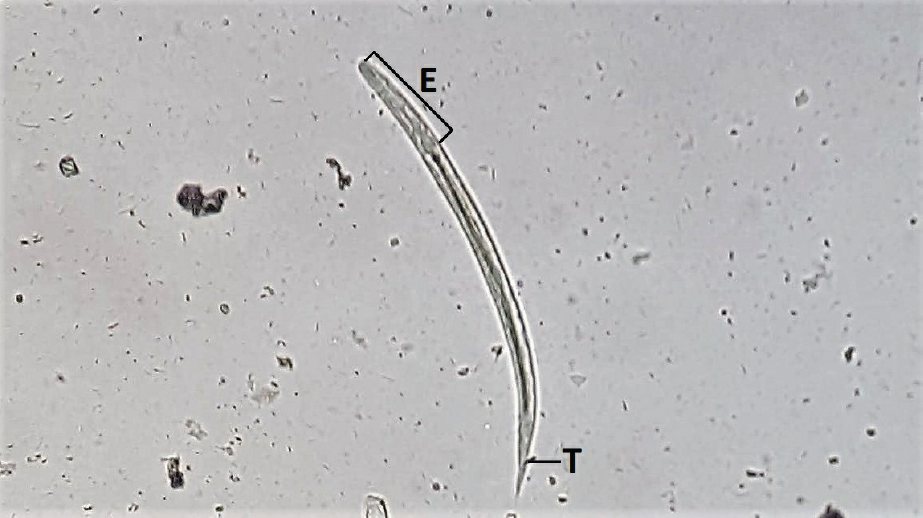
A six-month-old Pomeranian male dog, weighing 1.8 kg, was referred to the University Veterinary Hospital at the Faculty of Veterinary Medicine in Skopje in March 2017 with a history of unformed, soft faeces during the last month and mild weight loss. The dog was imported from Russia a week before the admittance to the Hospital. The dog was afebrile, with unchanged appetite and normal activity. Blood tests showed only mild anaemia (4.69x10¹²/l). A fresh faecal sample was collected, divided in 1 gram portions and analysed by direct faecal smear, Zinc sulphate flotation and the Baermann method (1, 7, 8, 10). The coprological analyses revealed nematode larvae (14 L1 per gram of faeces) with distinct rhabditiform oesophagus and length 200-300 µm. The oesophagus was well defined and obvious with length < 25% of the length of the larvae. All recovered larvae had straight, blunt pointed tails (Fig. 1). Based on the size of the larvae, morphology of the oesophagus and the tail, the L1 were identified as S. stercoralis (2, 11).
Figure 1. First-stage larva (L1) of Strongyloides stercoralis with well-defined rhabditiform oesophagus (E) and straight tail (T), 250x magnification
After identification of the larvae, the dog was initially treated once with a combination drug of praziquantel, pyrantel and febantel (½ Drontal® Plus Tablets for puppies and small dogs; Bayer; 1 tablet contains 22.7 mg praziquantel, 22.7 mg pyrantel base as pyrantel pamoate and 113.4 mg febantel). A clinical examination 12 days after the treatment revealed that the dog had gained weight (0.2 kg) and had normal and formed faeces. At the same time, a fresh faecal sample was collected again and the coprological analyses (all 3, as specified earlier) revealed 6 S. stercoralis L1 per gram of feces. The treatment was repeated for 3 consecutive days, and 14 days after the second treatment the 3 coprological analyses of 2 consecutive faecal samples were negative for S. stercoralis larvae. To confirm that the treatment was effective, we tested 3 consecutive faecal samples using the Baermann method, 3 months after the second treatment. All tested samples were negative for S. stercoralis larvae.
As a precautionary measure, the dog owner and the rest of the family members (three persons in total) were coprologically tested for S. stercoralis larvae (by all 3 methods in parallel with the dog’s second and third test). All tested samples (2 samples from each person; 6 in total) were negative.
This study reports the first observation of canine strongyloidosis in the Republic of Macedonia. The dog had mild, non-specific clinical signs and the disease was diagnosed by coprological analyses. The recovered nematode larvae were identified as L1 S. stercoralis. The dog was successfully treated by a repeated treatment with Drontal® Plus Tablets.
Canine strongyloidosis is frequent in kennels and shelters, with prevalence from 0 to over 50% in younger dogs (1, 9). There are numerous reports about the presence of the infection throughout the world, even in colder climates (3, 12). However, this is the first description of S. stercoralis in Macedonia, and this case was an imported dog from Russia where the infection is sporadically reported (13). Due to the non-specific clinical signs, the disease is undoubtedly underreported by veterinarians, and the prevalence therefore underestimated since parasitological laboratories regularly perform only standard faecal flotation methods which are known to have low sensitivity for detection of nematode larvae (1, 11, 12). In our case, we found larvae with all the test methods (direct smear, flotation and Baermann), but the definitive identification was made by examining larvae recovered using the Baermann funnel concentration method. This method is considered as one of the most reliable coproparasitological method for detection of S. stercoralis larvae (14).
The selection of Drontal® Plus Tablets as a treatment of choice was based on its broad spectrum anti-parasitic activity and the current availability. Although fenbendazole is the drug of choice for treatment of canine strongyloidosis (2, 9), we still do not have a commercially registered fenbendazole for veterinary use in Macedonia. The only alternative is febantel, a pro-benzimidazole that is metabolized to fenbendazole and oxfendazole which have the parasiticidal effect (15). Febantel is not available as a single formulation, and is commonly found in combined products with praziquantel and pyrantel (15). Our initial treatment was a single administration of ½ Drontal® Plus Tablets for puppies and small dogs which equals to 31.5 mg/kg bodyweight of febantel. The single treatment proved ineffective, and for complete elimination of the parasite, we repeated the treatment after 12 days with the same dosage for 3 consecutive days. Our treatment regime supports the findings of Itoh et al. (16) that, in order to be effective against Strongyloides sp. larvae, febantel needs to be administered at least at 15 mg/kg body weight for 3 consecutive days. Considering the migratory route, the prepatent period and the egg output that may last for 11 weeks (1), we performed a follow-up faecal examination of the dog 3 months after the second treatment. All samples tested negative, which further confirms the efficacy of the treatment regime.
The most concerning fact about canine strongyloidosis is the zoonotic potential of S. stercoralis (1). The infections may be shared between dogs and people (15), especially in areas with inappropriate sanitary conditions. Considering the potential danger for human infection, we also tested the owners of the dog for S. stercoralis and all samples from all individuals were negative. Nevertheless, as infected dogs pose a significant public health risk, the owners were advised to properly remove the dog’s faeces to avoid environmental contamination and possible spread of the infection.
The results of this study suggest that a repeated treatment with a commercial combination drug of praziquantel, pyrantel and febantel (Drontal® Plus Tablets) is effective against S. stercoralisin dogs and has no adverse effects.
This report should raise awareness among the veterinary practitioners about canine strongyloidosis, and that any case of diarrhoea or soft stools, especially in younger dogs, should be considered for S. stercoralis infection. There are no data about the prevalence of canine strongyloidosis in the Republic of Macedonia, and further studies should be carried out to evaluate the unknown epidemiological status of the disease.
©2017 Cvetkovikj A. This is an open-access article published under the terms of the Creative Commons Attribution License which permits unrestricted use, distribution, and reproduction in any medium, provided the original author and source are credited.
The authors declared that they have no potential conflict of interest with respect to the authorship and/or publication of this article.
Macedonian Veterinary Review. Volume 41, Issue 1, Pages 95-98, p-ISSN 1409-7621, e-ISSN 1857-7415, DOI: 10.1515/macvetrev-2017-0032, 2018