Mac Vet Rev 2018; 41 (1): 73 - 82
 10.2478/macvetrev-2018-0010
Received: 07 August 2017
Received in revised form: 05 December 2017
Accepted: 12 December 2017
Available Online First: 29 January 2018
Published on: 15 March 2018
Keywords: prevalence, gastrointestinal, parasitism, cattle, Bass Kabylie
In Algeria, the livestock sector observed a singular growth rate in recent years. The national herd, consisting of all types of ruminants, exceeds 34 million heads including 28.9 million sheep, 4.9 million goats and 1.9 million cattle (1). The larger part of the bovine population is concentrated in the northern region of the country, mainly in Kabylie (43.043 heads, 2.25 %).
Domestic ruminants raised on pasture are infested by gastrointestinal helminthes. The parasitic infection is a serious constrain to health and productivity of the livestock in cattle, sheep and goats (2, 3). The negative impact of helminthic infections on livestock productivity in some countries has been established (4). In dairy cattle, parasitic infections reduce milk yield by 1.2 to 2.2 kg milk/cow/day (5). In addition, reproductive performance, loss in body weight and digestive disturbances have been reported (6, 7).
Many studies reported that the GI parasite infections of ruminants are mostly caused by nematodes, cestodes and trematodes (8). However, the types of disease and parasites outbreaks among animal populations are greatly influenced by the geographic location and seasons (9). Moreover, climatic conditions like ambient temperature and rainfall patterns have great influence on the pasture and the food resources availability cycle throughout the year (9).
The epidemiology of gastrointestinal parasites infections (GIP) in cattle has been well documented in several countries which helped improve helminths control, animal performance and decrease in production losses (6, 9, 10).
Constituting a database of internal parasites and disease surveillance is extremely important in the management of cattle to achieve improved production. In Algeria, especially in the Bass Kabylie region, there are a few reports and unstructured data is available on the gastrointestinal parasite of bovine. In this study, we determine the prevalence, the species and the dynamics of gastrointestinal parasites during the humid and dry seasons in local cattle of different ages.
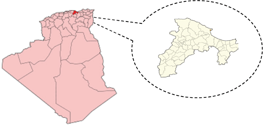
The study was carried out in the Province of Bejaia, Algeria (36°43’N, 5°04’W) (Fig. 1). It has an area of 326,826 kilometer square (km2) with a bovine population of 43,000. The topography of the area is predominated by mountains separated by the Soummam valley. With regards to the climate, it is part of the Mediterranean region. The winter rainfall in the region ranges from 600-1000 mm. The mean maximum summer temperature reaches 30.9 °C (September) and the mean minimum winter temperature falls to 8.8 °C (February). The vegetation is mainly composed of several species of trees and natural and cultivated herbs.
Figure 1. Map of the study of Bejaia province, Algeria
The study was conducted from December 2013 to June 2014. One hundred and forty-three fecal samples were collected from different cattle herds. Five grams of fecal samples were collected from the animal rectum directly into clean polyethylene disposal bags using a hand glove (11). Body condition scores (BCS) of animals were recorded. Scores were given by the same researcher based on a 1 to 5 scales (12). The age of the animals ranged between 2 months and over 3 years with mixed parity. Animals were selected randomly and they did not receive any anthelmintic treatment.
Parasitological examinations were carried out using standard methods at the Laboratory of Animal Biology, University of Bejaia. Fecal samples were visually examined then observed using flotation and sedimentation microscopic techniques (11).
Fecal eggs were counted in fresh fecal sample in a McMaster slide using saturated saline (NaCl) solution as flotation liquid (d=1.18). The number of eggs per gram of feces was obtained by multiplying the total number of eggs counted in the two squares of the counting chambers of the McMaster slide by the dilution factor of 50 (13). The results are expressed as eggs per gram of feces (EPG) or oocytes per gram of feces (OPG). Eggs were identified on the basis of their morphological features (14). Species identification and determination of worm were carried out according to standard procedures.
Statistical analyses were carried out using the XL Stat program (version 14.5.03). The prevalence was calculated as the ratio between the number of animals having the parasites and the number of surveyed cattle. The data (± SD) was expressed as percentage (%). The egg numbers were analyzed using gender (8 male and 135 female), age (13 young: ≤ 1 year aged; 130 old: > 1 years aged) and body corporal score (21 Thin: 1-2; 122 obese: 2.5-5) as factors of variation. The statistical analysis was performed using variance analysis (ANOVA). The values were statistically different when the P-value was < 0.05.
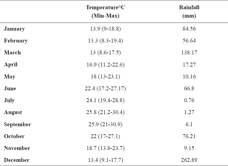
The monthly rainfall and mean temperature during the period of study are presented in Table 1.
Table 1. Annual rainfall and temperature data of the Bejaia area (Algeria) during the period 2013-2014
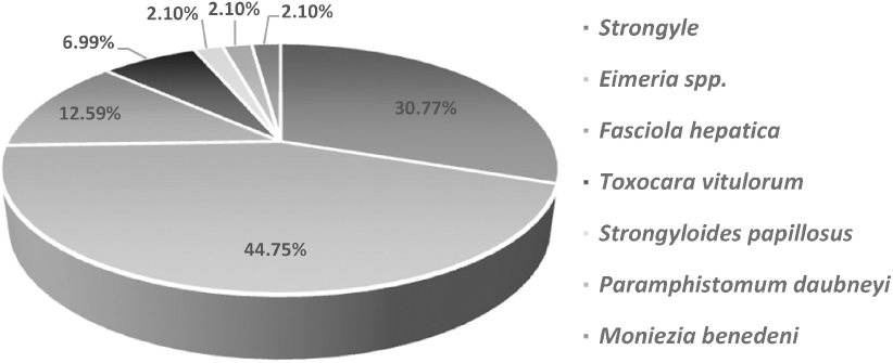
Sixty three percent of the 143 cattle examined were found positive with one or more gastrointestinal parasites. Species of gastrointestinal parasites (GI) are found after examination of fecal samples (Fig. 2). The microscopic observation revealed that the eggs of Eimeria spp. are predominant (43.87%) followed by Strongylus spp. (30.32%) and Fasciola hepatica (12.25%). Eggs of Strongyloides papillosus, Moniezia benedeni, Paramphistomum daubneyi and Toxocara vitulorum represent 1.29%, 1.93%, 1.93% and 6.45%, respectively.
Figure 2. Prevalence of various gastrointestinal parasites of cattle in the Bejaia province
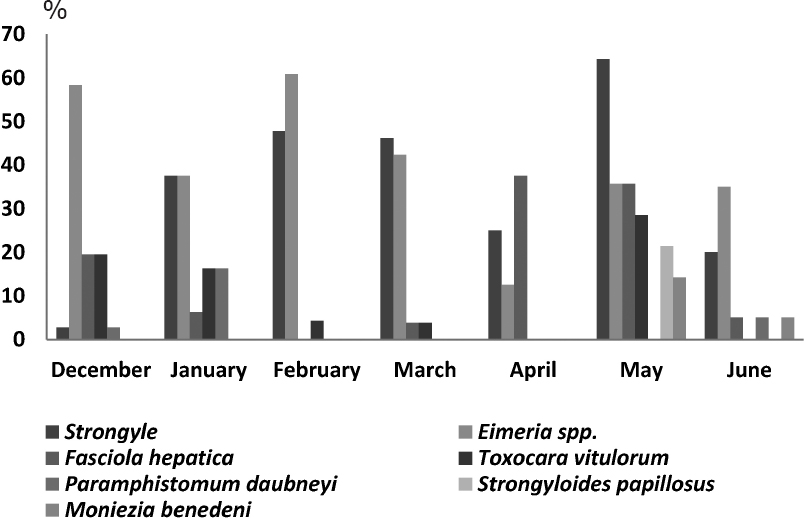
The monthly overall average eggs burden ranged from 2.78% and 0% in December to 64.28% and 21.43 in June for Strongyle spp. and Strongyloides papillosus, respectively (Fig. 3). Whereas, it varied from 64.28% in December to 2.78% in June for oocystes Eimeria spp. The highest burden was recorded in Fasciola hepatica in May 37.71% and the lowest in December and January. The highest burden for Toxocara vitulorum and Moniezia benedeni was observed in May (28.57% and 14.28% respectively), while for Paramphistomum daubneyi the only burden was found in June (5%).
Figure 3. Seasonal variation in proportions of gastrointestinal parasite of cattle during the humid and dry period
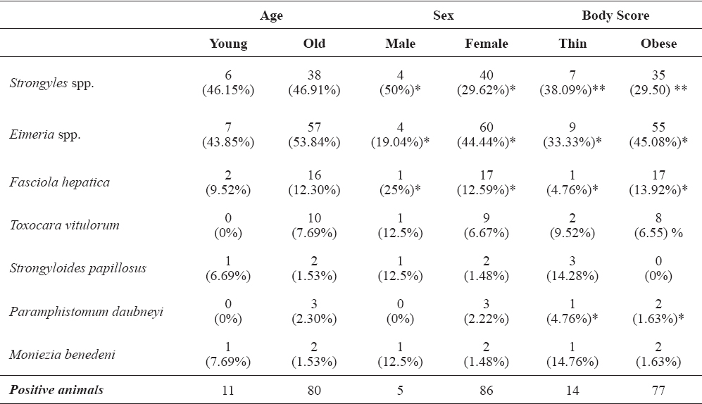
The results of relationships between age, sex, body score and GI parasites of cattle are shown in Table 2. Prevalence rates of Strongyle spp., Toxocara vitulorum, Fasciola hepatica, Strongyloides papillosus and Eimeria spp. in young animals (≤ 1 year old) was higher than in old animals (≥ 1 year old). Significant difference is recorded between the age of animal and the prevalence rate of two GI species namely Strongyle spp. and Eimeria spp. (P< 0.01). In contrast, prevalence rates of Moniezia benedeni and Paramphistomum daubneyi in young animals (≤ 1 year old) was lower than old animals (≥ 1 year old). No significant difference between prevalence rate and animal age was observed.
Table 2. Relationship between different factors (age, sex and body score) and gastrointestinal parasite of cattle in Bejaia province. Age (young: ≤ 1 year aged; old: > 1 years aged), sex (male and female), body corporal score (thin: 1-2; obese: 2.5-5). * Values with similar superscripts in the same factor differ statistically at the same parasite (P< 0.01)
Except the Eimeria spp. species, all prevalence rates of GI parasites in male cattle higher than female cattle. However, there is a significant difference between the sex of animal and the prevalence rate of Strongyle spp. and Eimeria spp. (P< 0.01).
As for the body condition score, there is a statistically significant (P< 0.01) difference between the prevalence rate of GI parasite and the nutritional status of cattle. Note that thin animals (under 2) were more infested with GI parasites than obese animals (above 2.5).
Helminth infections in ruminants are recognized as a major constraint to livestock production. Usually, infections are subclinical with significant economic losses due to both mortality and reduced productivity of animals (15, 16). The present study revealed that the cattle herds in the Bass Kabylie region (Algeria) suffers from a wide variety of GI helminth infestation. Sixty three percent of the cattle were infested with one or more GI parasite species. Prevalence of GI helminth has been reported ranging from 0.72 to 67 % in domestic animals from various parts of the world (10, 17). The epidemiology of GI parasites infections in livestock varies depending on the prevailing climatic conditions and management practices.
Proportion of gastrointestinal helminthic within the studied population clearly indicated heavy parasitic burden dominated by Strongles spp. Higher prevalence of helminth infections is probably caused by more frequent exposure to pasture contamination. The observed high prevalence in May might be due to favorable environmental conditions such as humidity and temperature. In contrast, the low prevalence in January may be due to hypobiosis. In our study the dry period is in June, and the egg excretion peak of digestive strongyles is reached in July and August. In 2012, Boucheikhchoukh (18) recorded prevalence of strongyles similar to our results (31.1%). Other researchers have reported a low prevalence of 17.7% (19) and a higher one of 50.75% (20). As mentioned before, other studies reported prevalence of GIP ranging from 0.72 to 67% (10, 17). The rate of helminth infestation in cattle varies in different country. This could be ascribed to a variety of factors like grazing habits, level of education of the farmers, livestock management and the anthelmintic used (21). Temperature and humidity constitute crucial factors which greatly influence the life cycle of the parasite and the transmission of endoparasites. Indeed, in agreement with our observations, Fuentes et al. (22) demonstrated that the season played a significant role in determining helminthes community species rate in cattle.
In general, the infestation intensity of strongyloidiasis and toxocariasis exhibit a low dominance in the area of our investigation. The prevalence of Strongyloides papillosus observed in cattle was very low compared to the results reported by Achi et al. (19) (7%). Toxocariasis is a very rare disease recorded in bovines because the parasitic disease often affects the very young (23). Our result is the only case of parasitosis indicated in Algeria (Bass Kabylie) with a rate of 6.45%. Strongyloidiasis remains discrete until May, and then a considerable prevalence of 21.43% is recorded in June. According to Alcaraz et al. (24), the most favorable conditions for the development of the parasite are a temperature above 20°C and a high precipitation extent ensuring high level of humidity. Note that a single case of strongyloidiasis was found in the humid highland (Boukhlifa). In contrast, toxocariasis is not really influenced by the climatic conditions, but it mainly affects young animals (25). However, we have few positive fecal samples from the young animals in presented study.
The results of the prevalence of fascioliasis are less significant compared to other reports (18, 19, 26). Furthermore, the results reported by Bendiaf (27) were similar (13.2%) to our findings. In contrast, a lower rate of infected animals (6.5%) has been recorded in the Northeast Algeria, case of Constantine area (28). The prevalence of fasciolosis has substantially progressed during the first months of the study to achieve a high rate of 35.71% in May. It is then significantly reduced to a rate as low as 8.33% in June. The epidemiology of fascioliasis is related to the exposure of cattle to the risk of ingestion of metacercariae (29). In our research, it corresponds to the pasturing and ingestion of metacercariae from cercariae released trans-wintering snails. This contamination in spring period is responsible for so-called summer Fasciolosis, which explains the presence of a high prevalence in April and May.
The prevalence of paramphistomosis in the Bass Kabylie region is highly comparable to those reported by Achi et al. (19) (12.1% vs. 1.93% and 6.4% vs. 1.93%, respectively). However, our results are very similar to those recorded by Mekroud et al. (28). This discrepancy in the prevalence could be related to many factors such as variable climatic conditions, habitats and age. This infection monitoring revealed two infestation peaks of 6.25% and 8.23% in December and March, respectively. In 2010, Lotfy et al. (30) observed the same epidemiological features. However, Paramphistomum daubneyi metacercaria leave the mollusks when the temperature drops (31). Therefore, the contamination with Paramphistomum daubneyi could have been developed earlier in spring and later in the end of grazing season (32).
As regards to the Moniezia benedeni, the overall prevalence found in our investigation was much lower than that reported by Borthakur and Das (33) and Boucheikhchoukh et al. (18) (19.6% vs. 1.93%. 11.1%. vs. 1.93%, respectively). Young and pastured animals are more sensible to the monieziosis (34).
In the present study, coccidiosis is considered a major parasitosis as indicated by the relatively high prevalence in the overall examined animals (43.87%). Achi et al. (19) reported a much lower prevalence of 9.9%. The results of this experiment showed a high rate of infestation during the investigation period. According to Brian (35), the oocysts are very resistant and are active up to 18 months after discharge, especially when they undergo sporulation.
The proportion of infection causes by different parasitic species varied in the two age groups of cattle. It is the highest in aged animals and the lowest in young cattle. Studies have showed that the susceptibility and the pathogenicity of nematode infections are greater in young animals than in mature animals (23, 36, 37). The immunity system of infected young animals is less developed at an early age (38). In contrast, the values of Moniezia benedeni and Paramphistomum daubneyi rates do not show a difference based on age, because the number of positive animal samples were too small to enable for an outstanding statistical analysis.
During the present study, a significantly higher proportion of young animals was infected with coccidian than was adult cattle (P < 0.01). This result agrees with many reports from Kenya (37), Nigeria (39), South Africa (40) and the Netherlands (41). It is well known that clinical coccidiosis occurs mainly in young cattle (42). Note that the breeding conditions play an important role in the appearance of the parasitosis. Indeed, overcrowding, the animal mixture of various origins and poor hygiene are the risk factors for parasite infestation (43).
In the present research, the sampling population was female (heifer and cow) dominated (94.4%) with only a few number of male cattle (5.6%). The overall prevalence of cattle infected with Strongyle spp. and Eimeria spp. clearly shows a significant difference (P<0.01) between male and females. During lactation, the immune response of the host to gastrointestinal nematodes is partially suppressed leading to an increase in the population of worms (44). Other studies revealed a higher prevalence of nematodes in lactating and pregnant cows compared to bulls and oxen (45, 46). Pfukenvi and associated (46) has concluded that lactating and pregnant cows might serve as a potential source of pasture contamination. In the same way, Barger (5) explained that acquired immunity to nematodes infection tends to be lost in late pregnancy and in lactation.
In the literature, a combination of poor nutrition, high stocking rate and lack of anthelmintic medication are considered as the probable causes of the development of heavy worm burden in communally grazed cattle (47). The results obtained from thin cattle infected with Strongyle spp. could be explained by a dysfunction in the digestive tract. Indeed, infestation by digestive Strongyle causes enormous damage to the stomach and bad nutrition absorption of the cow as a reduction in motility and decrease of acid secretion (48). In addition, a recent study reported that the intensity of the symptoms of strongyloidiasis depends on the infestation degree, the animal age and the body condition (49). Also, toxocariasis causes physiological disorders in animal infested such as hypoglycemia, mineral deficiency.
Immature flukes aggravate injuries causing severe anemia syndromes (50). That could explain the high rates of thin animals infected with Fasciola hepatica. Note that Paramphistomum daubneyi parasite have a negative impact on the animal infected including poor diet absorption, growth delays, weight loss and diminution of milk production (51). The coccidiosis disease causes malabsorption intestinal and reduced production performance in animals (52). This could justify the significantly higher prevalence in thin animals than in obese ones.
In conclusion, our study demonstrated a high prevalence and the abundance of the polyparasitism nature of the disease in the Bass Kabylie area. Also, there was a relationship between the distribution of GI parasitism in cattle and the factors analyzed (body condition score, age and sex). Therefore, the parasitic fauna of each species, when mapped out accurately in such agro-climatic zones (Bass Kabylie), elucidates fundamental information, depending upon which further control measures can be followed.
©2018 Moussouni L. This is an open-access article published under the terms of the Creative Commons Attribution License which permits unrestricted use, distribution, and reproduction in any medium, provided the original author and source are credited.
The authors gratefully acknowledge Mr. Z. Bouzid and all staff of CAZAL (Bejaia, Algeria) for the help during the sampling in the bovine farm. The authors thank also veterinary colleagues A. Belharet and S. Benidiri for their hospitality and reception during his farm visits. The authors thank Dr. C. Harrats (University of Mostaganem, Algeria) for the English correction.
The authors declared that they have no potential conflict of interest with respect to the authorship and/or publication of this article.
Macedonian Veterinary Review. Volume 41, Issue 1, Pages 73-82, p-ISSN 1409-7621, e-ISSN 1857-7415, DOI: 10.2478/macvetrev-2018-0010, 2018