Mac Vet Rev 2018; 41 (1): 99 - 105
 10.2478/macvetrev-2018-0011
Received: 17 March 2017
Received in revised form: 07 December 2017
Accepted: 11 December 2017
Available Online First: 29 January 2018
Published on: 15 March 2018
Keywords: Ehrlichia canis, doxycycline, clinical sings, hematology, biochemical parameters
Ehrlichia canis, a Gram negative obligate intracellular bacterium is the etiologic agent of canine monocytic ehrlichiosis (CME), a potentially fatal disease in dogs, mainly transmitted in Europe by the brown dog tick Rhipicephalus sanguineus. This disease can also be transmitted by blood transfusion (1). The bacterium Ehrlichia canis was first identified in 1935 in Algeria, when dogs infested with ticks revealed fever and anemia (2). Since then, it has been described in tropical, subtropical and Mediterranean areas all over the world (1, 3). In the last years this disease has been reported in our neighboring countries Albania, Serbia, Bulgaria and Greece (3, 4, 5, 6). In R. Macedonia studies about this disease have been published recently (7).
Infection occurs when an infected tick bites the dog and infects the biting site with its saliva (8). In the organism, E. canis enters in the monocytes and macrophages of the host and spreads through the Monocyte Macrophage System in the body. Incubation period is between 8-20 days, when the bacterium multiplies in macrophages and spreads through the body. Canine monocytic ehrlichiosis, the disease caused by E. canis, is a multisystem disease characterized by three stages: acute, subacute and chronic stage. Clinical signs of the disease are various, but most often depression, anorexia, pyrexia and bleeding tendencies are present (9). Physical examination typically reveals lymphadenomegaly, splenomegaly and hemorrhagic tendencies, usually dermal petechiae and ecchymoses, as well as epistaxis. The most common hematological and blood biochemical changes are: severe thrombocytopenia, mild to marked anemia and hypoalbuminemia (10). According to Neer at al. (11) clinical signs, clinicopathologic abnormalities, together with positive serology finding are sufficient for clinical diagnosis of ehrlichiosis.
Tetracyclines, especially doxycycline, are most commonly used antibiotics in the treatment of many bacterial infections transmitted by ticks (12). Doxycycline is an often prescribed medication due to its antimicrobial properties and pleiotropic functions, independent of its antimicrobial activity. There are other immunomodulatory and/or anti-inflammatory properties of this medication, associated with effects on blood leukocyte proliferation and function, cytokine synthesis and matrix metalloproteinase activity (13).
There are contradictory opinions about the efficiency of doxycycline, some authors reported persistence of infection after 7 to 85 days treatment (14, 15, 16), while others reported successful elimination of E. canis after 14 to 60 days treatment (17, 18, 19). Treatment duration seems to be more important than dosage or frequency of administration.
The usual treatment of canine ehrlichiosis includes antibiotic doxycicline 10 mg/kg/24h for 28 days and symptomatic therapy (fluids, vitamins, antipyretics etc.), and blood transfusion in severe cases (11). Clinical improvement usually appears before normalization of the hematologic and biochemistry values, as other authors already have noted (20). Among the laboratory findings, RBC and platelet counts are the parameters that first return to normal, often reaching reference values by the end of the treatment period, which indicates good response to therapy (21). Gradual resolution of hyperglobulinemia over 6–9 months also suggests therapeutic elimination of the organism. For this reason, monitoring hematology and biochemistry parameters could be considered a valuable prognostic aid in association with the clinical response.
The aim of this study was to analyze the most common changes in the hematological and blood biochemical parameters before and after treatment the naturally infected dogs with doxycycline.
This study covered 34 Erlichia canis positive dogs from the University Veterinary Hospital in Skopje, analyzed before and after treatment. Patients were admitted in the hospital for clinical examination due to different signs of the disease. Detailed anamnestic data was obtained and clinical examination was performed in all dogs. According to the anamnestic data, the owners confirmed the presence of tick infestation, despite using ectoparasitic prevention. Samples for biochemistry (serum samples) and hematology (whole blood) were taken by vein-puncture of cephalic or saphenous vein in tubes with anticoagulant (EDTA) and sera tubes.
Based on previous studies about laboratory changes in whole blood and sera from patient with CMA, haematology and biochemistry analyses were performed (22, 23).
Complete blood counts were performed on the automatic hematology analyzer Exigo EosVet (Sweden) with the following data: red blood cells (RBC), white blood cells (WBC), hemoglobin (Hb), hematocrit (HCT), platelet count (PLT) and differential leukocyte count. Serum biochemistry analyses were performed using the automatic analyzer ChemWell 2910 (Awareness Technology, INC, USA), and included alanine aminotransferase (ALT), aspartate aminotransferase (AST), urea, creatinine, albumin, total protein and globulin, alkaline phosphatase (ALP), with spectrophotometric kinetic and “end point” methods, according to the manufacturer’s instructions (Human, Germany).
Based on the anamnesis, hematology and biochemistry findings, there were indications about the presence of canine monocityc ehrlichiosis. Infection with E. canis was confirmed using a whole blood or sera with chromatographic immunoassay antibody snap test, according the manufacturer’s instructions (BioNote, Korea). The serum samples were also screened for detection of IgG antibodies against E. canis, using a micro-immunofluorescent assay (Biopronix Ehrlichia 96, AGROLABO S.p.A, Italy) according to the manufactures recommendation (optical density > 1 was considered as positive result). Erlichia canis positive dogs were a mixed population of ages, breeds, gender and origin. We used treatment protocol according to Neer et al. 2002, with doxycycline (10 mg/kg/day for 28 days) (11).
Statistical analysis was performed using STATISTICA for Windows 7, (StatSoft) software and significance of the differences was analyzed by Student’s t- test. Value of p<0.05 was taken for statistical significance.
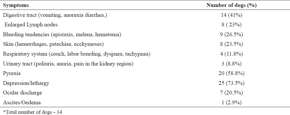
After collecting and processing all data (clinical and laboratory findings), comparisons between the results were made. All of the dogs that were positive on the snap test were confirmed by the serological analyses. Most common clinical signs during the clinical examination were depression (73.5%), pyrexia (58.8%), and digestive disorders (anorexia, vomiting or diarrhea) (41%) (Table 1). Clinical signs resolved after treatment. Average age of the patients was 4.3 years (youngest dog was 6 months old, oldest 11 years), while regarding the gender male dogs were more prevalent then female (70.6%).
Table 1. Clinical sings on physical examination of patients with CME
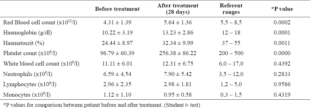
Hematological results revealed statistically significant difference regarding the RBC, HCT, Hb and PLT (Table 2) before and after treatment. Haemoglobin (g/L) and platelet count (x1012/L) showed a high statistically significant difference (p<0.0001) before and after treatment.
Table 2. Mean ± standard deviation changes in hematological parameters in dogs with canine monocytic ehrlichiosis (CME) before and after treatment with doxycycline
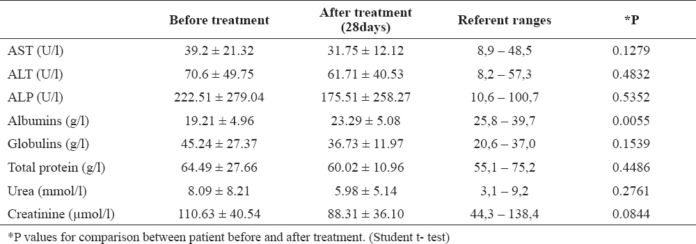
Blood biochemical parameters results have demonstrated statistical significance in the values for the albumin (p<0.005) (Table 3).
Table 3. Mean ± standard deviation changes in serum-biochemistry parameters in dogs with canine monocytic ehrlichiosis (CME) before and after treatment with doxycycline
Our study demonstrated that doxycycline treatment for 4 weeks resulted in increased platelet and erythrocyte number in CME affected dogs, as previously described (21, 24).
Canine monocytic ehrlichiosis is a tick borne disease and may be manifested by a variety of clinical sings. Similar to Nakaghi et al. (23), in our study the most common clinical sings were depression and lethargy (73.5%), pyrexia (58.8%), vomiting or anorexia (41%). These clinical signs are easy noticeable by the owners and clinical examination confirmed the condition of illness.
During the acute phase, when bacteremia occurs, severe haemo-pathological changes appeared as well as some biochemical alteration. In our study, the obtained results for hematological parameters (RBC, Hb, HCT and PLT) before treatment were significantly decreased from the reference ranges. The thrombocytopenia is a main marker of this disease (25). The mechanisms involved in the pathogenesis are increased destruction of the platelets due to inflammatory changes of the blood vessels endothelium and increased platelet sequestration in the spleen. These results are presented in significant difference in PLT (p ≤ 0.001) number before and after treatment (18). Villaescusa et al. (26) reported increase in PLT in healthy patients treated with doxycycline, claiming that besides antimicrobial activity it has effect on platelet proliferation. In contrast of this study Webb at al. (27) reported that there wasn’t any change in the platelet count or function in healthy dogs treated with doxycycline. Further studies on a larger group of dogs are necessary in order to clarify these findings. Hypergammaglobulinemia induced immune-mediated platelet destruction (8, 9). Abnormal hematology findings are present due to tropism for hematopoietic cells and bone marrow hypoplasia, such as suppression in erythroid, myeloid, and megakaryocytic cells. The anemia is non regenerative, as a result of progressive replication of Ehrlichia canis in the bone marrow and inhibition effect of colony forming units in the process of erythropesis and megacariopoesis (28). Our results are in accordance with this pathogenic mechanism, showed in the significant difference in hematology parameters such as RBC (p ≤ 0.001), HCT (p ≤ 0.01), and Hb (p ≤ 0.001), which is a common finding by other authors (29, 30). Regarding biochemical parameters before treatment, marked hypoalbuminemia was noted, followed by hyperglobulinemia. Similar results are reported by other authors (26, 30). Serum concentration of urea and creatinine were in reference ranges before and after treatment, but these parameters usually change in the chronic stage of the disease (31). Other biochemical parameters such as ALT, AST and ALKP, showed slight changes in serum concentration before and after treatment, but there was not a statistically significant difference. Some authors reported increase in ALP and ALT in dogs in naturally infected dogs (31, 32).
Thrombocytopenia, anemia and hypoalbuminemia are characteristic laboratory findings in patients with CME. Treatment protocol with doxycycline for 28 days, besides resolving the clinical signs, effects on increasing of the RBC, PLT, Hb, HCT and albumins in dogs naturally infected with E. canis.
©2018 Atanaskova Petrov E. This is an open-access article published under the terms of the Creative Commons Attribution License which permits unrestricted use, distribution, and reproduction in any medium, provided the original author and source are credited.
This research was part of a national project “Improved clinical and diagnostic approach of the prevalence of canine ehrlichiosis in R.Macedonia”, funded by the University Ss Cyril and Methodius, Skopje 2015/16 (02-640/23).
The authors declared that they have no potential conflict of interest with respect to the authorship and/or publication of this article.
Macedonian Veterinary Review. Volume 41, Issue 1, Pages 99-105, p-ISSN 1409-7621, e-ISSN 1857-7415, DOI: 10.2478/macvetrev-2018-0011, 2018