Mac Vet Rev 2018; 41 (2): 203 - 207
 10.2478/macvetrev-2018-0014
Received: 28 January 2018
Received in revised form: 23 March 2018
Accepted: 27 March 2018
Available Online First: 17 April 2018
Published on: 15 October 2018
Keywords: poisoning, dog, histopathology, welfare
Animal poisoning presents a worldwide problem (1, 2, 3) and is mostly encountered in dogs as the most frequent companion animals. Targets of poisoning are domestic and street dogs, whereas the poisoning can be either accidental or deliberate. Poisoned animals need rapid and proper treatment, which is why the veterinary doctor needs to be aware of the type of poisons commonly used in their environment. The types of poisons vary depending on the type of pesticides commonly used in the particular region and the animals exposed. Agents used most often for poisoning are insecticides, pesticides (carbamates, organophosphates), anticoagulant and non-aticoagulant rodenticides (4, 5). There is no information available on the prevalence of specific poisonings in dogs, which is why this retrospective study was undertaken.
The clinical findings depend on the poison type. Some of the agents are organ (system) specific, while some act on more than one organic system. Overt pathological effects of lead poisoning are mainly confined to the kidneys, brain, erythrocytes and haemsynthesis (6).
Intoxication with antifreeze has nonspecific clinical symptoms. Its acute form is characterized by digestive disorders, cardio-respiratory and nervous symptoms, while the subacute form is marked by nephrotoxic syndrome and renal failure (7). Carbamate pesticides (Carbofurane, Methomyl - Lannate) are acetylcholinesterase (AChE) inhibitors which lead to the accumulation of acetyl choline in the gap junction, causing hyperstimulation of cholinergic receptors. This results in a cholinergic crisis followed by muscarine, nicotine and central nervous signs such as: miosis, hypersecretion of exocrine glands, bradycardia, tonic and clonic convulsions (8). Necropsy findings include: congestion of gastric mucosa with or without petechial hemorrhages; congestion of viscera such aslung tracheae and bronchi, as well as edematous lungs (9, 10, 11). The liver, spleen, pancreas, adrenal glands and the urinary tract show degenerative changes (11). Anticoagulant rodenticides, especially coumarin show generalized hemorrhages in various organs (liver, kidneys, intestines, heart, and lungs) (12).
The diagnosis of poisoning is based on clinical, histopathological and toxicological data (13). There is scarce data in the literature on the type of histopathological changes that different poisons cause in animals, which is the goal of this work.
In this study, 31 cases of suspected intoxications from January, 2007 to May, 2017 were analised. 13 of the dogs were home kept, 7 were street dogs and 11 were of unknown origin. The age of the animals varied between one and nine years. The number of poisoned dogs for every city is presented in Table 1.
Table 1. Poisoned dogs per city
Table 2. Number of dogs with histopathological changes of different organs
All the animals were delivered post mortem, no longer than 24 hours after their death. Complete necropsy was performed on all of the corpses and tissue samples for histopathology and toxicology were collected. Tissue samples were also taken for viral, bacterial and parasite examination.
Samples from the following tissues were collected for histopathology: lung, spleen, kidney, liver, stomach, small and large intestine. The tissue samples were fixated in 10% neutral formalin, processed through series of alcohol and xylol and embedded in paraffin. Following this, 3-5 µm thick slides were cut using microtome and de-waxed prior to histochemical staining. Then, the slides were stained with Hematoxylin and Eosin (H.E.) for microscopic analysis.
For the purposes of toxicology examination, the complete stomach and intestines were collected along with their content. Samples from the intestines, liver, kidney, spleen and lungs were taken for microbiology examination, while for the parasite examination part of the intestines was taken.
According to the dogs’ owners, these are the most frequent symptoms prior to their death: vomiting, diarrhea, walking like drunk, abdominal cramping and convulsion, agitation, tachipnea and dyspnea, bradycardia, tonic and clonic convulsions, muscle tremors, seizures and coma. Most of the dogs died no longer than 24 hours after the initial signs.
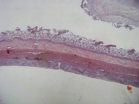
The most frequent findings during the necropsy were different forms of gastrointestinal inflammation. The wall of the stomach was thicker than normal, congested with great red areas. A small amount of dark red or brown liquid was most often the content of the stomach lumen. The intestines had severe diffuse hemorrhagic and necrotic inflammation. Among the histopathological changes of the gastrointestinal tract, necrotic gastroenteritis was the predominant finding in seven dogs (Fig. 1). Catarrhal gastroenteritis and catarrhal gastritis with peptic ulcus at the fundic part were also present in two dogs, with hemorrhagic gastroenteritis in one dog and pseudomembranous gastritis in one dog.
Figure 1. Necrotic enteritis (H.E; x2)
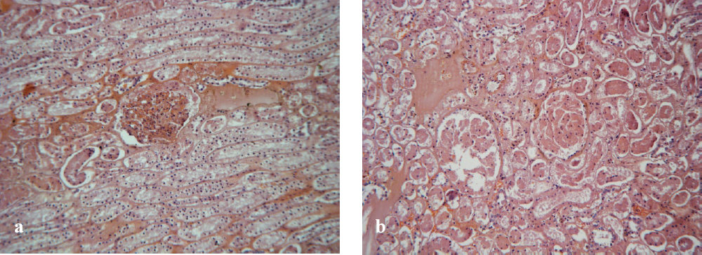
The kidneys were mostly pale with hemorrhagies in the cortex and the medula. Histopathologically, the renal changes were mostly in the form of renal hemorrhage found in 4 dogs. Hemorrhagic glomerulonephritis, necrotic glomerulonephritis (Fig. 4), glomerulonephritis with lymphocyte infiltration and necrosis and punctate hemorrhage were all found in one dog each, while hemorrhagic nephritis and vacuolar glomerulonephritis were found in 2 dogs and parenchymatose degeneration of the kidney in 3 dogs.
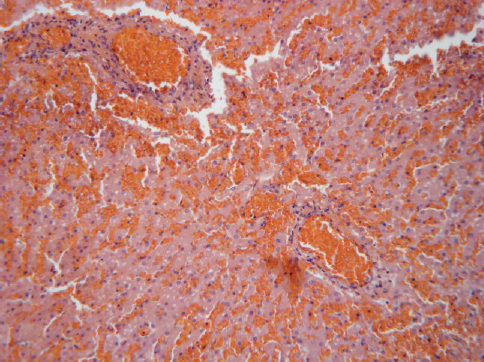
Figure 2. Hyperaemia and extravasation (H.E; x10)
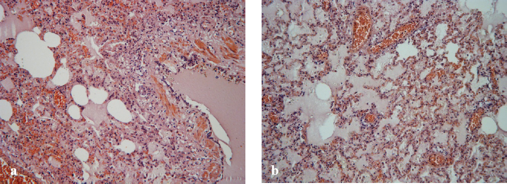
Figure 3. Necrotic and fibrinous pneumonia with alveolar edema (a, b). (H.E; x10)
Figure 4. Necrotic glomerulonephritis (a, b). (H.E; x10)
The viscera of the trachea was congested and the lumen of the trachea contained yelow or red foamy liquid. The lungs were mostly edematous, with areas of light or dark redish color. At the section surface, there was white-read foamy liquid. Hemorrhagic pneumonia with alveolar edema was the most frequent histopathological finding in 4 dogs. Among the other types of lung changes, alveolar edema and alveolar edema with atelectasis were found in 3 dogs, while necrotic and fibrinous pneumonia with edema was detected in one of the dogs (Fig. 3).
Very often the spleen and liver were enlarged in length and weight with darker red color, whereas the heart was usually discolored and dilated. Histopathologically, most of the spleen changes were in the form of hemorrhage. Furthermore, with regards to the changes of the liver, adipose degeneration was the predominant finding in 5 dogs. Other liver changes were in the form of adipose degeneration with hemorrhage of the liver, cirrhotic and necrotic liver, or only necrotic liver, hemorrhagic and necrotic liver, hemorrhage of the liver (Fig. 2) and passive edema of the liver found in one dog each. Also, two of the dogs had parenchymatous degeneration with miliar necrosis of the liver. Finally, two of the dogs had myofibrilar degeneration of the miocard and acute hemorrhagic pancreatitis.
Stomach and intestine, along with their contents from the last three dogs, were sent for poison analysis at the Institute for Forensic Medicine. All of them were positive for lanate (Methomyl), which is a carbamate pesticide and is legally distributed in the Republic of Macedonia. It is an acetylcholinesterase (AChE) inhibitor and historically has a bad reputation, as it has been extensively misused for dogs and cats poisoning. The necropsy and histopathological findings for the last three dogs and most of the other dogs were characteristic for this type of poison.
However, the investigation showed that poisoning was not the cause of death in all of the dogs. Several cases did not have any histopathological signs of poisoning. Instead, they had a viral, bacterial or parasitic diagnosis.
Most of the dead dogs with suspected poisoning that were brought to the Laboratory of Pathology at the Faculty for Veterinary Medicine in Skopje were clinically, morphologically and histologically positive for poisoning. For the last three cases, the poisoning was confirmed by poison analysis at the Institute of Forensic Medicine. The majority of cases had clinical symptoms that included abdominal cramping and convulsion, agitation, tachipnea and dyspnea, bradycardia, tonic and clonic convulsions, muscle tremors, seizures and coma, all typical for lanate (Methomyl) (8) which was confirmed by the poison analysis. The necropsy and histopathological findings such as: thickening and congestion of the gastric mucosa, congestion of the trachea and edematous lungs, degeneration, hemorrhage and necrosis of the liver, spleen, pancreas, lungs, adrenal glands and the urinary tract are also characteristic for poisoning with lanate (9, 10, 11).
There is no record or prohibition of the use of lanate in the Republic of Macedonia. Anyone can purchase it from the agricultural pharmacies in the form of insecticide Methomil, which is 90% lanate. This pesticide was prohibited in the European Union in 2009.
The practice of dog poisoning is unethical and hurts the animals’ welfare. According to DEFRAs (Department for Environment, Food and Rural Affairs) Section 9 of the Animal Welfare Act 2006 (the Act), every dog’s need must be properly met. This includes the need for suitable environment and suitable diet, its need to be able to exhibit normal behaviour patterns, any need it has to be housed with, or apart from, other animals and its need to be protected from pain, suffering, injury and disease (14).
The Law on Animal Welfare states that all the animals should be treated as conscious creatures which needs must be met. According to this law, the city government has the authority to capture unregistered dogs, as well as registered dogs which are not accompanied by their owners. This capturing of dogs can only be performed by qualified personal in a manner which will not cause unnecessary suffering to the dogs (15).
Dog poisoning directly violates DEFRAs act and The Law on Animal Welfare because it causes pain and suffering to the poisoned dogs. In the Republic of Macedonia, there is a constant increase in the number of pet and street dogs and only a few shelters. In Skopje, the capital city of the Republic of Macedonia, there is only one shelter for stray dogs and it has limited capacity, which is not enough to provide efficient care for all the dogs on the supposed territory. In the urban areas, people complain of problems associated with stray dogs such as: bite injuries, road accidents, spreading of diseases, as well as pollution from feces and the noise they make. That is why certain individuals carry out illegal spreading of poison which affects street, but also domestic dogs, and is potentially hazardous for the people. This is an inhumane method and should be avoided by using well planned strategy for the control of the population of stray dogs, such as the World Society for the Protection of Animals framework protocol which encompasses: legislation, registration and identification, garbage control, neutering owned animals, neutering unowned animals, control of breeders/sales outlets and education.
This study shows that the practice of dog poisoning is used on the whole territory of the Republic of Macedonia, with most of the dogs being poisoned by lanate (Methomyl). Dog poisoning is illegal and violates the welfare of animals. Greater attention should be devoted to developing strategies for control of the dog population, as well as to education of people about the risk to their own health from using poisons.
© 2018 Gjurovski I. This is an open-access article published under the terms of the Creative Commons Attribution License which permits unrestricted use, distribution, and reproduction in any medium, provided the original author and source are credited.
The authors declared that they have no potential conflict of interest with respect to the authorship and/or publication of this article.
Macedonian Veterinary Review. Volume 41, Issue 2, Pages 203-207, p-ISSN 1409-7621, e-ISSN 1857-7415, DOI: 10.2478/macvetrev-2018-0014, 2018