Mac Vet Rev 2018; 41 (2): 195 - 201
 10.2478/macvetrev-2018-0022
Received: 28 December 2017
Received in revised form: 19 June 2018
Accepted: 07 July 2018
Available Online First: 19 September 2018
Published on: 15 October 2018
Keywords: static ovary, metabolic profile, P4, E2, ultrasound, cow
The early resumption of ovarian cyclicity following parturition has a great impact on the reproductive efficiency in dairy cows. Achieving high reproductive efficiency generally requires early onset of ovarian activity, insemination and conception within 90 days after calving, leading to once a year calving (1). Indeed, early re-establishment of ovarian activity derives maximum economic benefit to the farmers. In that respect, Ambrose et al. (2) reported that cows that resume ovarian cyclicity and had their first ovulation within 3 weeks after calving were more fertile at first service than cows that ovulated for the first time after 9 weeks post-partum (46% versus 23% conception rate), respectively. Nevertheless, there are still large proportions of postpartum dairy cows (6–59%) that have not resumed cyclicity until day 60 after calving, traditionally classified as non-cycling (anoestrus or anovular) cows, (3). Among the factors that contribute to non-cycling status, such separated ovarian disease (4), persistent corpus luteum (1), post-partum uterine disease (5), static ovaries (hereinafter in the text: SO) that stand as one of the possible underlying reasons (6). Cessation of cyclicity caused by SO could transpire in cows that are either experiencing severe nutritional restriction (7) or are in a negative energy balance – NEB (8). The latter occurs during the transition period due to the differences between dietary energy intake and requirements for milk production, resulting in mobilization of body fat reserves and increased blood serum non-esterified fatty acid (NEFA) and β-hydroxybutyric acid (β-HBA) concentrations (9). Indeed, some studies have showed that anovular cows (non-cycling cows) have a reduced feed intake (between 2.5 kg and 3.6 kg per day) compared to cycling cows (10), and hence more progressive NEB status and significantly increased plasma NEFA and β-HBA concentrations in comparison to the ovulatory (cycling) cows (11). It was assumed that the NEB status negatively affects LH secretion necessary for resumption of the follicular growth, causing a certain delay in the re-establishment of cyclicity (12). Therefore, as soon as cows overcome the NEB status, they could achieve earlier onset of cyclical ovarian activity and possibility of conception (13).
In addition, parity also has been shown to play an important factor that contributes to non-cycling status (3, 4). Several studies showed that PP cows are more susceptible to metabolic stress, to experiencing more severe NEB during the transition period (8, 14), to having extended recovery from NEB and hence longer intervals to first ovulation in comparison to MP cows. A reduced capacity of PP cows for food intake, greater demands for nutrients for their own growth and increased requirements for their first lactation have been assumed as possible underlying reasons for their prolonged NEB recovery (15).
Since PP cows generally have a higher incidence of being non-cycling (bearing SO) after day 60 postpartum than MP cows, we hypothesized that the metabolic profiles and hormonal statuses between non-cycling PP and MP cows bearing SO are different. Therefore, the objective of the present study was to compare the metabolic profiles and the hormonal status between the non-cycling PP and MP cows diagnosed with SO.
The study was conducted during the period between January 2010 – December 2012 at two dairy farms (Farm A and B) located at north most (Farm A) and southeast part (Farm B) of Republic of Macedonia. On Farm A, the cows were housed in free-stall barns with cubicles, fed a standard TMR ration based on corn silage, chopped alfalfa, straw and a 16 % protein concentrate-mineral mix and milked twice daily with an average 305 d-milk production level of 6100 kg. On Farm B, the cows were housed in tie-stall barns on deep straw bedding, milked thrice daily with an average 305 d-milk production level of 6500 kg. The cows were fed a corn silage, grass silage, alfalfa, brewers’ grain and concentrate-mineral mix (16 % protein), offered twice daily according to the stage of lactation, milk production and reproductive status of the animals in amounts of between 9-12 kg per day.
One hundred and twenty one animals that either did not express signs or were not seen in oestrus by farm personnel by day 60 after parturition were included in the study. The animals were grouped by parity PP (n=58) and MP (n=63) cows, blood sampled and examined using transrectal ultrasonography. The blood sampling was done prior to the ultrasound examination. Cows were classified as non-cycling (bearing SO) if no luteal tissue (corpus luteum, CL) and no follicles larger than 8 mm were detected concomitantly with a serum progesterone (P4) concentration < 0.5 ng/mL (16, 17). If a luteal tissue was observed along with serum P4 > 1 ng/mL, the cows were classified as cycling; cystic (presence of follicular cyst – follicular like structure larger than 25 mm without presence of luteal tissue and P4 level <0.5 ng/ml); and cows in heat (presence of follicle between 16-18 mm, no luteal tissue, P4 < 0.5 ng/mL and fluid within the uterus).
Ultrasonographic examination of the ovaries was done with a B-mode scanner Aloka SSD 500, (Tokyo, Japan), equipped with a 7.5 MHz linear-array transducer for intra-rectal use. The examination was done as described previously (17). Briefly, before insertion of the lubricated transducer, the rectum was emptied, and the ovaries were first manually located. After insertion, the size of the ovaries and the diameters of the follicles were obtained from two linear measurements taken at perpendicular angles by means of electronic callipers located on the ultrasound device and using the images on which the diameters of the ovaries and follicles were maximal.
Blood samples for estradiol (E2) and P4 analysis were collected from the jugular vein into glass tubes (without anticoagulant) and transported at +4°C within 3 hours after collection. The samples were centrifuged (2500 RPM x g 5 minutes), and after serum extraction were stored at – 20°C until assayed for E2 and P4 using enzyme-immune assay (EIA). The assay was done at the Faculty of Veterinary Medicine – Skopje (Macedonia), using commercially available kits (HUMAN, Progesterone and Estradiol ELISA Test - Germany) on Immuno-scan BDLS reader. The intra-assay CV averaged 7.2% and 9.3%; while the inter-assay CV was 8.6% and 9.2% for P4 and E2 respectively.
The metabolic profile analysis was done at the Faculty of Veterinary Medicine in Skopje. The glucose, total protein, albumin, cholesterol, triglycerides, NEFA and β-HBA concentration analyses were performed by enzyme-colorimetric determination with an end point method using commercially available kits. For glucose, total protein, albumin, cholesterol - Human (Germany), for triglycerides-Sentinel (Italy) and for NEFA and β-HBA - Randox (UK), all in accordance with the IFCC, on semiautomatic photometer Stat-Fax 3300 (Inc., Awareness Technology, USA).
The data for the tested parameters on the individual and farm level were subjected to descriptive statistics and analysed for normality distribution using Shapiro–Wilk test, setting the level at p<0.05. Comparison of tested parameters between parity groups was done by Student’s T-test and Mann–Whitney U test depending on the data distribution. The results are presented as mean and standard deviation values (mean ± SD). The statistical analysis was carried out in STATISTICA (data analysis software system), version 8.0, Stat Soft, Inc. (2007).
In total, 70 out of 121 cows (57.8%) were diagnosed as non-cycling (bearing SO). From these cows, 42 (72.4 %) were PP and 28 (44.4 %) MP cows. In the remaining cows, ovarian cysts were detected in 6.6%, CL in 25.2 %, and 10.3% of the cows were in heat. The length of the ovaries in both parity groups ranged between 16.0 mm to 19.0 mm, while the width ranged between 10.0 mm to 11.0 mm. The follicle diameters in both groups of cows ranged between 2 mm to 6 mm.
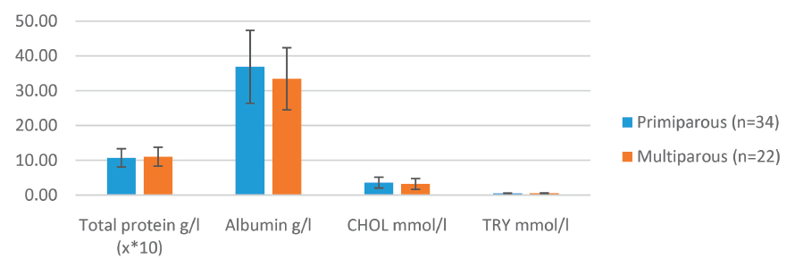
The P4 (0.24 ± 0.15 ng/ml and 0.30 ± 0.16 ng/ml) and E2 (2.86 ± 0.79 pg/ml and 2.49 ± 1.05 pg/ml) concentrations did not differ between the PP and MP cows, respectively (p>0.05). The total protein, albumin, cholesterol, and triglycerides concentrations did not significantly differ between the parity groups (Fig. 1). Similarly, the glucose concentration was not significantly different between the PP (1.43 ± 0.59 mmol/l) and MP cows (1.69 ± 0.71 mmol/l).
Figure 1. Comparison of blood serum parameters between PP and MP cows diagnosed with SO* *Animals not observed in oestrus until d 60 postpartum were grouped by parity (primiparous vs. multiparous), bled and examined using trans-rectal ultrasonography. The metabolic profiles were compared between the PP and MP cows that bear SO; abbreviations: SO- static ovaries, PP- primiparous, MP- multiparous, CHOL-cholesterol, TRY-triglycerides
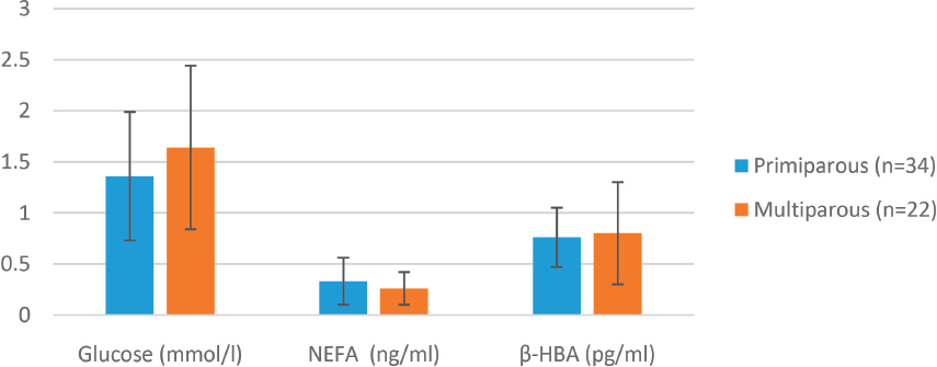
In addition, no differences were observed between the PP and MP cows regarding the NEFA (0.39 ± 0.26 mmol/l and 0.33 ± 0.22 mmol/l) and the β-HBA concentrations (0.83 ± 0.34 mmol/l and 1.03 ± 0.60 mmol/l, p>0.05, Fig. 2), respectively.
Figure 2. Metabolic parameters (glucose, NEFA and b-HBA) between primi- and multiparous cows
The present study intended to compare the metabolic profiles and hormonal status between the non-cycling PP and MP cows diagnosed with static ovaries. Based on the gathered results, we could not find any differences between neither the metabolic profiles nor hormonal status of both parity groups of cows; therefore, we have rejected our hypothesis. However, the present study revealed several findings.
Firstly, - the clarification of the non-cycling status of the cows using a single ultrasound examination. In order to clarify the non-cycling status of the cows, we included cows that did not express signs of oestrus by day 60 post-partum. Using an ultrasound accompanied with the serum P4 and E2 examination, we have classified 57.8 % of the examined cows as non-cycling. Similarly, Silva et al. (18) and Stevenson et al. (19), using the same method (ultrasound and P4) have classified the cows as cycling or non-cycling. It should be noted that, in the present study, we have performed a single ultrasound examination to clarify the non-cycling status of the cows while vast majority of the studies use two sequential ultrasound examination 7 to 14 days apart (17). Indeed, when a single ultrasound examination is performed (as opposed to serial) there are limitations to making conclusions, since cows in pro-oestrus, oestrus and first days of met-oestrus will have no visible CL and P4 concentration <0.5 ng/mL. Nevertheless, cows in pro-oestrus or oestrus will have at least one dominant or preovulatory follicle, respectively, and cows in met-oestrus a growing CL (except for the first two days of met-oestrus) that distinguishes them from non-cycling cows. In fact, detection of non-cycling cows at a strategic time in the postpartum period (day of first injection of GnRH in the breeding Ovsynch protocol) when a single ulrasonographic examination was performed and compared with P4, resulted in misdiagnosing of 21 % (37/174) cycling cows, that were incorrectly classified as non-cycling (19). Nevertheless, when implemented in detecting non-cycling cows, this method showed an accuracy, sensitivity, and specificity of 87.3 %, 85.7 % and 87.7 %, respectively (18). Therefore, from a practical point of view and according to the results of the present study, implementation of a single ultrasound examination is a reliable method for diagnosis of non-cycling cows (16).
Secondly, - the hormonal status of PP and MP non-cycling cows. Regarding the hormonal status, our results have shown that non-cycling PP and MP cows have low concentrations of both P4 and E2without any significant differences. The latter is somehow expected, since both parity groups of cows lacked a CL and had very small antral follicles. It is interesting to note, that in both groups, the cholesterol, as a substrate for P4 and E2 production, ranged within its physiological values. It seems that these small antral follicles (present on the ovaries) are not capable of producing higher amounts of E2, thereby leading to lower concentration of E2 (3). Decreased E2 concentrations are sufficient to block the pulsatility of GnRH and LH, thus impeding the growth of the follicle leading to non-cyclicity (3, 20). In contrast, CL-absent cows (low P4 concentration) have been shown to have a higher E2 concentration than CL-present cows (high P4 concentration). which in turn leads to a higher LH pulse frequency that enhances the follicular growth (21). Nevertheless, it should be emphasized that, for cows diagnosed as non-cycling, the major underlying factor for compromised follicular development could be the low LH pulse frequency, although, other metabolic hormones like IGF-1, insulin, growth hormone (GH) that are involved and crucial for normal follicular development should not be avoided (3).
Thirdly, - the metabolic profiles between the PP and MP cows; our results have shown that PP cows have glucose concentration similar to that of MP cows. Nevertheless, both groups had glucose concentrations less than the normal physiological concentrations (2.3 - 4.1 mmol/l) (22) i.e. in the state of hypoglycaemia. Hypoglycaemia has been shown to have a negative impact on the process of resumption of the follicular growth postpartum (23). In that respect, it has been reported that glucose together with insulin were the most likely molecules that exert an effect on GnRH secretion in post-partum dairy cows (24). As long as glucose remains low (state of hypoglycaemia), insulin remains low. Decreased plasma concentration of insulin reduces the androgen and E2 production and therefore compromise the ability of follicles to acquire LH receptors (25) necessary for the resumption of the follicular growth. When the glucose concentrations are increasing, then the insulin and IGF-1 (later on) starts to increase. The latter has been shown to represent a ‘metabolic signal’ of the resumption of ovarian function (10). Elevated insulin concentrations affect the GnRH secretion thereby causing the cows to release more GnRH, which in turn stimulates the LH pulsatility (23). Additionally, an increased insulin concentration recouples the GH/IGF-1 axis causing substantial increases in plasma concentrations of IGF-1 (26). Increased insulin and IGF-1 concentrations have been shown to enhance the androgen production in theca cells (as a substrate for E2), which in turn causes increased follicular E2 production (27) that stimulates the LH pulse frequency and hence supports and sustains follicular growth (28).
The remaining biochemical parameters were similar between the groups. The protein status (total proteins and albumins) were not significantly different between the parity groups. The latter implies that cows in both groups have a normal equilibrium in anabolic and catabolic protein metabolism during this stage of lactation. Although the energy status is affected by hypoglycaemia in both parity groups, the lipid parameters did not present liver failure development, since serum concentrations of triglycerides and cholesterol did not reveal significant differences between the groups. Therefore, all biochemical parameters revealed a normal alimentary supply of the metabolic requirements necessary for this stage of the productive cycles.
Finally, our results have shown similar non-significant NEFA and β-HBA concentrations between the parity groups. The serum NEFA concentration together with β-HBA and glucose concentration serves as indicators of the energy status of the animals (29). In both groups, the NEFA and β-HBA concentrations ranged within the normal values (0.10-0.90 mmol/l, and 0.03-1.20 mmol/l), (22), respectively. Since the cows were 60 days post-partum, these results suggest that cows have passed the period of negative energy balance that usually diminishes around 60 days postpartum. Therefore, we are assuming that, in the present study, the non-cycling status of both PP and MP cows was not influenced, at the time of sampling, by the negative energy status.
In summary, our results showed a similar metabolic profile and hormonal status between non-cycling PP and MP cows diagnosed with SO. Therefore, it can be assumed that one possible underlying reason for compromised follicular development in both PP and MP non-cycling cows could be hypoglycaemia that occurs in the postpartum period due to increased demands for glucose in milk production.
© 2018 Jasari B. This is an open-access article published under the terms of the Creative Commons Attribution License which permits unrestricted use, distribution, and reproduction in any medium, provided the original author and source are credited.
The authors declared that they have no potential conflict of interest with respect to the authorship and/or publication of this article.
Macedonian Veterinary Review. Volume 41, Issue 2, Pages 195-201, p-ISSN 1409-7621, e-ISSN 1857-7415, DOI: 10.2478/macvetrev-2018-0022, 2018