Mac Vet Rev 2018; 41 (2): 177 - 186
 10.2478/macvetrev-2018-0024
Received: 06 March 2018
Received in revised form: 12 May 2018
Accepted: 04 July 2018
Available Online First: 16 September 2018
Published on: 15 October 2018
Keywords: Biovar, Brucella spp., inhibition, isolation, selective medium
Brucella spp. causes Brucellosis, which is one of the most common zoonotic diseases and which brings about important problems related to health and economy (1, 2, 3). Brucellosis causes economic loss in husbandry; in addition, it poses a risk to public health as it is transmitted to people and causes infections through dairy products. It is possible to trace the roots of this disease in the 5th plague of Egypt around 1600 BC (4, 5). It is defined as a chronic contagious disease causing necrotic inflamatory infections and complications such as abortion, infertility, arthritis, orchitis and mastitis in susceptible hosts (6). According to the World Health Organization (WHO hereafter), there are 500,000 reported Brucellosis cases annually worldwide (7, 8). Due to the transmission of Brucella species via aerosol way, it is classified as a potential bioteror agent as well (1, 2). Moreover, Brucella organisms are described as belonging to risk group 3 microorganisms in the manual of WHO laboratory biosecurity (9, 10, 11).
For the diagnosis of Brucellosis, isolation of bacteria is regarded as the gold standard (10). Test-and-slaughter and vaccination are important activities being implemented as part of eradication programs against Brucellosis (12). Furthermore, the investigation of the epidemiological source of the disease is as important as these implementetions (13). Isolation and identification of the etiological agent is necessary for this investigation, which can determine the source and the spread of the infection. As the number of contaminant organisms growing fast is big in the diagnostic material, using a selective medium for the isolation of Brucella spp. is necessary. (14, 15).
There is a great variety of selective media types including different basal media, antibiotic mixture, and concentration (16). Marin et al. (17) and Vicente et al. (18) contend that every medium has got a specific effect on Brucella species, its biovar and contaminants owing to the differences in media. After the first selective medium was created, new species and strains were found out in a variety of hosts and they were included in the Brucella genus; and this led to the extension of the ecological range of the Brucella genus. (1, 3, 19). For this reason, selective media, which have a significant role in isolation, are undeniably important for bacteriological isolation as a gold standard.
In this context, this study investigates the success of Brucella spp. isolation by using different selective media.
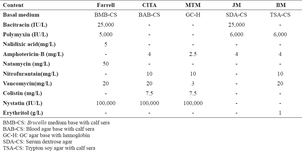
This study was carried out in the Pendik Veterinary Control Institute between 2014 and 2015. 51 organs and 7 fetal stomach content of abortion cases were utilized. The media included in this study involves 4 different selective media and the Tryptic Soy Agar (TSA) as a non-selective medium. The content of Farrell (20), CITA (21), Modified Thayer Martin (14), and Jones and Morgan (22) as selective media is illustrated in Table 1. In addition to these, one medium with antibiotics was used to extend the range of results. This medium is called ‘Brucella medium’ and labelled as BM in the following sections of this study.
Table 1. The contents of the selective media
Amphotericin-B has been preferred instead of natamycin or cycloheximid, which is part of the antimicrobial content of the JM medium. It is considered to be one of the antifungal agents suggested for the selective media for the first isolation of Mycobacterium spp. (23), Campylobacter spp. (24) and Brucella spp. (21).
For this study, the basal medium required for the selective media was prepared and sterilized by autoclaving (121°C ± 3°C, 15 minutes). Antibiotics and sterile new born calf sera were added to the media at about 56°C depending on their contents (25). Sterility controls of media were conducted after they were incubated at 37ºC for 48 hours (26). The organ suspensions were prepared from organ samples diluted 1/10 in a phosphate buffered saline in a biosafety cabinet (18, 27).
Organ suspensions and fetal stomach content were inoculated to media and incubated in 37°C, 5-10% CO2 condition for 5-8 days. Biovar identification of isolates was implemented according to CO2 requirement, H2S production, growth in media containing thionin (20µg/ml), basic fuchsin (20µg/ml), safranine (100µg/ml), penicillin, streptomycin, and i-erythritol sensitivity, lysis with Tibilisi (TbØ 104 RTD) and R/C phages and agglutination with monospecific A and M antisera. Media including streptomisin (2,5 µg/ml), penicillin (5 IU/ml) and i-erythritol (1 mg/ml) were used in the identification of vaccine strains. In addition, we have observed the growth level of contaminant microrganisms.
The classification categories for the inhibition ability of the media against contaminant microorganisms in this study were total inhibition (TI) and partial inhibition (PI). These categories were formed regarding the diffuseness of the contaminant growth through counting the colony forming units (cfu) (26). When the contaminant colony counts were taken into account, the media’s inhibition ability was listed by focusing on the range of the contaminant burden. We have classified the ranges into 4 groups; namely, 1 total inhibition group without any contaminant colonies and 3 partial inibition groups with less than 10, ones between 10 and 100, and ones with more than 100 colonies (26, 28, 29). Pearson Chi-Square Test in SPSS18.0 program was used to evaluate the results of this study.
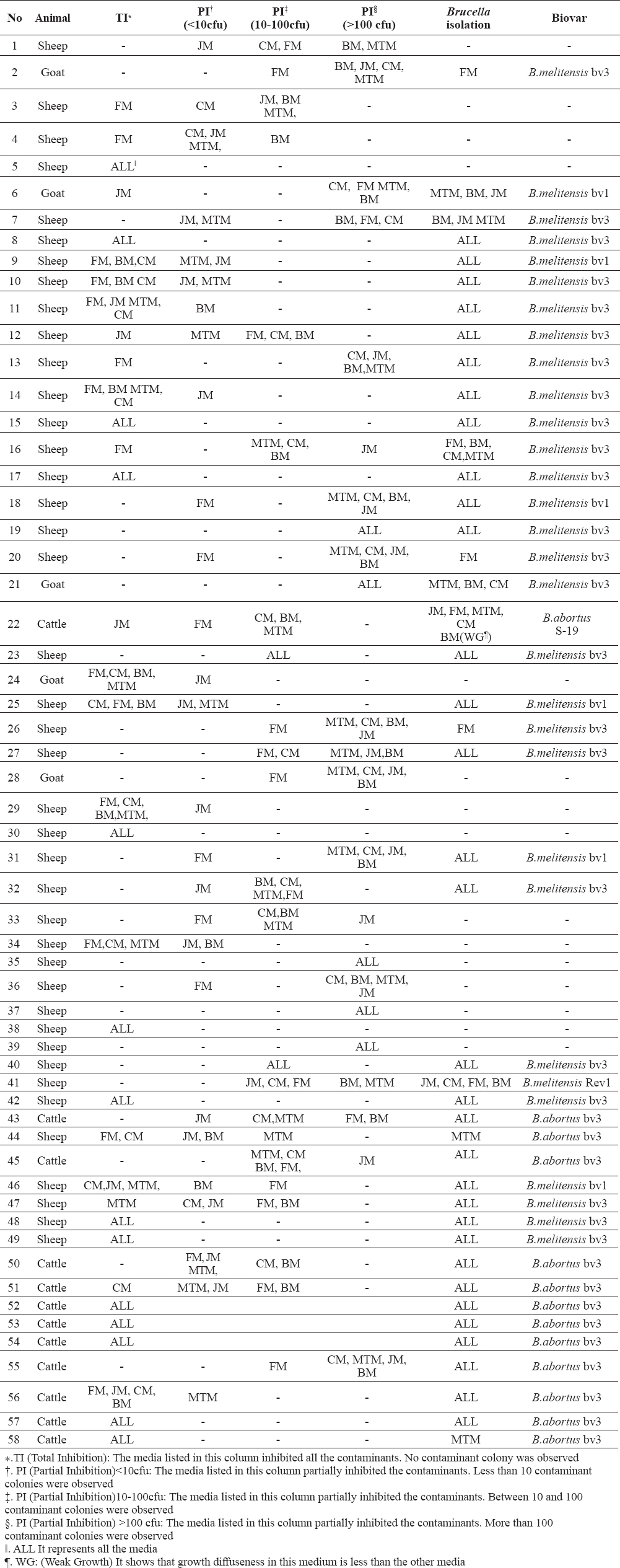
Biyotyping results of the isolates and the media in which isolation was carried out are illustrated in Table 2. The table also shows the inhibition level of the contaminants for each medium in every single sample.
Table 2. The isolation and inhibition results of selective media for each sample
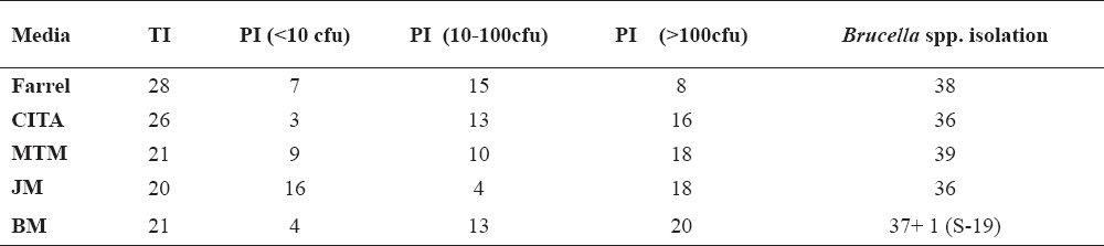
According to the aforementioned results, isolations could not be carried out in every medium. Moreover, selective media had different performance levels in terms of inhibition ability against contaminants. In addition to these, even though B. abortus S19 was isolated in Brucella medium after inoculation of sample No. 22, the growth of strain was at weak growth (WG) level and the growth diffuseness was clearly lower than the other media. The detailed results in Table 2 are summarized in Table 3 and Table 4. The numbers of Brucella isolation and the distribution of inhibition abilities are listed in Table 3.
Table 3. The number of Brucella spp. isolations and the distribution of inhibition ability
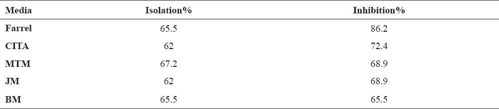
Table 4. The isolation and the inhibition ability of the media in percentages
Table 3 shows that MTM medium’s isolation percentage is the highest one with 39 Brucella spp. isolates when we consider all 58 samples. Moreover, Farrell medium’s performance is a lot better than the other media regarding the inhibition ability.
We have listed the inhibition and isolation ability of the media as percentages in Table 4 so as to clarify the numbers in Table 3. The percentages of the inhibition ability in Table 4 was found by taking the sum of TI and PI inhibition abilities without including the PI (>100 cfu). Even if two of them (PI <10 cfu, PI=10-100 cfu) show partial inhibition ability, they are considered to be sufficient inhibition ability as they make the isolation of Brucella spp possible.
The media’s isolation and inhibition ability illustrated in Table 4 was analyzed statistically. The media’s isolation percentages are similar and they are not statistically significant. There are far more differences in the media’s inhibition ability against contaminants than their isolation ability. The statistical analysis of these differences is outlined in Table 5.
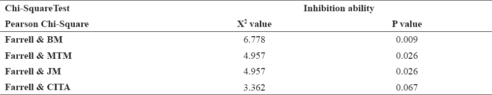
Table 5. The results of the statistical analysis related to the media’s inhibition ability
In Table 4, all the media except the Farrell medium have similar inhibition ability percentages. Therefore, in Table 5 statistical analyses were carried out only between the results of Farrell, which has the highest inhibition percentage, and the other media. The p value between Farrell and the other media except CITA medium is smaller than 0.05; therefore, the difference between Farrell and CITA is statistically insignificant but the difference between Farrell and the other media is statistically significant.
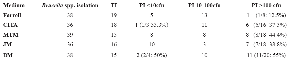
In order to investigate the correlations between isolation and inhibition, the inhibition ability of the media was examined in the samples in which Brucella spp. isolation occurred. The distribution of the media’s inhibition ability for these samples is listed in Table 6.
Table 6. The distribution of the media’s inhibition ability in the samples where Brucella spp. was isolated
Based on the distribution results, it can be stated that the highest Brucella spp. isolation for each medium was obtained in the samples in which all the contaminants were totally inhibited (TI). In addition, in Table 3, the distribution of the media’s inhibition ability was made based on 58 samples regardless of Brucella spp. isolation. In this table, on the other hand, the media’s inhibition ability was calculated for the samples in which Brucella spp. was isolated. When the values of these two tables were evaluated, the isolation percentage of the samples in which the contaminants were partially inhibited at the level of PI >100 was lower than the other levels (TI, PI <10, and PI 10-100), except for two results shown in italics. These low percentages of isolation were given in the last column of Table 6.
Regarding the number of isolations, MTM has the highest and Farrell medium has the second highest isolation percentage. Marin et. al. (17) obtained a higher isolation sensitivity for Brucella melitensis in MTM than they did in Farrell, which was actually developed for Brucella abortus isolation. In this study, it was found out that using these two media simultaneously could increase the isolation percentage up to 74.1% with 43 successful isolations (15). In OIE Cattle Brucellosis Chapter, too, using two media simultaneously to be able to augment the isolation sensitivity is recommended (17, 11). Similarly, Ferreira et al. (30) suggested using more than one selective medium to enhance the isolation sensitivity.
When the inhibition ability is taken into consideration, the Farrell medium has the highest percentage while CITA has the second highest percentage. Even though the difference between the Farrell medium and the CITA medium is statistically insignificant, the difference between the Farrell medium and the other media is statistically significant. It is pointed out that the Farrell medium is able to inhibit most of the contaminants; thus, it is the most common selective medium for the bacteriological diagnosis of Brucellosis (21). In the study by Vicente et al. (18) where CITA and Farrell were compared and contrasted, both media were found to be similar in their inhibition abilities against the contaminants and they showed good results when they were used together. De Miguel et al. (21) stated in their study, in which they developed the CITA medium, that CITA could inhibit most of the contaminants and it had better isolation sensitivity than those of MTM and Farrell. In a similar study, Brucella agar, the Farrell and CITA media were compared and contrasted and despite the same number of isolations in each medium, Farrell had the highest inhibition ability against contaminants and it is regarded as the best selective medium for microbiological diagnosis (31).
According to the results, it can be stated that the highest Brucella spp. isolation for each medium was obtained in the samples in which all the contaminants were totally inhibited (TI). When all the 58 samples were evaluated, the number of the samples where all the contaminants were totally inhibited is bigger in Farrell and CITA than in the other media. In his study, Farrell classified the growth levels of contaminants as 1+, 2+ and 3+ (20). He stated that most of the Brucella spp. isolations were obtained in the samples with contaminant growth at 1+ level. The findings of this study, too, indicate that the increase in the inhibition ability of the selective media plays an important role in enhancing isolation sensitivity. It was also pointed out that although the antibiotics added to the media could decrease contamination, B. abortus colonies might be masked by the excessive amount of contaminant growth due to the length of incubation period (29). For this reason, in such samples, the isolation rate can decline in the selective media which are not as effective as Farrell and CITA in terms of contaminant inhibition.
Brucella bacteria are fastidious microorganisms and require a longer period of incubation when we compare them with contaminants growing fast in the samples (14, 15, 25). The generation period of Brucella organisms, which is 2.5-3.5 hours, is considered as a long duration, too (32). It was also pointed out that it may take some more days to observe colonies on the selective media as compared to the usual incubation period on non-selective media (25). In our study, as well, we found out that when we passage the Brucella suspect colonies one day later, we could not identify Brucella colonies due to the contaminants which hid the Brucella colonies. For this problem, Alton et al. (25) recommended that Brucella suspect colonies should be passaged before the contaminants spread on the media’s surface and they should be checked three days after the incubation. Using solid medium is considered to be the most satisfactory method for isolation as it can facilitate the isolation of Brucella colonies and it can also minimize the risk of mixing Brucella colonies with the other fast growing microorganisms (11, 25).
In the samples in which the contaminants were partially inhibited at the level of PI >100, the isolation percentage was the lowest or close to the lowest. The reason behind the low isolation percentage might be predicted as the contaminants covering the medium’s surface. In a study by Stack et al. (15), it was stated that they could not spot the Brucella bacteria colonies in some of the artificially infected milk samples. They contend that the reason behind it was that contaminants in the milk samples disguised the Brucella colonies. Based on the findings of this study, it is possible to say that an increase in the inhibition ability of the media may facilitate Brucella spp. isolation and it may lead to an increase in the isolation sensitivity. In this sense, following and improving the inhibition abilities of the media according to the samples with different contaminant microflora might increase the isolation rate.
Her et al. (27) developed a selective medium which includes indicator neutral red for the isolation of Brucella abortus strains. This medium can facilitate the observation of the Brucella spp. colonies more easily by differentiating them from contaminants. A novel approach might be passaging the slow-growing Brucella bacteria with the help of the indicators before they are masked by contaminants. This approach may be a useful tool to increase the low isolation rate in samples where contaminants are inhibited inadequately. In our study, as well, lower inhibition ability level led to a lower isolation percentage; therefore, it might be a good idea to focus on the development of such kind of media. That kind of development and modification in the Brucella selective media as well as the findings of this study indicate the significance of contaminant inhibition for a better isolation percentage.
The medium with antibiotics, which was labeled as BM in this study, includes erythritol which stimulates the growth of Brucella strains. BM is composed of fewer antimicrobial agents compared to the media with high inhibition ability. Although BM has the lowest inhibition ability, it does not have the lowest isolation rate. This fact might be interpreted as the positive effect of the erythritol component it includes. The isolation rate can decrease in the samples in which contaminant organism burden increases qualitatively and quantitatively, while the number of the target bacteria decreases. In the development of media, components such as erythritol provoking growth and antimicrobial agents providing inhibition can be added to the media.
In the Modified Brucella selective (MBS) medium developed by Her et al. (27) for B. abortus strains, too, erythritol was used to provoke and improve the delayed growth of strains among antibiotic mixtures. Erythritol is also mentioned as a sugar alcohol which is effective in the tissue tropism of Brucella bacteria in ruminants (27, 33, 34). However, it is believed that erythritol does not stimulate (35, 36) but inhibits the growth of the S19 strain (34, 37). Conversely, S19 isolation took place in BM as weak growth in one of the samples (Table 2, no:22). Alton et al. (25) suggested that the mutation level of S19 strains against erythritol tolerance was high. They also stated that even though some suspected S19 isolates resembled S19 in other tests, they could grow in erythritol. It is pointed that the reason behind this weak growth in this sample might be what Alton et al. (25) suggested above. In light of the findings of this study, it might be stated that adding erythritol component to the selective media will provoke growth of Brucella strains except S19 and it might be recommended as a way of increasing isolation sensitivity.
To sum up, using two different media with high inhibition ability like Farrell simultaneously might be helpful while choosing the appropriate selective media. In the process of developing media, on the other hand, adding components that will provoke Brucella spp. growth should be considered. Moreover, checking the performance of media repeatedly will be beneficial for obtaining better isolation rates. In these repeated controls, qualitatively and quantitatively different microbiol burden of the field samples should also be taken into consideration.
© 2018 Karagul M.S. This is an open-access article published under the terms of the Creative Commons Attribution License which permits unrestricted use, distribution, and reproduction in any medium, provided the original author and source are credited.
This article was originated from part of a PhD thesis by Mustafa Sencer Karagul. The research was approved by the Local Ethics Committee for Animal Experiments, Istanbul University, Istanbul, Turkey (Ethics Committee Decision No: 2014-84).
The authors declared that they have no potential conflict of interest with respect to the authorship and/or publication of this article.
Macedonian Veterinary Review. Volume 41, Issue 2, Pages 177-186, p-ISSN 1409-7621, e-ISSN 1857-7415, DOI: 10.2478/macvetrev-2018-0024, 2018