Mac Vet Rev 2019; 42 (1): 43 - 49
 10.2478/macvetrev-2018-0028
Received: 13 February 2018
Received in revised form: 27 May 2018
Accepted: 16 August 2018
Available Online First: 27 November 2018
Published on: 15 March 2019
Keywords: calves, pathology, pneumoenteritis, bovine coronavirus
Coronaviruses are causative agents of digestive, respiratory and nervous illnesses in mammals and birds (1). The genus belongs to the family Coronaviridae and its members are enveloped, 100 to 120 nm in diameter and with single-strand positive-sense RNA. They are divided into three groups depending on the source, nucleotide sequence and serological groups (2).
Coronaviruses inducing disease in large ruminants belong to group 2 – Bovine coronaviruses (BCoV). They are able to replicate both in gastrointestinal enterocytes, as well as in the epithelium of the upper airways. BCoV with tropism to both the respiratory and the digestive system was detected by virological studies in cases of pneumoenteritis syndrome in growing calves (3). The virus is defined as pneumoenteritic, infecting the upper and lower airways, as well as the proximal and distal gastrointestinal compartments (4).
Bovine Corona Virus – BoCV is outlined as an etiological agent in three clinical syndromes: gastrointestinal diseases in newborn calves a.k.a. calf diarrhoea (CD), winter dysentery (WD) in adult cattle and infections of the respiratory and digestive systems of calves known as pneumoenteritis syndrome (PES). There is evidence that BoCV is involved in the etiology of the bovine respiratory disease complex (BRDC) affecting calves from 6 to 10 months of age (5).
According to Kanno et al. (6) calves from 1 to 3 months of age are most susceptible to coronavirus infection. Other researchers reported clinical signs of disease in 2-8-week-old animals (7). Calves are infected via the faecal-oral route as well as aerogenically, via inspired air. It is affirmed that the virus is shed with nasal discharge as early as the 3rd day, before appearing in faeces (8).
Clinical signs of disease could be observed as early as 24 hours postpartum to 5 months of age in colostrum-deprived calves. The general prevalence of the disease varies from 15% to 70% (9).
Infected calves shed extensively the virus (about 1 billion of viral particles per 1 mL faeces daily) throughout 14 days. After recovery, calves continue to shed the virus in the environment. BCoV could be found in the faeces of both diseased (8% - 69%), and healthy caves (about 24%) (10).
Viral particles have been detected through electron microscopy in the faeces of calves day or two before the onset of diarrhoea. After recovery, animals are immune and still could release the virus in the environment through nasal discharges and faeces (11).
Fulton et al. (12) affirmed that signs of intestinal and lung infection were simultaneously present in 30% of coronavirus infections of calves aged from 2 to 16 weeks.
The analysis of literature reports on gastrointestinal and respiratory diseases in newborn and juvenile calves confirms their wide prevalence and importance at cattle farms. The aim of the present study was to investigate the etiological and pathomorphological aspects of coronavirus-induced pneumoenteritis syndrome in newborn and growing cattle.
The study comprised 370 calves from the Black-and-White dairy breed. The age of the animals was from 24 hours to 25 days. Clinical and epidemiological studies were carried out with newborn and growing calves in all farms.
For detection of pathogens, rapid pentavalent antigenic test strips were used - Rainbow calf scour 5 BIO K 306 Detection of Rota, Corona, E.coli F5, Crypto and Clostridium perf. in bovine stool (BIOX Diagnostics, Belgium). Eighteen carcasses of calves dead after manifestation of enteritis and respiratory signs were submitted to gross anatomy study. Tissue samples (1 cm x 1 cm) were collected for histopathological examination from lungs and bronchial lymph nodes, as well as from affected proximal and distal gastrointestinal compartment: abomasum, duodenum, jejunum with mesenteric lymph nodes, ileum, caecum colon, rectum − all with 2,5 cm length. Specimens were fixed in 10% neutral buffered formalin for 48–72 h and embedded in paraffin. Cross sections, 4 μm thick were cut from paraffin blocks with a Leica RM 2235 microtome and stained with haematoxylin-eosin (H/E). Furthermore, tissue samples were obtained from lungs and the ileum for immunofluorescence studies. A monoclonal conjugated antibody Monoclonal Antibody anti-bovine Coronavirus FITC conjugated) 0,5 ml (20X), BIO 023, (BIOX Diagnostics, Belgium) was used to this end. Protokol for IFA: After fixation with acetone, rinse thoroughly with PBS and drop the FITC Conjugated Monoclonal Antibody (FITC conjugated) 0.5 ml (20X), BIO 023 (BIOX Diagnostics, Belgium). The infiltrated lamellae and deparaffinated FITC orthotics were kept in a humid chamber at 37ºC for 1 hour. After 1 hour, remove FITC and wash 3 times in 10 minutes with PBS. Contrast for 10 minutes with elongated blue at a concentration of 1:10,000 for historesis and 1: 100,000 for cell culture lamellas. The final step is drying the lamellas and histosrets, fitting with PBS and glycerol in a ratio of 9:1 and observed under a fluorescence microscope.
Epidemiological surveys in the six farms revealed that intestinal and respiratory infections in newborn and growing calves were the main health issue. Two-thirds of calves between 1 and 15 days of age exhibited clinical signs of digestive disorders. In the other 25% of calves of this age group (mainly 4-10 days of age), respiratory signs were present along the gastrointestinal ones.
The etiological agents in 75% of cases of gastroenteritis were bovine rotaviruses and cryptosporidiae. In complicated cases (25%) with simultaneous manifestation of gastrointestinal and respiratory signs (pneumoenteritis), only bovine coronaviruses were detected in the stool.
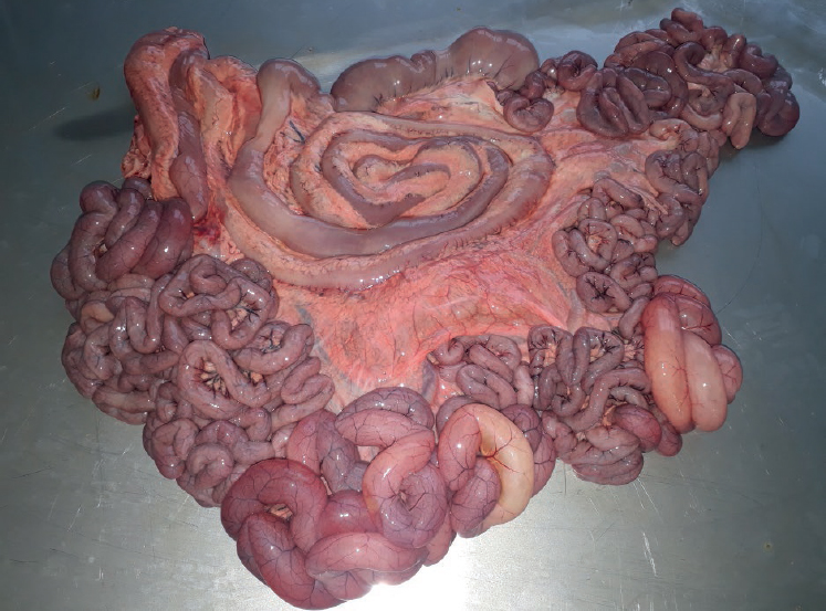
Eighteen calves with signs of pneumoenteritis were necropsied. The exterior examination showed catarrhal purulent nasal discharge and strongly hyperaemic nasal mucosa. Bilateral conjunctivitis was present, and 6 of the calves had enophthalmos. The carcass was dehydrated and emaciated, the perianal area and tail region were stained with greenish diarrhoeic faeces. Changes specific for catarrhal or catarrhal haemorrhagic enterocolitis were present. Mesenteric lymph nodes were juicy and enlarged, and blood vessels – hyperaemic. The intestinal mucosa was hyperaemic, with yellow-green intestinal content mixed with gas bubbles and non-digested food particles (Fig. 1).
Figure 1. Serosal hyperemia and gases in the small and large intestines, calf with pneumoenteritis syndrome
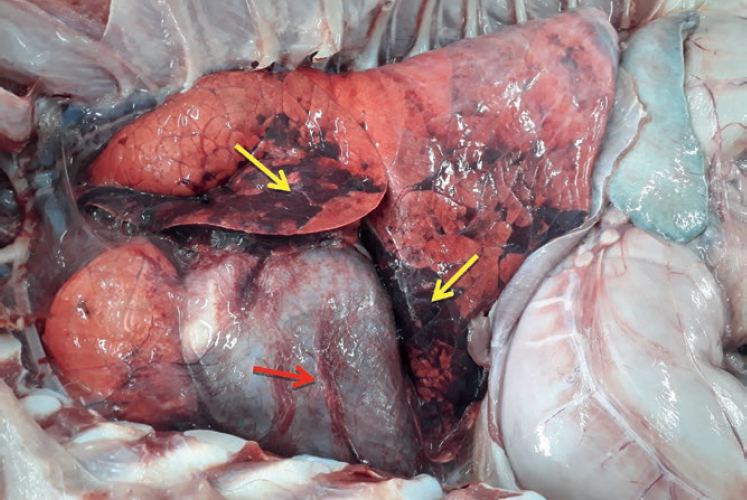
After dissection of the thoracic cavity, various yellowish fluid collections, sometimes up to 1000 ml were observed, while the lungs exhibited pneumonic areas (Fig. 2). Bronchial lymph nodes were hyperaemic and enlarged. The dissection of the trachea revealed non-transparent foamy fluid and hyperaemic mucosa. The pericardial sac contained increased amount of yellowish fluid. The laryngeal mucosa was swollen and diffusely reddened. The nasal cavity mucosa was spattered with haemorrhages, hyperaemic and lined with catarrhal purulent exudate. Similar changes were also found out in the paranasal sinuses.
Figure 2. Pneumonic foci in the lung (yellow arrows) and pericarditis (red arrow) in a calf with pneumoenteritis syndrome
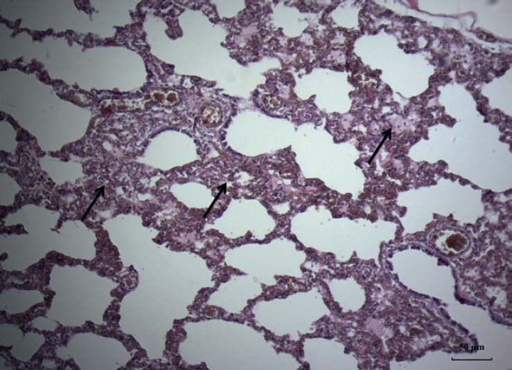
Microscopic lesions were established in the lungs and the distal small intestinal compartment (ileum). In the pulmonary interstitium, round-cell infiltration comprising of lymphocytes and macrophages, vascular hyperaemia and haemorrhages – signs specific for interstitial pneumonia – were demonstrated (Fig. 3). The epithelium lining the bronchial mucosa was affected by necrosis of bronchial epithelial cells with desquamation of necrotic cells.
Figure 3. Interstitial pneumonia (arrows) in the lung of a calf with pneumoenteritis syndrome (H&E; Bar=10μm)
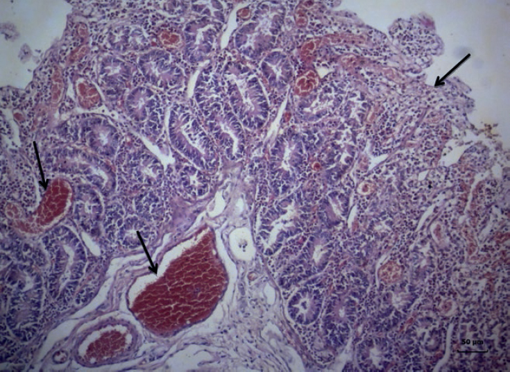
The microscopic changes in the ileum consisted in vascular hyperaemia in the mucosa and submucosa, submucosal oedema and dilated crypts. The villi were atrophied and fused, infiltrated with lymphocytes, while epithelial cells exhibited degeneration and necrobiotic changes resulting in desquamation (Fig. 4).
Figure 4. Atrophy of intestinal villas (arrows), intensive vascular hyperaemia of the mucosa and the submucosa (arrows) in the ileum of a calf with pneumoenteritis syndrome (H&E; Bar=10μm)
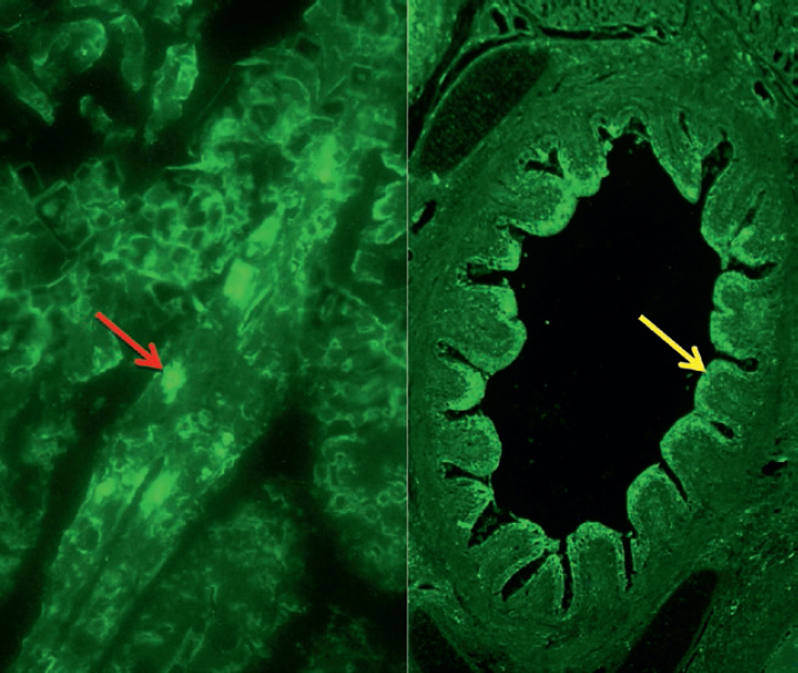
The used direct immunofluorescence test evidenced the presence of bovine coronavirus antigens in the ileal and pulmonary tissue cross sections (Fig. 5). Immunofluorescence data confirmed the field antigenic tests in calves with pneumoenteritis.
Figure 5. Direct immunofluorescence test with positive result for BCoV from the ileum (left, red arrow) and lung (right, yellow arrow) in a calf with pneumoenteritis syndrome
The results from the performed clinico-epidemiological, etiological and pathomorphological studies among newborn and growing calves in surveyed cattle farms confirmed that coronaviruses (BCoV) were the causative agents of the pneumoenteritis syndrome in this animal category at an early age.
We agree with the common belief that PES occurrence was mainly associated to the lack of adequate immunoprophylaxis of pregnant cows, as well as to faults in the rearing technology of newborn calves (13). Our results allowed us to affirm that BCoV induced pathological changes occurred simultaneously in the respiratory and gastrointestinal tract of newborn and growing calves in line with earlier reports (14). Furthermore, we assume that the involvement of other viruses and bacteria affecting the respiratory and digestive systems could exacerbate the clinical signs and aggravate the course of the disease (15).
The nature of the observed gross and macroscopic lesions in the lungs (interstitial pneumonia), as well as the cell infiltration of lymphocytes and macrophages in the distal small intestine (ileum) is associated with the viral etiology of the disease as also confirmed by other researchers (16). We confirmed that the observed interstitial pneumonia was of viral etiology unlike bronchopneumonia which is bacterial (Escherichia coli, Mannheimia haemolytica, Pasteurella multocida, Mycoplasma bovis and Histophilus somni in some possible causes). Our results allowed also to substantiate that microlesions due to coronaviral interstitial pneumonia were characterised with the absence of intracytoplasmic inclusion bodies in bronchial epithelial cells, which are present in infections with bovine respiratory syncytial virus (BRSV) and bovine parainfluenza 3 virus (BPI3V) (9).
We agree that unlike rotaviral enteritis characterised with degenerative and necrobiotic changes in the middle part of the small intestinal and large intestinal villi, coronaviral enteritis affected the surface part of jejunal and ileal villi resulting in superficial catarrhal desquamative inflammation (8, 15, 16).
In conclusion, the used express antigenic and virological diagnostic tests were reliable and accurate for detection of pneumoenteritis in calves. The macro- and micro lesions in the lung and the ileum of calves affected by PES are relevant with regard to the differential diagnosis of the syndrome and its differentiation from respiratory (IBR, BVD, BRSV, M. haemolytica etc.) and intestinal (Cryptosporidium parvum, bovine rotaviruses, bovine coronaviruses and Escherichia coli K99 (F5) diseases in this category of animals.
© 2018 Kalkanov I. This is an open-access article published under the terms of the Creative Commons Attribution License which permits unrestricted use, distribution, and reproduction in any medium, provided the original author and source are credited.
The authors are grateful to their colleagues serving the private cattle farms from different regions of the country for cooperation and collaboration.
The authors declared that they have no potential conflict of interest with respect to the authorship and/or publication of this article.
Macedonian Veterinary Review. Volume 42, Issue 1, Pages 43-49, p-ISSN 1409-7621, e-ISSN 1857-7415, DOI: 10.2478/macvetrev-2018-0028, 2018