Mac Vet Rev 2019; 42 (1): 35 - 42
 10.2478/macvetrev-2018-0029
Received: 02 March 2018
Received in revised form: 22 June 2018
Accepted: 22 August 2018
Available Online First: 18 November 2018
Published on: 15 March 2019
Keywords: goats, ultrasonography, fetal number, progesterone
Fetal number determination is an important step of reproduction management in goats. The classification of herds based on fetal numbers would result in a different technological regimen, feed cost and reproductive management optimization (1, 2). The determination of fetal numbers via blood progesterone concentrations has been reported in sheep (3) and goats (4).
Ultrasonography offers clear advantages for real-time determination of fetal numbers compared to other techniques (5). It is the most commonly used method as it allows for a simultaneous determination of the number, vitality and gestational age of fetuses. For this purpose, precise criteria and optimum time of the exam should be chosen depending on the equipment, age and breed of animals (6, 7, 8, 9).
The number of embryos/fetuses could be identified until the 100th gestational day, but the period with ultimate accuracy and visualization of the uterus without overlapping of fetuses is between the 40th and the 70th gestational day (5, 10, 11, 12). To minimize false positive and negative diagnoses, some authors recommend preliminary fasting of animals, and lifting of the abdominal wall during the exam (12, 13).
Using a transabdominal approach with probe frequency of 5 MHz in Alpine does, researchers differentiated single, twin and triplet pregnancies with precision of 44%, 73% and 67% respectively during the 5th gestational week (1). Suguna et al. (8) reported that the distinction between single and twin pregnancy was possible at the earliest by the 35th gestational day via transrectal approach and by the 42nd day with the transabdominal approach. The authors suggested that the period between the 5th and 7th gestational weeks was the most appropriate for differentiation between single and twin pregnancy in local goat crosses. Others determined fetal numbers in Saanen goats using the transvaginal approach with a 5-7.5 MHz probe and demonstrated 17% accuracy during the 3rd week and 60% during the 8th week of gestation, also confirming that the transvaginal approach was not the best for determination of multiple pregnancy in goats (14).
According to numerous researchers, the type of pregnancy (single or multiple) influences substantially the precision of the method, so it is difficult to distinguish among twins, triplets and quadruplets at each stage of the gestation in both goats and sheep (3, 15, 16). Increased locomotor activity of the embryo impedes the differentiation between twin and multiple pregnancies especially until the 40th gestational day. Counting twice an embryo is another error, which could be avoided by B/B scan regimen to differentiate the fetuses (9), and the operator should scan slowly the area from left to the right at the same level to localize the uterus cross-section. Then the direction of scanning should be perpendicular to the previous one to include successively the entire area (12).
Although many available ultrasonographic studies of fetal counting were performed in goat breeds in different regions of the world, investigations on the effect of pregnancy type, breed, ultrasonography method and equipment in local goats have not been performed yet. For these reasons, the purpose of the present study was to determine the fetal number in Bulgarian local goats by use of hormonal and ultrasonographic examinations.
The experiments were performed with 106 Bulgarian local goats with an average body weight 35-52 (mean 43.5) kg, 1.5 to 7 (mean 4.25) years of age. Some of animals were reared in the Experimental Farm of the Trakia University, Stara Zagora (n=24), and the other goats were from different private farms in Losenets, Sliven region (n=54) and Marinka, Burgas region (n=28). The goats were both stall-fed and pasture-grazed and also received additionally concentrate. Roughage, drinking water and slat licks were offered ad libitum. All goats were given antiparasitic and immunoprophylactic treatments before the experiments. The experiments were approved by the Animal Ethics Committee to the Faculty of Veterinary Medicine, Trakia University – Stara Zagora, in compliance to Ordinance No. 20 of the Ministry of Agriculture and Food from 1 November 2012.
For this purpose, 24 pregnant animals were used. Serum progesterone levels were assayed between 21 – 63 days of gestation at 7-day intervals and until the 133rd day at 14-day intervals using a commercial ELISA kit and ELISA Huma Reader (HUMAN, Germany). After parturition, the animals were divided into 3 groups depending on the number of offspring (one, two or more) and the gestational stage, accounting also for abortions and stillborn fetuses. The mean serum progesterone concentrations in the different groups were calculated and analyzed.
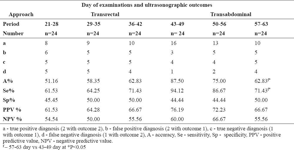
Ultrasound scans were performed on 82 goats. All animals were examined once at 7-day intervals using the transrectal approach between gestational days 21-28, 29-35, 36-42 and by the transabdominal approach between 43-49, 50-56, and 57-63 days of pregnancy. For this purpose, goats were fixed in a standing position by an assistant. Fetal numbers were determined after visualization of an embryo/fetus with or without cardiac activity in the anechoic uterine lumen section, using the B and B-B working modes of the equipment.
Serum samples for hormonal analysis were collected from the vena jugularis externa after previous fixation. Disposable 18G needles and closed-system serum tubes (with pearls) 16×75 mm, 10 ml (Biosigma, Italy) were used to obtain 5 ml from each animal. After being transported to the lab, blood was centrifuged at 3000g for 15 min. Enzyme activity was detected at a wavelength of 450 nm on a Huma Reader (HUMAN, Germany). Optic densities of samples were read as progesterone concentrations (ng/ml) against a standard curve.
Ultrasonography was done with a SonoScape A5v (SonoScape, China) ultrasound machine equipped with linear multifrequency linear transducer (5.0-12.0 MHz) after preliminary application of contact gel (Eco-Ultra gel, Milano, Italy).
The number of fetuses (embryos) was determined as per Gearhart et al. (17). For a more convenient counting and avoiding errors, the B-B working mode of the equipment was used. After the kidding data became available, the true positive (a), false positive (b), true negative (c), false negative (d) diagnoses were determined, as well as positive (PPV) and negative (NPV) predictive values. The accuracy specificity and sensitivity of the method, PPV and NPV were calculated (18).
The data was analysed using the statistical software StatSoft (Statistica 7, Microsoft Corp. 1984-2000 Inc.) by means of ANOVA, LSD test for multiple comparisons, non-parametric comparisons of proportions using the t – criterion of Student. The analysis of variance determined the statistically significant effects of the studied factors. Results were presented as mean ± standard deviation (SD), relative share (%) and levels of significance. The level of significance was set at P<0.05.
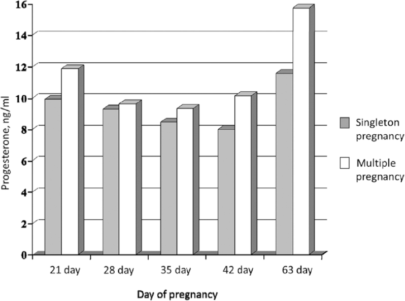
The animals were classified according to the number of offspring at parturition. Then they were separated into two principal groups. The first group comprises of goats that gave birth to one kid (single pregnancy, n=8) and in the second – those giving birth to two or more kids (multiple pregnancy, n=16). Mean serum progesterone concentrations (Fig. 1) in goats with multiple pregnancies were 13.88±3.28 ng/ml on day 21 vs 9.92±4.91 ng/ml in those with single pregnancy. By the 63rd gestational day, the attained levels were 15.73±5.42 ng/ml and 11.58±4.67 ng/ml, respectively. There were statistically significant differences between both groups of goats on gestational days 21 (P<0.01) and 63 (P<0.05) depending on the pregnancy type.
Figure 1. The mean serum progesterone concentrations (ng/ml) in single and multiple pregnancies
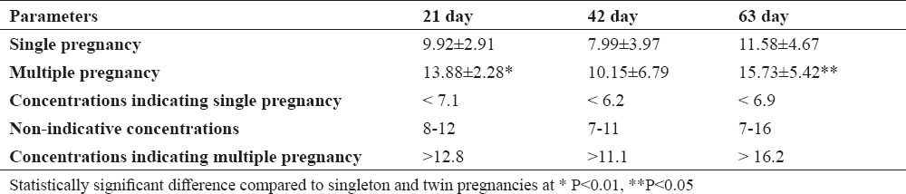
Indicative progesterone concentrations for goats with single pregnancy during the different periods were < 7.1 ng/ml, < 6.2 ng/ml, < 6.9 ng/ml, while for multiple pregnancies: >12.8 ng/ml, >11.1 ng/ml, > 16.2 ng/ml (Table 1). The accuracy of fetal number determination in the different periods was 51%, 58%, 69% and 47%, 52%, 62% for single and multiple pregnancies, respectively.
Table 1. Average, indicative and non-indicative serum progesterone concentrations (ng/ml) in single and multiple pregnancies
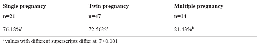
Fetal numbers were determined in 82 goats at a different gestational age. They were divided into 3 groups – with single pregnancy (n=21), with twin pregnancy (n=47) and carrying triplets or more (n=14). After the parturition, 16 out of 21 diagnoses were confirmed and 5 were false. With regards to twin pregnant goats detected ultrasonographically, 34 diagnoses were confirmed and 13 of them were false. In the group with 3 or more fetuses, 3 true and 11 false diagnoses were detected. In summary, 53 of all 82 diagnoses were confirmed after the parturition and 29 of them were found erroneous.
The accuracy of ultrasonography with respect to fetal number was the highest in single pregnancies (76.18%), followed by twin (72.56%) and the lowest in multiple pregnancies – 21.43% (Table 2).
Table 2. Accuracy of ultrasonographic fetal counting results depending on pregnancy type
Within the 21-28 gestational days (Fig. 2), the accuracy of ultrasonography for fetal number determination was 51.16% with sensitivity of 61.53%. Between the 29-35 (Fig. 3) and 36-42 days of pregnancies, the accuracy and sensitivity of the method were higher. During the next gestational periods, a substantial increase in accuracy for transrectal fetal number determination was established. The highest accuracy of 87.50%, sensitivity of 94.12% and positive predictive value of 76.19% were found out between the 43rd and 49th gestational day using the transabdominal approach (Table 3). It was considerably higher than that registered during the subsequent periods (P<0.05), and showed a tendency towards reduction as the gestational age advanced.
Table 3. The accuracy, sensitivity, specificity and prognostic value of ultrasonographic fetal counting results depending on gestational age and sonographic approach
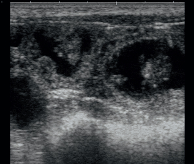
Figure 2. Ultrasonographic image of twin fetuses at 28th day of pregnancy
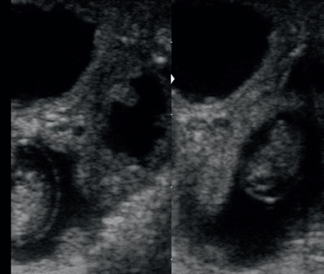
Figure 3. Ultrasonographic image of twin fetuses at 35th day of pregnancy (B-B mode)
Fertility is among the most important economic traits in small ruminants, especially in goats. The subsistence of millions of people in poorly developed rural regions depends on goat breeding results.
The possible methods for fetal number determination are measurement of blood progesterone concentrations and B-mode ultrasonography. The application of a hormonal test for this purpose depends on the ability for differentiation of circulating progesterone levels during the luteal stage in non-pregnant animals, and during the different gestation stages in pregnant ones, as well as whether it is single or multiple (10, 19).
The determination of the fetal number via blood progesterone assay has been mainly carried out in sheep (3, 20, 21, 22) and recently, it is increasingly applied in goats (4, 23).
The serum progesterone data obtained in studied goats with different fetal numbers are similar to data reported with various goat breeds (24, 25, 26). They are opposite to results of Sousa et al. (27), which stated that blood progesterone concentrations were not different in goats with single or multiple pregnancy. The differences in plasma progesterone in sheep carrying one, two or three fetuses are due to the production of higher amounts of progesterone by the multiple placenta apart from the corpora lutea (CL), unlike goats, in which the CL could be the primary source of progesterone for maintenance of pregnancy (28).
The ultrasonography offers a clear advantage for real-time identification of fetal numbers compared to other techniques (5, 6). It is the most commonly used method, as it allows for simultaneous determination of fetal number, fetal vitality and gestational age.
In this study, the detection of single or multiple pregnancy through ultrasound imaging indicated that the type of pregnancy had a substantial effect on the accuracy of the method (P<0.05). For single pregnancies the accuracy was 76.18%, and in twin pregnancies – 72.6%. Lavoir and Taverne (11) published comparable results. A better accuracy was reported by Dawson et al. (8) - 44%, 73% and 67% on the 35th day up to 83%, 89% and 100% on the 49th day of gestation, respectively. Amer (29) reported lower percentages in Egyptian dairy goats (accuracy of 45.7% for single and 54.3% for twin and triplet pregnancies). The lower accuracy for twin pregnancies is attributed to the higher number of false diagnoses, due to increased fetal moving, counting the same fetus twice or embryonic death occurring in later stage.
The lowest accuracy (21.43%) was detected in goats carrying three or more offspring in our study. This result was even lower than values reported by Dawson et al. (1) on the 35th day – 67%, but comparable to 25% accuracy in Saanen goats with 2 or more fetuses investigated by Abdelghafar et al. (30). This unsatisfactory result can be explained by the fact that the precision of the technique depends on the gestational age, the equipment, the probe frequency, study position and the experience of the operator, which has been shown in a previous study (13).
In the period between 21st-28th days, the accuracy of fetal counting by transrectal ultrasonography was 51.16% with sensitivity of 58.35%. In one research for determination of fetal numbers in goats, Karen et al. (6) established a total accuracy of 76% between post mating 24-36 days using the transrectal approach, which is considerably higher than the accuracy in our studies. As the gestational age increases, the possibility for precise determination of fetal numbers using this approach decreases, especially after the 42nd day. The possible causes are the overlapping of ultrasound images of fetuses and impossibility of ultrasonic waves to reach the pregnant uterus (6).
The highest accuracy of 87.50%, sensitivity 94.12% and positive predictive value 76.19% of the imaging method were demonstrated between gestational days 43-49th using the transabdominal approach. Medan et al. (7) reported accuracy of 67% on the 40th day compared to 84% on the 50th day and 92% on the 60th day. The cited results confirm the accuracy established by us between the 42nd and the 49th day.
The accuracy of the method could also be influenced by the technique of examination and parturition number, especially in uterine dislocations and examinations performed after the 49th day. In support of this thesis, Karen et al. (31) reported accuracy of 69% between the 43-56 day and 71.6% between 76-87 gestational days via transabdominal ultrasonography in sheep. In a later research in goats, Karen et al. (6) established a statistically significant difference in accuracy of imaging between young and old animals (93.8% vs 56.3%, P<0.05). This difference is attributed to the possibility for scanning of the entire uterus in young does between the 24-29th day and at the same time; it is much easier to evaluate the number of fetuses than in the advanced stages of pregnancy. The authors concluded that the transrectal approach (6-8 MHz) could be utilized for determination of fetal numbers between gestational days 24-36 with accuracy similar to that between 39-51 days and transabdominal approach (3.5-5 MHz) without statistically significant influence of the age of goats.
According to Lavoir and Taverne (11), the most appropriate period for fetal number determination in goats is between the 40th and 70th gestational days using the transabdominal approach. Later studies of Suguna et al. (8) with Indian goat crosses established that transrectal ultrasonography for fetal number determination should be done between the 35th and the 42nd days, whereas transabdominal imaging – between the 42nd and the 49th day.
In conclusion, analysis of our results shows that the fetal numbers in local goats should be determined between gestational days 36-42 by transrectal approach and between days 43-49 with the transabdominal approach. The most appropriate time for determination of fetal number of goats using ultrasonography is between gestational days 42-49.
© 2018 Karadaev M. This is an open-access article published under the terms of the Creative Commons Attribution License which permits unrestricted use, distribution, and reproduction in any medium, provided the original author and source are credited.
The authors are grateful to the staff of the goat private farms for their assistance during experiments.
The authors declared that they have no potential conflict of interest with respect to the authorship and/or publication of this article.
Macedonian Veterinary Review. Volume 42, Issue 1, Pages 35-42, p-ISSN 1409-7621, e-ISSN 1857-7415, DOI: 10.2478/macvetrev-2018-0029, 2018