Abstract
A fast and sensitive HPLC method was validated in order to analyze doxycycline in plasma and milk of sheep and in plasma of rabbits. The samples were processed with trifluoroacetic acid (TFA). After the centrifugation step, a supernatant containing extracted doxycycline and internal standard oxytetracycline was injected into the HPLC system with PDA detection. The method showed linearity in the range of 0.125 - 2.5 μg/mL for ovine plasma, 0.125 – 5.0 μg/mL for ovine milk, and 0.125 – 1 μg/mL for rabbit plasma. The inter-assay precision varied between 5.69 – 13.55 %. Values for intraassay precision were between 0.62 – 8.67 %. Accuracy was higher than 90% in all of the tested concentrations in the three types of biological matrices. The mean extraction recovery was higher than 90 % for all matrices. In order to handle only with free drug concentrations, microfiltration of standard solutions with low (0.25mg/mL), medium (0.5mg/mL) and high (1.0mg/mL) concentration was performed. A percentage for correction of the quantified doxycycline was calculated. The most significant adjustments should be made at the low concentrations. The correction for rabbit plasma is 24.63±5.03%, for ovine plasma is 20.10±8.01% and for milk –16.68±0.04 %. This method can be used for routine determination of doxycycline concentrations for pharmacokinetic studies and further dosage adjustment.
Keywords: free doxycycline concentration, HPLC analysis, sheep plasma and milk, rabbit
INTRODUCTION
Tetracyclines have been widely used in veterinary medicine for treatment of farm and pet animals. This is mainly due to their broad spectrum of activity, high efficacy, and relatively low risk of adverse effects. Doxycycline is a second generation semisynthetic tetracycline antibiotic with an activity against Gr+ and Gr– aerobic and anaerobic bacteria, including some intracellular pathogens (
1). Its bacteriostatic activity is due to the blocking of the 30S ribosomal subunit and thereby the microbial protein synthesis (
1). In practice, it is mainly used as doxycycline hyclate. Doxycycline has advantages over other tetracyclines. It penetrates well in tissues, has a higher bioavailability after oral administration, better antimicrobial activity, and lower affinity for Ca2+ (
2). Doxycycline has the longest elimination half-life compared to other tetracyclines, as well as the highest protein binding (
3). Therefore, it is retained in the body longer than the other antibiotics from the group. Doxycycline is more stable in water solution, which makes it suitable for administration with the drinking water (
4).
The presence of antimicrobial resistance represents a significant problem in veterinary and human medicine. Resistance to doxycycline has been observed in some microorganisms such as Staphylococcus aureus, Enterococcus spp., Salmonella spp., Pasteurella spp. (
5,
6,
7). In order to limit the selection and spreading of resistance, precise choice of antibiotic treatment and evidenced based adjustment of the dosage regimen of doxycycline are needed. For this purpose, not only MIC value determination is required, but also knowledge of accurate in vivo free drug concentrations. Only free, protein unbound doxycycline levels are effective concentrations in the body and the calculation of the pharmacokinetic parameters should be based on the free plasma concentrations found in the biological fluids. A simulation, predicting mean plasma concentrations in dogs, shows that the unbound level of the drug is remarkably lower than the total plasma concentrations, which influences the success of the therapy (
8). So far, a lot of the pharmacokinetic studies have not been corrected for binding to plasma or milk proteins (
9,
10). Information about free doxycycline levels in small ruminants is needed because of its importance for control of bacterial infections and because it was increasingly common in last years tick-borne rickettsioses. The data about doxycycline in small ruminants and rabbits is insufficient. It is not clear whether the methods used in other animal species are applicable, and whether those methods exclude utmost the bound part of the drug (
10).
The aim of the current study was firstly to validate an HPLC method for determination of doxycycline concentrations in ovine plasma and milk, and in rabbit plasma, which would permit the analysis of a large number of samples for clinical studies and settings. The second goal was to purify the samples and to determine the eventual fraction which is bound to blood proteins. The calculated percentage for correction of the quantified amounts can be used in pharmacokinetic studies and future adjustments of dosing regimen.
MATERIAL AND METHODS
Chemicals and reagents
The reagents used in the current study were HPLC grade. Doxycycline hyclate with purity ≥ 98% (TLC, Sigma D9891, Lot#/BCBF9827V) and oxytetracycline hydrochloride ≥95% crystalline (Sigma, HPLC, 05875, Lot#BCBG9599V), were used as reference compounds and for preparation of standard solutions. Acetonitrile CHROMASOLV®, HPLC grade, ≥99.9% purity (Sigma-Aldrich,), methanol ≥99.8% purity HiPerSolv CHROMANORM® for HPLC isocratic grade (VWR BDH PROLABO®), oxalic acid 98 % purity (Sigma Chemical Co., St. Louis, MO, USA), ethylenediaminetetraacetic acid disodium salt dihydrate 99.0 – 101.0% (Na2H2EDTA x 2H2O, Sigma-Aldrich) and trifluoroacetic acid ReagentPlus®, 99% purity (Sigma-Aldrich) were used for sample preparation and for the mobile phase.
Standard solutions
Stock solutions of doxycycline hyclate and oxytetracycline hydrochloride were prepared in deionized water at concentration of 10 mg/mL. They were used within one month. Oxytetracycline solution was used as an internal standard (IS) at concentration of 11 µg/mL. Standard working solutions were freshly prepared on the day of analysis. They were obtained by dilution of the stock solution of doxycycline hyclate with drug-free ovine plasma or milk, and with drug-free rabbit plasma at concentrations of 5, 2.5, 1, 0.5, 0.25 and 0.125 µg/mL. Blood samples were obtained from lactating sheep (n=6) and rabbits (n=7) according to the Bulgarian legislation for welfare of experimental animals (Regulation No 20/09.11.2012). Standard solutions were prepared in deionized water at the same concentration range as described above for plasma and milk.
Extraction procedure
Plasma samples (400 µl) were spiked with 40 µl IS and 52 µl trifluoroacetic acid (TFA). They were vortexed for 1 min and centrifuged for 10 min at 10 800 g at 22°C. The supernatant was carefully placed in HPLC vials and 20 µl of each concentration were injected into the HPLC system. Milk samples (900 µl) were mixed with 90 µl IS and 117 µl TFA. After vortexing for 1 min and centrifugation for 10 min at 10 800 g at 22°C, the supernatant was transferred to another tube. These samples were centrifuged again for 5 min at 10 800 g at 22°C and then filtered through standard filter paper (grade 600 pore size 10-20 mm, Fiorony Filters). The filtrate was placed in a vial and 20 µl of each concentration were injected into the HPLC system.
Protein purification
Standard solutions of doxycycline in ovine plasma and milk and in rabbit plasma with low (0.25 µg/mL), medium (0.5 µg/mL), and high (1.0 µg/mL) concentrations were further purified, before injecting them into the HPLC. For this purpose Ultrafree® - MC Centrifugal Filters with hydrophilic PTFE membrane and 0.45 µm pore size were used. The purification step was performed for each concentration in each matrix in triplicate. The samples were centrifuged for 4 min at 10 800 g at 22°C. 20 µl of each filtrate were injected three times into the HPLC system. The percentage for protein binding was calculated according to manufacturer instructions and the following equation:
% = [(conc NF-conc F)/conc NF]*100
where conc NF is the measured concentration before microfiltrationandconc Fistheconcentration after microfiltration.
HPLC analysis
All analysis was performed on a HPLC system comprising Hypersil Gold column (5μM, 150 x 4.6 mm), Surveyor LC Pump Plus, PDA detector Surveyor and a Surveyor Auto sampler Plus (Thermo FisherScientific Inc., USA). The column was used at room temperature. The mobile phase consisted of acetonitrile, methanol, 0.02 M oxalic acid and 0.02 M Na H EDTA x 2H2O (20 : 15 : 64 : 1, v/v/v/v). The f low rate was 1.0 mL/min. The detection wavelength was set at 345 nm. Peak area integrations were measured by the ChromQuest Chromatography Data System (Thermo Fisher Scientific Inc., USA).
Calibration curve
Standard solutions were prepared and analyzed as described above. The peak area ratio of doxycycline to the IS was plotted against the spiked concentrations to obtain the calibration curve. The curves were characterized by linear regression. To test the validity of the linear regression model, a statistical test for lack-of-fit was performed (Statistica for Windows 7, Stat Soft Inc., USA). Regression coefficient, slope and intercept were calculated.
Limit of detection (LOD) and Limit of quantitation (LOQ)
The LOD of doxycycline in plasma and milk samples was determined as the lowest amount in a sample that can be detected but not necessarily quantified (
11). The LOQ is defined as the lowest amount of the analyte in the samples that can be determined with acceptable precision and accuracy (
11). Calculations for the LODs were based on the standard deviation (SD) of y-intersepts of the regression lines and the slope (S), using the following equation: LOD = 3.3 (SD/S). The LOQs were calculated by the equation LOQ = 10 (SD/S) (
11). procedure was repeated also on different days (n=3) (
11). The accuracy was determined by comparison of the value of measured concentration to its true value (
11).
Extraction recoveryThe recovery of doxycycline was determined by comparing its concentration in standards, prepared in plasma or milk after extraction, to those prepared in deionized water. The analyses were performed on low (0.25 µg/mL), medium (0.5 µg/mL) and high concentrations (1.0 µg/mL).
Accuracy and precision
Inter-assay and intra-assay precision of the method was assessed. The different concentrations of doxycycline in each biological matrix were analyzed on the same day in triplicate.The procedure was repeated also on different days (n=3) (
11). The accuracy was determined by comparison of the value of measured concentration to its true value (
11).
Interferences
The analysis of blank ovine plasma and milk, and rabbit plasma, obtained from different individuals did not show interfering peaks at the retention time of doxycycline. The analysis of standard working solutions of doxycycline in plasma (from sheep and rabbits) and in ovine milk showed no interfering peaks at the retention time of doxycycline.
RESULTS
The parameters of standard curves for doxycycline are shown in
Table 1. The goodness- of-fit of linear regression model for standard curves was evaluated according to the value of r2 (coefficient of determination).
It varied between 0.9923 and 0.9989 for the three different matrices. Additionally, a test for lack of fit was performed. It showed that the standard curves were linear in the range of 0.125 - 2.5 µg/mL for ovine plasma (p = 0.99), 0.125 – 5.0 µg/mL for ovine milk (p = 0.99), and 0.125 – 1.0 µg/mL for rabbit plasma (p = 0.08). Interfering peaks did not appear at the retention times of the investigated compounds: doxycycline and the internal standard
Table 1. Retention time, linearity of standard curves, limit of detection (LOD) and limit of quantification (LOQ) of doxycycline determined by HPLC Method with PDA detector
Figure 1. HPLC chromatogram of doxycycline in ovine plasma spiked with 1 µg/mL doxycycline hyclate and 11 µg/mL oxytetracycline hydrochloride (IS). The detection wavelength was set at 345 nm
Figure 2. HPLC chromatogram of doxycycline in ovine milk spiked with 1 µg/mL doxycycline hyclate and 11 µg/mL oxytetracycline hydrochloride (IS). The detection wavelength was set at 345 nm
Figure 3. HPLC Chromatogram of doxycycline in rabbit plasma spiked with 1 µg/mL Doxycycline hyclate and 11 µg/mL oxytetracycline hydrochloride (IS). The detection wavelength was set at 345 nm
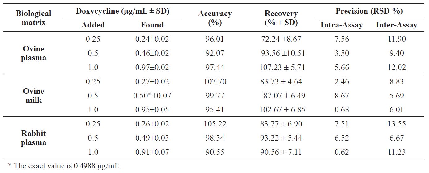
Table 2. Data for accuracy, recovery and precision for HPLC method with PDA detection for determination of doxycycline in different biological matrices. Each concentration was run three times daily, on three different days
oxytetracycline. The run times were less than 7 minutes and allowed a high sample throughput on a daily basis. Representative chromatograms are shown in
Fig. 1 for ovine plasma, in
Fig. 2 for ovine milk. A chromatogram for rabbit plasma is shown in
Fig. 3.
Table 2 presents the results for the accuracy and precision. The mean values of accuracy for doxycycline in ovine plasma were between 92.07 and 97.44 %, and for rabbit plasma they were in the range of 90.55-105.22 %. The same values for ovine milk were between 95.41 and 107.70 %. The intra-day precision of the ovine and rabbit plasma was lower than 8%, and lower than 9% for the ovine milk. The values for inter-day precision were with minor variations for the ovine milk – lower than 9 % and for ovine and rabbit plasma lower than 13 % and 14%, respectively. Mean values of recovery for the tested concentrations were close to 90% (
Table 2).
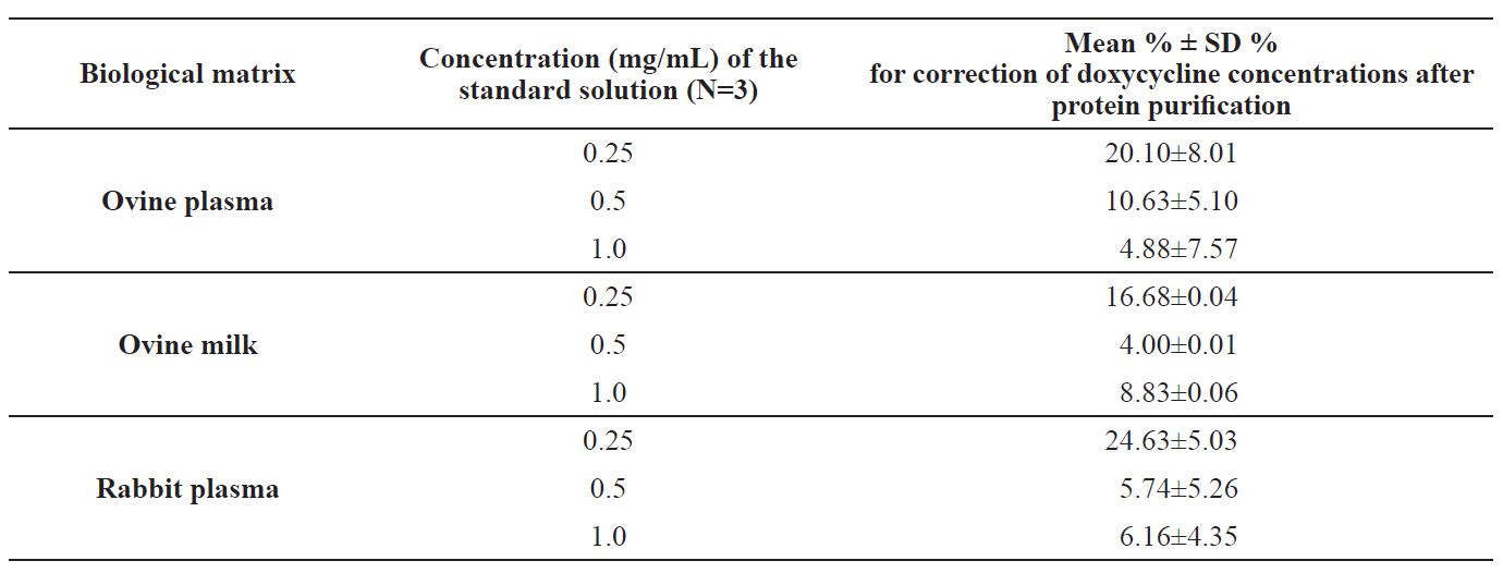
Sample processing was performed by using ultrafree microfilters. The additional step was done in order to further purify the samples and to determine the free doxycycline concentration in the range of concentrations that can be found in vivo. After microfiltration, a percentage for correction of the quantified doxycycline in standard solutions in low, medium, and high concentrations was calculated. The results are shown in
Table 3. It appears that even after protein precipitation with TFA, a small percentage of bound doxycycline remains in the sample. The highest correction is necessary for the lowest concentrations in the three biological matrices.
Table 3. Percentage bound doxycycline at the tested concentrations in ovine plasma and milk and in rabbit plasma after protein precipitation with TFA (ReagentPlus®, 99% purity (Sigma-Aldrich) and sample purification with Ultrafree® - MC Centrifugal Filters (PTFE membrane, pore size 0.45 µm)
DISCUSSION
The developed HPLC method is intended for future pharmacokinetic studies and dosing regimen adjustments. It allows detection of doxycycline in plasma and milk of sheep and in plasma of rabbits.
The data about accuracy, precision and recovery cover the criteria according to other researches (
11,
12). The limitations of the method are related to the linearity range between 0.125 – 1 µg/mL of the standard curve in rabbits’ plasma. Pharmacokinetic studies in rabbits showed that maximum plasma concentrations depend on the drug dosage form: doxycycline hyclate solution versus microcapsule suspension (
13). After oral administration of doxycycline hyclate at a dose rate 20 mg/kg, higher C
max were obtained after administration of solution is significant especially when the substance has (1.74±0.00 µg/ml) in comparison to suspension (1.10±0.00 µg/ml) (
13). The rabbits in the cited study were treated with 5 to 8-fold higher dose than the recommended and widely accepted therapeutic doses of 2.5 - 4 mg/kg. These data suggest that the accepted dosage regimen would result in much lower concentrations and the linearity range would be wide enough and will allow the determination of plasma concentrations without dilution of the samples. When higher doses are used, plasma samples will need to be diluted. The linearity of the standard curves for ovine plasma from 0.125 to 2.5 µg/mL and for ovine milk from 0.125 to 5.0 µg/mL cover a sufficient range of drug concentrations. The published pharmacokinetic data about doxycycline in ovine plasma indicates that it is broad enough to allow analysis of the concentrations that can be detected after using therapeutic doses of 5-10 mg/kg, but when higher doses of 20 mg/kg are applied, higher C
max concentrations of 2.18 ± 0.95 μg/mL can be expected (
14). In these cases, a dilution of the samples will be required. Since the use of doxycycline in lactating animals is prohibited when the milk is used for human consumption, it is not possible to refer the sensitivity of the current method to the quantification of residues in milk.
The recovery rate, LOD, LOQ, intra-, and inter- assay precision are similar to those reported by other authors (
9,
10,
13). Values of LOD, achieved from the three different matrices, were higher than those of the original method published by Laczay et al. (
10). Apart from the different HPLC column used in the cited method, which can be discussed as a reason for lower LOD, the samples were subjected to solid- phase extraction. The last technique ensured better clean-up of the sample matrix and concentration of analyte which can explain the higher sensitivity of the validated method for doxycycline determination in the plasma of broiler chickens.
The extraction procedure, described by Lagzay et al. (
10) was used after some modification. TFA was applied to precipitate the proteins in samples. As a strong acid, it leads to extreme decreasing of pH (
15) and thus to denaturation and subsequent protein precipitation. In pharmacokinetic studies, it is very important to determinate only the unbound fraction of antibiotics because it is pharmacologically active. It is also important to calculate the dosing regimen on the basis of free antibiotic concentration. MIC are determined in broth and their values represent free drug concentrations. However, in many of the published studies over the years a correction for protein binding had not been done (
9). The difference is significant especially when the substance has high protein binding such as doxycycline (
16). TFA showed great precipitation efficiency, but still а low amount of bound doxycycline was left after processing of blood samples with the used method of extraction. A mean percentage for correction was calculated in order to be used in future studies. The most substantive adjustments need to be made at the lower concentrations. It is probably due to concentration-dependant protein binding, observed in many other drugs (
17). Less free, respectively higher percentage of bound doxycycline was detected at lower concentrations, when equilibrium dialysis of mice plasma was applied (
18).
CONCLUSION
In conclusion, the presented HPLC method with PDA detection can be used in pharmacokinetic studies and future adjustments of dosing regimen. It is simple and fulfills the criteria for accuracy, precision, recovery, and sensitivity. The assay is neither time-consuming, nor expensive. Screening for the presence of doxycycline in ovine plasma and milk, and in rabbit plasma can be performed within a short time by analysis of a single sample. The method allows processing numerous samples per day because of the short run time and simple preparation procedure. A calculation of mean percentage for correction for protein binding was performed and it can be used in future pharmacokinetic studies.
CONFLICT OF INTEREST
The authors declared that they have no potential conflict of interest with respect to the authorship and/or publication of this article.
ACKNOWLEDGEMENTS
The research was financially supported by Trakia University, Stara Zagora, Bulgaria (Grant № 07/2018).

 10.2478/macvetrev-2019-0016