Abstract
The aim of the research was to establish the morphology of the vascular bodies of the encephalon ventricles of cow (Bos taurus taurus). Methods used: thin anatomical preparation, histological method of examination, transmission electron microscopy. Given the relationship, structure and general origin of the vascular bodies, they were divided into bodies III, IV and the lateral ventricles of the encephalon. This unit has topographical nature. Functionally, vascular bodies are an indivisible organ whose main function is the secretion of the cerebrospinal fluid, which maintains the constancy of the central nervous system internal environment. Three types of capillaries, differing in their morphology, were found in the composition of the vascular bodies’ villi in Bos taurus taurus.
Keywords: bovine, central nervous system, encephalic ventricular system
INTRODUCTION
The vascular bodies of the cerebrum’ ventricles are derivatives of the pia mater. They consist of a large number of blood vessels and nerve endings (
1). They are represented by the bodies of the lateral ventricles, the III and the IV ventricles (
2). These formations have a villus structure and are covered with a continuous layer of epithelial cells outside (
3). A loop-shaped curved capillary underlies each villus (
4,
5,
6). It consists of fenestrated endothelium, pericytes, and basal membrane (
7).
The functional state of the vascular bodies determines the changes in liquor dynamics and the state of blood supply to the cerebrum in pathological conditions (
8). The pathogenesis of hydrocephalus, schizophrenia, epilepsy and Alzheimer’s disease is based on atrophy of the vascular plexus epithelium (
9,
10,
18).
However, sources of literature do not contain information about the morphology of the animals’ vascular bodies. In this regard, the purpose of this work was to establish the morphology of the vascular bodies of the encephalon ventricles of cow (Bos taurus taurus).
MATERIAL AND METHODS
The material for this study were encephalon’ preparations from seven adult black-and-white cows. They were obtained during the planned slaughter at OJSC «Velikolukskiy myasokombinat».
The extraction of the brain was produced by the developed method (
11). The study was performed using the method of anatomical dissection and histological method. The material was fixed in a 4.0% neutral formaldehyde solution for 24 hours (
13). The selected fragments of the material according to the standard technique were embedded in paraffin (
14). Then cuts were made 5-7 µm thick. They were stained with hematoxylin and eosin and according to the method of Van-Gieson (
14,
15).
The analysis of histological preparations was carried out using a Carl Zeiss Axio Scope A1 (Germany) optical microscope at magnifications of 25, 50, 100, 200 and 400. Microphotography was performed using an AxioCam ICc 1 digital camera. For a transmission electron microscopic study of the vascular plexuses of the ventricles of the encephalon and hematologic patterns barrier, tissue fragments were selected from the middle part of the lateral ventricle of the cerebrum with a volume of not more than 2 mm³. Samples were fixed in a solution of 2.0% glutaraldehyde on cacodylate buffer (pH 7.2-7.4) for 2 hours, washed in three portions of the same buffer. Post fixed in a 1.0% solution of osmium tetroxide (prepared on cacodylate buffer, pH 7.2- 7.4) - 1 hour. After they were dehydrated in alcohols of upward concentration and absolute acetone. Filling was carried out in epon-812 according to the standard technique (
12,
16). Ultrathin sections were obtained on an ultramicrotome (LKB-III - Sweden), contrasted with a 2.0% aqueous solution of uranyl acetate and a solution of plumbum citrate (
17). Afterwards, photographed in a Jem-1011 electron microscope (JEOL, Japan) at magnifications of 2500–30000. The study was conducted taking into account the Directive 2010 / 63UE on the protection of animals used for experimental purposes.
RESULTS
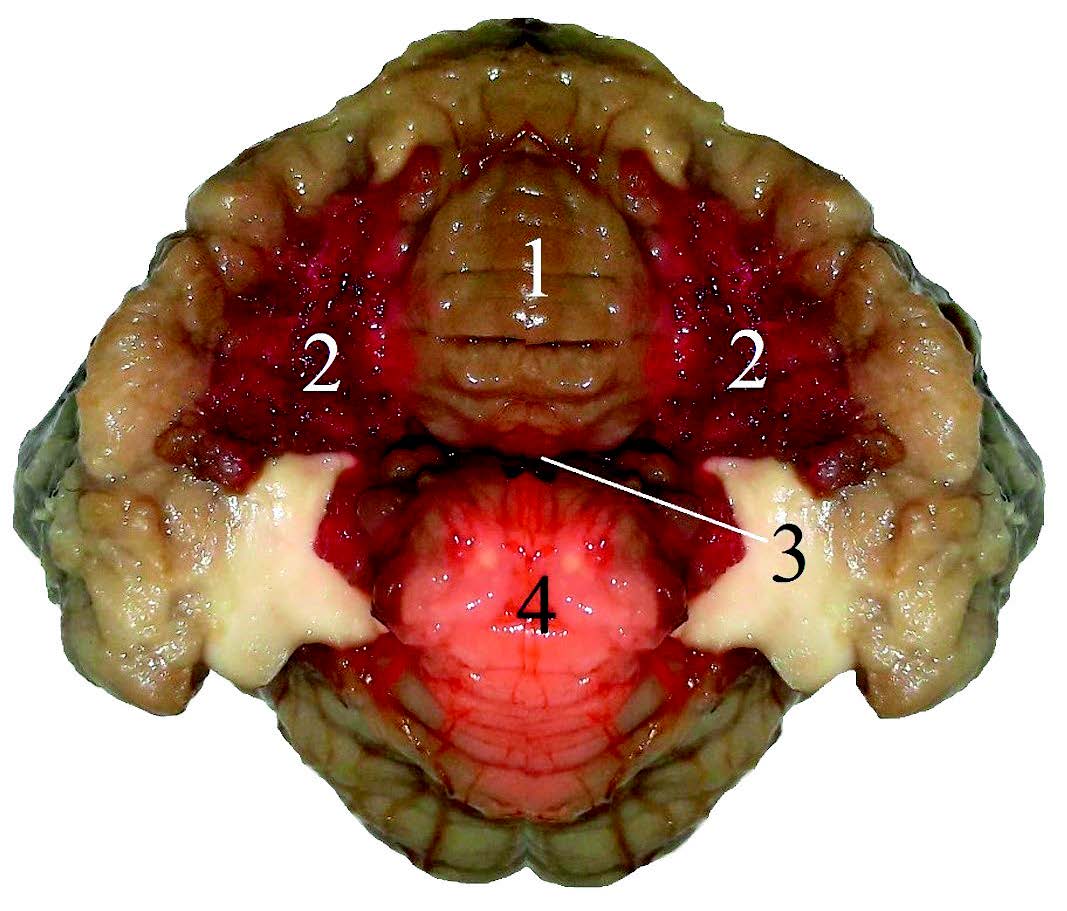
The study found that the vascular bodies are formed from the soft (vascular) membrane of the encephalon in the studied animals. The vascular body of the IV ventricle (
Fig. 1) has the appearance of a triangular plate, adjacent to the caudal medullary velum.
Figure 1. Vascular body of the encephalon IV ventricle. 1- lobus caudalis; 2 – tela choroidea ventriculi quarti; 3 – tegmen ventriculi quarti; 4 – lobus rostralis
The apex of the triangle is directed to the caudal angle of the rhomboid fossa, and triangle’s base follows dorsally. Together with the medullary velum, the vascular body forms the dorsal wall of the IV ventricle. Branches that depart from the cerebellar arteries take part in the formation of the vascular body of the fourth ventricle.
The vascular body of the III ventricle (
Fig. 2) is part of the hypothalamus and lies in the form of a thin plate under the crest of the cerebrum between the optic thalamuses. Through the interventricular foramens, it connects with the vascular bodies of the lateral ventricles (
Fig. 2).
 Figure 2.
 Vascularbodiesoftheencephalonventricles (III ventricle and lateral ventricles). Longitudinal section of the encephalon. 1 – tela choroidea ventriculi tertii; 2 – ventriculus lateralis; 3 – talamus; 4 – tela choroidea ventriculi lateralis
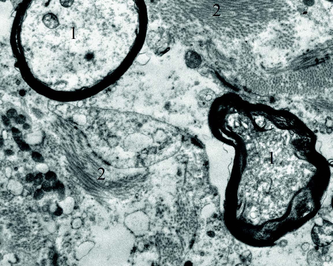
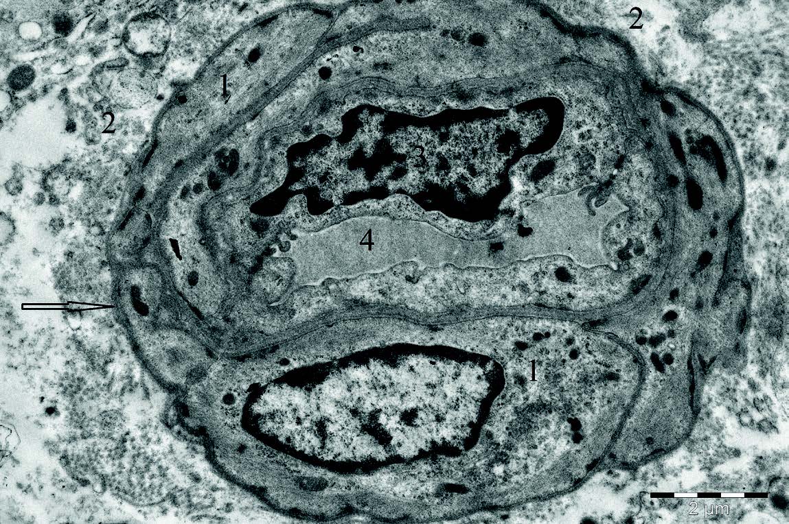
Figure 3. Ultrastructure of the vascular body’s stroma of the lateral ventricle of the cow’s cerebrum. Transmission electron micrograph. 1 - myelin nerve fiber; 2 - collagen fibers
The vascular bodies of the lateral ventricles are essentially a continuation of the III ventricle’vascular body. This vascular body occupies a central position, entering the temporal horn in the composition of the lateral ventricle.
Histologically, the vascular bodies of the ventricles in animals are a specialized organ of the encephalon. The basis of the cerebrum in the studied animals is connective tissue (
Fig. 3).
This tissue contains collagen and nerve fibers which form the stroma. The stroma contains a dense vascular network (
Fig. 4). The simple epithelium covers the vascular bodies outside. Its cells have cubic and prismatic shapes.

Figure 4. Section of the vascular body of the lateral ventricle of the cow’s cerebrum. (H&E; X100)
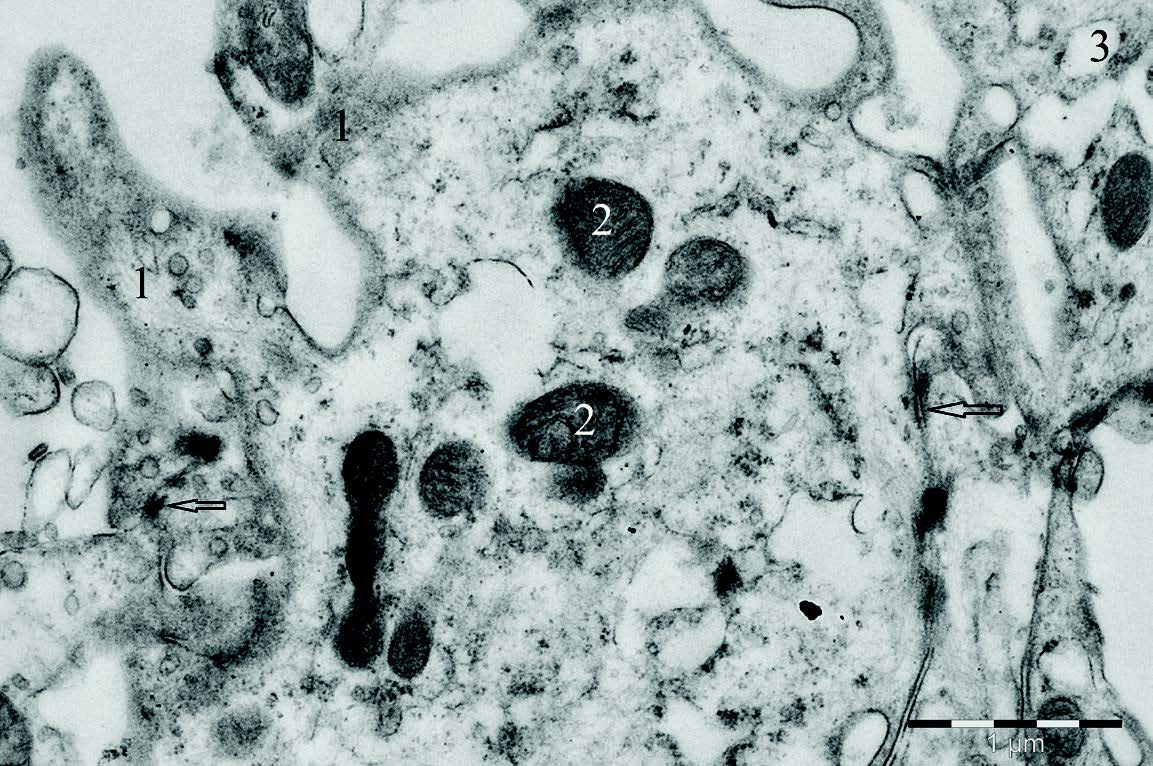
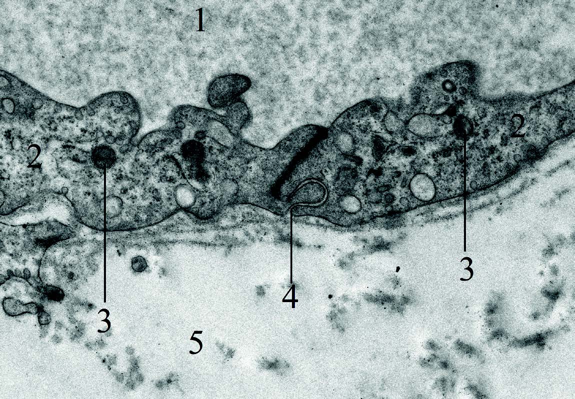
Figure 5. Ultrastructure of the apical surface of vascular body’s epithelial cell in lateral ventricle of the cerebrum. Transmission electron micrograph. 1 - microvilli; 2 - mitochondria; 3 - ribosomes; ↑ - desmosomes
A large number of microvilli are detected on the apical surface of epithelial cells (
Fig. 5). In the activity phase, the epithelial cells covering the plexus increase in height. In certain places, near their apical ends, one can see the liquor produced by them, located at the rim of microvilli. The cells covering the vascular bodies lie on the basement membrane. The membrane separates them from the underlying connective tissue.
In the composition of the connective tissue of the vascular bodies around the links of the hemomicrocirculatory channel, accumulation of smooth myocytes is sometimes found. Five components of the hemomicrocirculatory channel were found in the composition of the encephalon’s vascular bodies. Thus, the small arteries that bring blood to their sites are divided into arterioles. In turn, arterioles - give rise to precapillaries. The precapillaries are divided into capillaries. By merging the capillaries, postcapillaries are formed, forming the venules that pass into the veins.
As part of the vascular bodies’ villi, there are several types of capillaries, differing in their morphology and lined with fenestrated endothelium. The first type is thin-walled capillaries, not accompanied by pericytes (
Fig. 6). The diameter of their lumen is about 2-4 microns, and the wall consists of a single layer of light elongated endothelial cells that are in contact with each other using tight contacts. In certain places, tight contacts become contacts of the “lock” type (
Fig. 7). Endothelial cells outside are surrounded by a weakly expressed fine fibrillar basement membrane (about 10–20 nm thick).

Figure 6. Ultrastructure of the vascular body’s capillary of the lateral ventricle. Transmission electron micrograph. 1 - erythrocyte; 2 - vessel lumen; 3 - endothelial cell; 4 - surrounding connective tissue; 5 - “lock” type contact; ↑ - basement membrane
The second type is capillaries surrounded by a layer of elongated pericyte cells (
Fig. 8). There is a thin basement membrane between the pericytes and endothelial cells. However, the latter gets more developed than in the capillaries of the first type.
Figure 7. Ultrastructure of the vascular body’s capillary of the lateral ventricle. Transmission electron micrograph. 1 - vessel lumen; 2 - endothelial cell; 3 - mitochondria; 4 - tight contact type “lock”; 5 - surrounding connective tissue
Figure 8. Ultrastructure of the vascular body’s capillary of the lateral ventricle. Transmission electron micrograph. 1 - endothelial cell; 2 - surrounding connective tissue; 3 - red blood cells; 4 - pericyte; 5 - vessel lumen; ↑ - basement membrane
The third type are the capillaries surrounded by a rather thick “clutch” from several pericytes immured into the cleavage of the thick basement membrane (
Fig. 9). This creates the impression of layering pericytes or their processes on each other. Endotheliocytes are in contact with tight contacts and lay on a thick dense basement membrane.
Figure 9. Ultrastructure of the vascular body’s capillary of the lateral ventricle, covered with a dense pericyte clutch. Transmission electron micrograph. 1 - pericyte; 2 - surrounding connective tissue; 3 - endothelial cell; 4 - vessel lumen; ↑ - basement membrane
DISCUSSION
The vascular plexus lies in the composition of the ventricular system of the encephalon and is covered with a single layer of epithelial cells located on the basement membrane. The apical end of the cell forms microvilli. The similar structure of the vascular plexus epithelium in humans is described in the literature (
18,
19). The vascular components of the vascular bodies are composed of connective tissue forming their stroma.
Such an organization is characteristic of the vascular bodies of the human cerebrum (
20). The endotheliocytes of the blood vessels are connected to each other by means of tight contacts, which is also characteristic of humans (
21).
As a result of the study, we discovered the presence of the vascular bodies of capillaries of three types in the composition, not described in the available literature. The first type is thin-walled capillaries, not accompanied by pericytes. Their wall consists of a single layer of light elongated endothelial cells that are in contact with each other using tight contacts. In places, tight contacts become contacts of the “lock” type. The endothelial cells are surrounded by a weakly expressed fine fibrillar basement membrane. The second type are capillaries surrounded by a layer of elongated pericyte cells. There is a thin basement membrane between the pericytes and endothelial cells, which is more developed than in the capillaries of the first type. The third type are the capillaries, surrounded by a rather thick “clutch” of several pericytes bricked into the cleavage of the thick basement membrane. This creates the impression of layering pericytes or their processes on each other. Endotheliocytes of these capillaries are contacted using tight contacts and lay on a thick dense basement membrane.
CONCLUSION
In our opinion, given the relationship, structure and common origin of vascular bodies, their division into bodies III, IV and the lateral ventricles of the encephalon, may have topographic and cognitive character. Functionally, the cerebral vascular bodies of the brain are an indivisible organ, the main function of which is to excrete the cerebrospinal fluid that maintains homeostasis of the central nervous system.
In the composition of the vascular bodies of the encephalon of Bos taurus taurus, we found three types of capillaries with different morphological characters. The basis of their subdivision into morphological types is the presence and number of pericyte cells accompanying the capillary involved in the formation of its wall.
CONFLICT OF INTEREST STATEMENT
The authors declared that they have no potential conflict of interest with respect to the authorship and/or publication of this article.
ACKNOWLEDGEMENTS
The studies were conducted on the base of the Department of Anatomy of St. Petersburg State Academy of Veterinary Medicine and the Laboratory of Comparative Morphology of St. Petersburg.

 10.2478/macvetrev-2020-0011