Original Scientific Article
Evaluation of the corrected QT interval with Bazett’s Method in Cavalier King Charles Spaniel dogs with myxomatous mitral valve disease
Hande Sağoğlu ,
Remzi Gönül * ,
Lora Koenhemsi ,
Emine Merve Alan ,
Suzan Murat ,
Ashkan Seddigh Nia ,
Mehmet Erman Or

Mac Vet Rev 2023; 46 (1): 61 - 67

10.2478/macvetrev-2023-0014

Received: 29 August 2022

Received in revised form: 18 January 2023

Accepted: 21 February 2023

Available Online First: 01 March 2023

Published on: 15 March 2023

Correspondence: Remzi Gönül, gonul@iuc.edu.tr
PDF

Abstract

Myxomatous mitral valve disease (MMVD) is one of the most common heart diseases in dogs. The disease progresses faster in Cavalier King Charles Spaniel (CKCS) dogs and occurs at an earlier age. QT interval length reflects abnormalities in ventricular repolarization which may predispose to the formation of fatal arrhythmias such as torsades de pointes. A fast and accurate assessment is therefore essential. The study aimed to examine the changes in QT duration in MMVD cases of CKCS and to calculate the corrected QT durations with Bazett’s formula in various stages of the disease. The study included 20 CKCS dogs of both genders, various ages and weights, and different stages of MMVD (n=6 in B1 stage, n=6 in B2 stage, and n=8 in C stage), and 5 healthy CKCS which were included in the control group. Clinical, radiological, hematological, biochemical, echocardiographic, and electrocardiographic examinations were performed. The corrected QT interval duration in the MMVD group was longer than the control (p<0.05). However, there was no significant difference between B1, B2, and C. It was concluded that the corrected QT interval can give a significant distinction between healthy and MMVD CKCS dogs.

Keywords: Cavalier, myxomatous, mitral valve, QT

INTRODUCTION

Myxomatous mitral valve disease (MMVD) is one of the most common cardiac diseases in small and medium-sized dog breeds (1, 2). In dogs of the Cavalier King Charles Spaniel breed, the disease progresses more rapidly and occurs at an earlier age, and genetic reasons are thought to be responsible (3, 4). The average age of MMVD onset in CKCS is 6.25 years, whereas in other dog breeds 12 years (5). The clinical findings range from mitral regurgitation to exercise intolerance, respiratory distress, cyanosis, ascites, and syncope, depending on the severity of the disease. The severity of the systolic murmur detected on auscultation is directly proportional to the severity of mitral regurgitation (6, 7). In humans and dogs with MMVD, the histopathology findings on the mitral valve included nodular thickening, lipid deposition, and mucoid degeneration of fibrotic tissue (8).
The QT interval, which reflects the total time for depolarization and repolarization of the ventricles, comprises the interval between the onset of QRS conduction and the end of the T wave (9). The duration of the QT interval, measured as a ventricular activation time, is one of the cardiac parameters commonly used to describe cardiac abnormalities and assess drug safety (10). The QT interval is inversely proportional to the R-R interval and heart rate. For proper evaluation of the electrocardiogram, the lower and upper limits of the normal QT interval must be known (11). The QT interval should be less than half of the R-R interval. In dogs, QT>0.25 mm/second is defined as interval prolongation and <0.15 mm/second as interval shortening. QT prolongations may occur as a result of hypocalcemia, hypokalemia, quinidine toxicity, ethylene glycol intoxication, central nervous system problems, hypothermia, myocardial ischemia, left ventricular hypertrophy, right or left bundle branch block, myocarditis, and pericarditis (12). Variations in the heart rate can affect the QT and can lead to misinterpretation. QT interval length reflects abnormalities in ventricular repolarization which may predispose to the formation of fatal arrhythmias such as torsades de pointes. Therefore, fast and accurate assessment is essential (13). The corrected QT interval (QTc) is often used in clinical applications by utilizing Bazett, Fredericia, Framingham, and Hodges formulas which correct for the heart rate (14, 1516). Henry Cuthbert Bazett’s formula, published in 1920, is most commonly used for humans and dogs, (14, 15, 17).
The study aimed to investigate the changes in QT duration in MMVD of CKCS dogs and to differentiate the corrected QT by Bazett’s formula in different MMVD stages (B1, B2, and C).

MATERIAL AND METHODS

Twenty CKCS dogs diagnosed with MMVD were included in the survey group and five CKCS healthy dogs were included in the control group. The dogs in the control group were classified as group A because of their predisposition to myxomatous mitral valve disease. The age and sex of the dogs in both groups were recorded.
All examinations were performed after obtaining consent from the patient’s owner and were approved by the Ethics committee of the Veterinary Faculty, Istanbul University-Cerrahpaşa (March 16, 2021, No 2021/15). Clinical, radiological, hematological, biochemical, echocardiographic, and electrocardiographic examinations were performed on MMVD and control groups.
Dogs with MMVD were classified into A (n=5), B1 (n=6), B2 (n=6), and C groups (n=8) according to the American College of Veterinary Internal Medicine (ACVIM) standards. Group D patients with severe respiratory distress, cyanosis, ascites, and syncope were not included in the study because of the difficulty of ECG imaging. Laterolateral chest radiographs were obtained using a digital radiography unit SMS-CM -N (EcoRay, Korea). The vertebral heart score (VHS) of each patient was measured using the technique established by Buchanan (18). Pulmonary edema was graded according to severity as 0, +1, +2, and +3. The echocardiographic and Doppler echocardiographic examination was performed on the right parasternal long axis, right parasternal short axis, right parasternal heart base acoustic windows, left apical 4 chambers, and apical 5 chambers. It was performed on the SIUI Apogee 3500V model Doppler ultrasound machine (Shantou Institute of Ultrasonic Intstruments, China). Systolic and diastolic measurements of LA/Ao (left atrial to aortic root ratio), IVS (interventricular septal thickness), LVID (left ventricular internal dimension), LVPW (left ventricular posterior wall thickness), FS (fractional shortening), and EF (ejection fraction) were calculated by the Teicholz method in M-mode echocardiography. Pulmonary artery Doppler measurements were performed at the base frame of the right parasternal heart. Mitral measurements were performed in the apical fourchamber view on the left side. Echocardiographic measurements were performed according to ACVIM standards (19).
Electrocardiographic recordings were performed on conscious animals in the lateral position according to the technique described by Edward (9). Alcohol and electrolyte gel was applied to the area during the placement of the electrocardiogram clips to ensure conductivity. Electrocardiograms were recorded with the CONTEC ECG600G class I electrocardiography machine (Contect Medical System, China) at paper speeds of 25 to 50 mm/s and an amplitude sensitivity of 10 mm=1 mV. Patients with sudden changes in respiratory rhythm and ECG waveforms with artifacts, such as a shift in the isoelectric line, were not included in the study. Measurements of P, PR, QRS, RR, QT, and T durations and P, Q, R, S, and T amplitudes were made manually from an ECG trace of the lead II. Mean values were obtained from five consecutive ECG waveforms with steady beats. The QT range was measured from the beginning of the Q wave to the end of the T wave. Areas of arrhythmia were not evaluated in the QT interval measurements. The Bazett formula (QTcb=QT/√RR) was used to determine the corrected QT interval.
Blood was sampled (3 mL) from the cephalic vein. A complete blood count was performed using the Idexx ProCyte Dx Model blood scanner (IDEXX Laboratories, USA). Biochemical analyses were reviewed using the Idexx Catalyst One instrument (IDEXX Laboratories, USA).
Statistical analyses of the data obtained at the end of the study were performed using the SPSS statistical software package (version 28 Windows, IBM Corporation, NY). The One-way ANOVA was used for the comparative assessment of normally distributed parameters, and the chi-square test was used for cross-group comparison of nonparametric qualitative data.

RESULTS

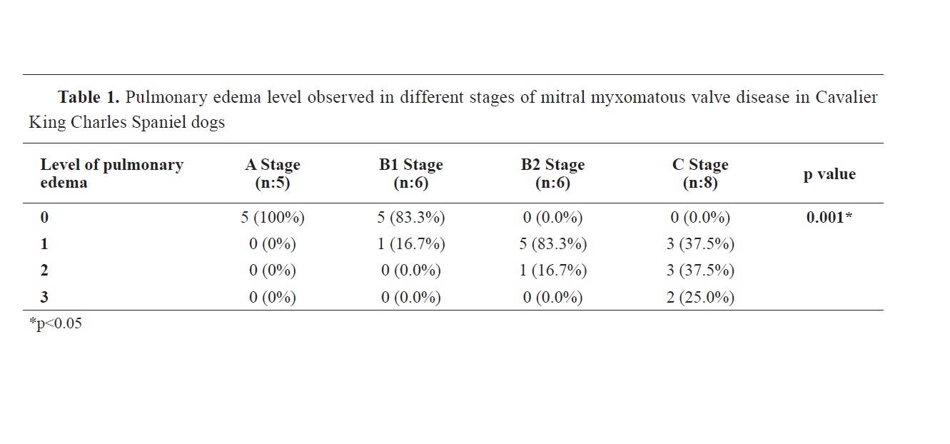
There were two female (40%) and three male (60%) dogs in the A group. In the MMVD groups, seven (35%) were female and thirteen (65%) were male. The mean age was 8.26 years in A, 5.66 years in B1, 7.66 years in B2, and 9 years in C group. Significant differences were observed for the level of pulmonary edema between the groups (p=0.001). B2 and C groups had a significantly higher probability of pulmonary edema compared to A and B1 (p<0.05) (Table 1).


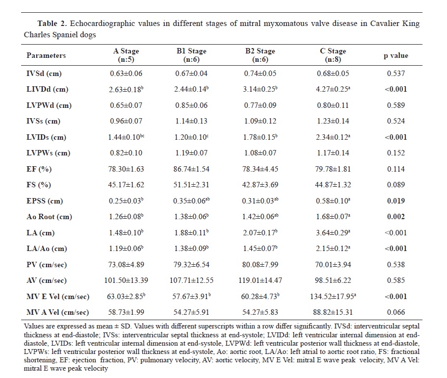
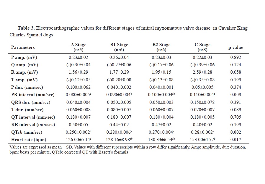
VHS was higher in the C group compared to the other groups (p<0.001). The VHS was 10.28 in group A, 10.70 in group B1, 11.11 in group B2, and 12.17 in group C. The echocardiographic values LA, LA/AO, LVIDd, LVIDs, EPSS, and MV E Vel were significantly higher in patients with stage C heart disease compared to the other groups (p<0.001) (Table 2). The heart rate was higher in group C than in groups A, B1, and B2 (p<0.05) (Table 3). While sinus rhythm was noted in almost all healthy (group A), B1, and B2 dogs, sinus tachycardia was noted in only one dog from the B1 group. In the C group, 2 had sinus rhythm, 2 had sinus tachycardia, 1 had atrial tachycardia, 1 had sinus arrhythmia, and 2 had SA block.
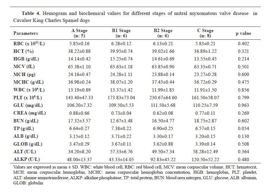
The corrected QT duration was longer in the MMVD groups than in the control (p<0.05) (Table 3), however, there was no significant difference between them. No significant differences were observed for the RR interval, blood count, and biochemistry (Table 3 and 4).

DISCUSSION

MMVD is the most commonly diagnosed cardiovascular disease in dogs and accounts for more than 70% of all cardiovascular diseases in dogs (1). The studies conducted in the CKCS breed have reported a prevalence of 56.6% in dogs older than 4 years (5, 20). The data from our study shows that the disease occurs in animals older than 5 years and the stage of the disease increases with age. Several studies have reported that clinical signs occur at an earlier age in CKCS due to genetic predisposition (4, 21). Due to the long preclinical phase, it can lead to death in the early stages before congestive heart failure occurs (22).
Increased ALT and creatine levels may be observed in congestive heart failure (23, 24). The current research observed a slight increase in ALT levels at different stages of MMVD similar to other reports.
QT prolongation is associated with myocardial ischemia, cardiomyopathies, and hypertension (25). It is a marker of myocardial electrical instability and is associated with sudden death in people with congestive heart failure (10). Because the QT interval depends on the heart rate, the values obtained at different heart rates are used as the “corrected” QT interval (QTc) in the analysis of patients’ electrocardiographic data (26). Universal QT - correction methods, such as the Bazett method, are widely used in clinical and preclinical safety studies because of their ease of use and broad support in the literature (27). Studies have reported that QTc prolongation is associated with malignant arrhythmias and a high risk of mortality in cardiac patients (27, 28). In our study, arrhythmias were found to be more common in patients with QTc prolongation, especially in stage C.
Bazett’s formula is most applicable in dogs with high heart rates (12, 29). In another study (30), a significant prolongation of the QTc value calculated by the Bazett method was observed with the progression of heart failure. In our study, although there was no significant prolongation of QT duration between the groups, an increase in QTc value was observed in MMVD groups compared to the control. It has been suggested that the Fridericia and Hodges formulas can be applicable in patients with rapid heart rates (25). Although many authors (31, 32, 33) reported that the Bazett formula was not sufficient to correct the QT interval in dogs, Patel et al. (29) reported that it is more appropriate in cases where the heart rate exceeds 120. Similarly, we concluded that Bazett’s formula is suitable for distinguishing MMVD from healthy dogs with high heart rates.
The limitations of Bazett’s formula are well known and may lead to over- or under-correction in slow and fast heartbeats. Nevertheless, it remains the most important parameter for the evaluation of cardiac electrical function because of its high diagnostic value (34). In the current study, the use of Bazett’s formula proved that it can be useful in distinguishing MMVD from healthy animals, especially in the more advanced stages of the disease when the heart rate is increasing.

CONCLUSION

Although the QTcb value was not significantly different among B1, B2, and C groups, it can be used in cases with high heart rates. Bazett’s formula correction of the QT interval can discriminate between healthy and MMVD-affected CKCS dogs when pulmonary edema, cough, and exercise intolerance have not yet occurred or when echocardiography cannot be performed. In conclusion, we believe that long-term studies with a larger number of patients are needed to obtain more reliable results.

CONFLICT OF INTEREST

The authors declare that they have no potential conflict of interest with respect to the authorship and/or publication of this article.

ACKNOWLEDGMENTS

This study was supported by the Scientific Research Found of the Istanbul University-Cerrahpaşa with project number: TSA-2021-35735. The authors are also grateful to Dr. Pembe Dilara Keçeci from the Department of Animal Breeding and Husbandry for her help with the statistical analysis.

AUTHORS’ CONTRIBUTIONS

RG designed the study, HS, EMA, SM and ASNA collected the patients, made ECGs, and echocardiography, LK and HS wrote the study, LK made the translation from Turkish to English, MEO has decided if the patients is stuitable for study.

References

1.Parker, H.G., Kilroy-Glynn, P. (2012). Myxomatous mitral valve disease in dogs: does size matter? J Vet Cardiol. 14(1): 19-29. https://doi.org/10.1016/j.jvc.2012.01.006 PMid:22356836 PMCid:PMC3307894
2. Madsen, M.B., Olsen, L.H., Häggström, J., Höglund, K., Ljungvall, I., Falk, T., Wess, G., et al. (2011). Identification of 2 loci associated with development of myxomatous mitral valve disease in
Cavalier King Charles Spaniels. J Hered. 102(Suppl 1): S62-S67. https://doi.org/10.1093/jhered/esr041 PMid:21846748
3. Markby, G.R., Macrae, V.E., Corcoran, B.M., Summers, K.M. (2020). Comparative transcriptomic profiling of myxomatous mitral valve disease in the cavalier King Charles spaniel. BMC Vet Res. 16(1): 350. https://doi.org/10.1186/s12917-020-02542-w PMid:32967675 PMCid:PMC7509937
4. Lewis, T., Swift, S., Woolliams, J.A., Blotta, S. (2011). Heritability of premature mitral valve disease in Cavalier King Charles spaniels. Vet J. 188(1): 73-76.  https://doi.org/10.1016/j.tvjl.2010.02.016     PMid:20347358
5. Beardow, A.W., Buchanan, J.W. (1993). Chronic mitral valve disease in cavalier King Charles spaniels: 95 cases (1987-1991). J Am Vet Med Assoc. 203(7): 1023-1029.
6. Summers, J.F., O’Neill, D.G., Church, D.B., Thomson, P.C., McGreevy, P.D., Brodbelt, D.C. (2015). Prevalence of disorders recorded in Cavalier King Charles Spaniels attending primary-care veterinary practices in England. Canine Genet Epidemiol. 2, 4. https://doi.org/10.1186/s40575-015-0016-7 PMid:26401332 PMCid:PMC4579365
7. Ljungvall, I., Ahlstrom, C., Höglund, K., Hult, P., Kvart, C., Borgarelli, M., Ask, P., Häggström, J. (2009). Use of signal analysis of heart sounds and murmurs to assess severity of mitral valve regurgitation attributable to myxomatous mitral valve disease in dogs. Am J Vet Res. 70(5): 604-613. https://doi.org/10.2460/ajvr.70.5.604 PMid:19405899
8. Pomerance, A., Whitney, J.C. (1970). Heart valve changes common to man and dog: a comparative study. Cardiovasc Res. 4(1): 61-66. https://doi.org/10.1093/cvr/4.1.61 PMid:5416844
9. Edwards, N.J. (1987). Balton’s handbook of canine and feline electrocardoigraphy (2nd ed.). Philadelphia: W.B. Saunders Co 10. Barr, C.S., Nass, A., Freeman, M., Lang, C.C., Struthers, A.D. (1994). QT dispersion and sudden unexpected death in chronic heart failure. Lancet. 343(8893): 327-329. https://doi.org/10.1016/S0140-6736(94)91164-9 PMid:7905146
11. Viskin, S. (2009). The QT interval: too long, too short or just right. Heart Rhythm. 6(5): 711-715. https://doi.org/10.1016/j.hrthm.2009.02.044 PMid:19389656 
12. Gonul, R., Koenhemsi, L., Yildiz, K., Or, M.E. (2019). Determination of corrected QT interval in Kangal breed dogs. Pak Vet J. 39(1): 86-90. https://doi.org/10.29261/pakvetj/2018.115 
13. Oliveira, M.S., Muzzi, R.A.L., Muzzi, L.A.L., Cherem, M., Mantovani, M.M. (2014). QT interval in healthy dogs: which method of correcting the QT interval in dogs is appropriate for use in small animal clinics? Animal Morphophysiology. Pesq Vet Bras. 34(5): 469-472. https://doi.org/10.1590/S0100-736X2014000500014 
14. Phan, D.Q., Silka, M.J., Yueh-Tze, L., Lan, Y.T., Chang, R.K. (2015). Comparison of formulas for calculation of the corrected QT interval in infants and young children. J Pediatr. 166(4): 960-964. https://doi.org/10.1016/j.jpeds.2014.12.037 PMid:25648293 PMCid:PMC4380641
15. Cobos Gil, M.A. (2013). A new, simpler and better correction formula for the QT interval. J Am Coll Cardiol. 61(10): E294. https://doi.org/10.1016/S0735-1097(13)60294-6 
16. Molnara, J., Weiss, J., Zhang, F., Rosenthal, J.E. (1996). Evaluation of five QT correction formulas using a software-assisted method of continuous QT measurement from 24-hour Holter recordings. Am J Card. 78(8): 920-926. https://doi.org/10.1016/S0002-9149(96)00468-7 PMid:8888666
17. Bazett, H.C. (1920). An analysis of the time-relations of electrocardiograms. Heart 7, 353-370. 
18. Buchanan, J.W. (2000). Vertebral scale system to measure heart size in radiographs. Vet Clin North Am Small Anim Pract. 30(2): 379-393. https://doi.org/10.1016/S0195-5616(00)50027-8
19. Keene, B.W., Atkins, C.E., Bonagura, J.D., Fox, P.R., Häggström, J., Fuentes, V.L., Oyama, M.A., et al. (2019). ACVIM consensus guidelines for the diagnosis and treatment of myxomatous mitral valve disease in dogs. J Vet Intern Med. 33(3): 1127-1140. https://doi.org/10.1111/jvim.15488 PMid:30974015 PMCid:PMC6524084
20. Häggström, J., Hansson, K., Kvart, C., Swenson, L. (1992). Chronic valvular disease in the cavalier King Charles spaniel in Sweden. Vet Rec. 131(24): 549-553.
21. Birkegard, A.C., Reimann, M.J., Martinussen, T., Häggström, J., Pedersen, H.D., Olsen, L.H. (2016). Breeding restrictions decrease the prevalence of myxomatous mitral valve disease in Cavalier King Charles Spaniels over an 8- to 10-year period. J Vet Intern Med. 30(1): 63-68. https://doi.org/10.1111/jvim.13663 PMid:26578464 PMCid:PMC4913653
22. Borgarelli, M., Haggström, J. (2010). Canine degenerative myxomatous mitral valve disase: natural history, clinical presentation and therapy. Vet Clin Small Anim Pract. 40(4): 651-663. https://doi.org/10.1016/j.cvsm.2010.03.008 PMid:20610017 
23. Cho, E.J., Han, K., Lee, S.P., Shin, D.W., Yu, S.J. (2020). Liver enzyme variability and risk of heart disease and mortality: a nationwide populationbased study. Liver Int. 40(6): 1292-1302. https://doi.org/10.1111/liv.14432 PMid:32153096
24. Nicholle, A.P., Chetboul, V., Allerheiligen, T., Pouchelon, J.L., Gouni, V., Tessier Vetzel, D., Lefebvre, H.P. (2007). Azotemia and glomerular filtration rate in dogs with chronic valvular disease. J Vet Intern Med. 21(5): 943-949. https://doi.org/10.1111/j.1939-1676.2007.tb03047.x PMid:17939547
25. Isbister, G.K., Page, C.B. (2013). Drug induced QT prolongation: the measurement and assessment of the QT interval in clinical practice. Br J Clin Pharmacol. 76(1): 48-57. https://doi.org/10.1111/bcp.12040 PMid:23167578 PMCid:PMC3703227
26. Gralinski, M.R. (2003). The dog’s role in the preclinical assessment of QT interval prolongation. Toxicol Pathol. 31: 11-16. https://doi.org/10.1080/01926230390174887 PMid:12597426
27. Ether, N.D., Jantre, S.R., Sharma, D.B., Leishman, D.J., Bailie, M.B., Lauver, D.A. (2022). Improving corrected QT; Why individual correction is not enough. J Pharmacol Toxicol Methods. 113, 107126. https://doi.org/10.1016/j.vascn.2021.107126 PMid:34655760 
28. Dekker, J.M., Crow, R.S., Hannan, P.J., Schouten, E.G., Folsom, A.R. (2004). Heart rate-corrected QT interval prolongation predicts risk of coronary heart disease in black and white middle-aged men and women: the ARIC study. J Am Coll Cardiol. 43(4): 565-571. https://doi.org/10.1016/j.jacc.2003.09.040 PMid:14975464
29. Patel, S., Bhatt, L., Patel, R., Shah, C., Patel, V., Patel, J., Sundar, R., et al. (2017). Identifiction of appropriate QTc Formula in beagle dogs for nonclinical safety assesment. Regul Toxicol Pharmacol. 89, 118-124. https://doi.org/10.1016/j.yrtph.2017.07.026 PMid:28751260
30. Koyama, H., Yoshii, H., Yabu, H., Kumada, H., Fukuda, K., Mitani, S., Rousselot, J.F., et al. (2004). Evaluation of QT interval prolongation in dogs with heart failure. J Vet Med Sci. 66(9): 1107-1111. https://doi.org/10.1292/jvms.66.1107 PMid:15472475
31. Batey, A.J., Doe, C.P.A. (2002). A method for QT correction based on beat-to-beat analysis of the QT/RR interval relationship in conscious telemetred beagle dogs. J Pharmacol Toxicol Methods. 48(1): 11-19. https://doi.org/10.1016/S1056-8719(03)00009-1 PMid:12750037 
32. Chiang, A.Y., Holdsworth, D.L., Leishman, D.J. (2006). A one-step approach to the analysis of the QT interval in conscious telemetrized dogs. J Pharmacol Toxicol Methods. 54(2): 183-188. https://doi.org/10.1016/j.vascn.2006.02.004 PMid:16567113
33. Andršová, I., Hnatkova, K., Šišáková, M., Toman, O., Smetana, P., Huster, K.M., Barthel, P., et al. (2021). Infuence of heart rate correction formulas on QTc interval stability. Sci Rep. 11(1): 14269. https://doi.org/10.1038/s41598-021-93774-9 PMid:34253795 PMCid:PMC8275798
34. Kmecova, J., Klimas, J. (2010). Heart rate correction of the QT duration in rats. Eur J Pharmacol. 641(2-3): 187-192. https://doi.org/10.1016/j.ejphar.2010.05.038 PMid:20553920


Copyright

© 2023 Sağoğlu H. This is an Open Access article distributed under the terms of the Creative Commons Attribution-NonCommercial License (http://creativecommons.org/licenses/by-nc/4.0/), which permits unrestricted non-commercial use, distribution, and reproduction in any medium, provided the original work is properly cited.

Conflict of Interest Statement

The authors declared that they have no potential lict of interest with respect to the authorship and/or publication of this article.

Citation Information

Macedonian Veterinary Review. Volume 46, Issue 1, Pages 61-67, e-ISSN 1857-7415, p-ISSN 1409-7621, DOI: 10.2478/macvetrev-2023-0014, 2023