Short Communication
Immunohistochemical diagnostic characteristics of parvovirus infection in dogs
Ivica Gjurovski * ,
Spiro Bozinovski ,
Emilija Murdzevska ,
Todor Novakov ,
Mehmet Fatih Bozkurt ,
Trpe Ristoski

Mac Vet Rev 2023; 46 (2): 193 - 198

10.2478/macvetrev-2023-0024

Received: 10 February 2023

Received in revised form: 21 June 2023

Accepted: 10 July 2023

Available Online First: 24 July 2023

Published on: 15 October 2023

Correspondence: Ivica Gjurovski, igjurovski@fvm.ukim.edu.mk
PDF

Abstract

Thе current study goal was to compare the results from the histopathological and immunohistochemical findings in dogs that have previously been clinically diagnosed with parvovirus infection. The Canine parvovirus enteritis (PVE) is a highly contagious disease mostly affecting dogs below six months of age. The canine parvovirus (CPV) belongs to the parvoviridae family within the feline parvovirus sub group of the genus Parvovirus. This investigation was performed on twenty dogs 2 to 12 months old with previous clinical diagnosis for PVE, and it included necropsy, histopathology, and immunohistochemistry. The necropsy findings included severe hemorrhagic enteritis and enlarged mesenteric lymph nodes as most frequent and prominent changes. The histopathological changes were also most significant in these organs in the form of villus atrophy and lymphoid depletion, respectively. These areas revealed the highest expression of CPV antigen. The histopathological and immunohistochemical methods provide strong base for a reliable CPV diagnosis.

Keywords: diagnosis, necropsy, histopathology, immunohistochemistry, canine parvovirus

INTRODUCTION

Canine parvovirus enteritis (PVE) is a highly contagious disease (1) being observed for the first time in the late 1970s as a worldwide infectious disease mostly affecting puppies (2). The causative agent belongs to the parvoviridae family (1). Canine parvovirus (CPV) is almost identical to feline panleukopenia virus (FPLV), as it is within the feline parvovirus sub group of the genus Parvovirus (3). Currently, there are three familiar strains of canine parvovirus throughout the world: CPV-2a, CPV-2b, and CPV-2c. All ages of dogs have been shown to be susceptible to CPV, with the highest deathrate of 67.6% in dogs below six months of age and declining with the rise in age (4). The disease is prevalent in unvaccinated dogs. The high susceptibility in puppies is due to lack of immunity from maternally derived antibodies, unsuitable time of vaccination, and/or from ineffective responses to vaccinations (5).
The major route of transmission between dogs is indirect, via the fecal-oral route (6, 7). However, the virus can also be transmitted directly (8). There are two clinically distinguished canine parvoviral forms: gastroenteritis in puppies 6-20 weeks of age which happens in the period when the maternal antibody protection declines and vaccination has not yet produced enough protection against infection (9, 10), and myocarditis in young puppies, especially in the early neonatal period (8). Usual clinical findings of PVE infection include vomiting, bloody diarrhea with specific odor, dehydration,anorexia, hypovolemic shock, hypothermia or fever, marked thrombocytopenia and leucopenia, as well as tachycardia, and hypotension (11). Palpation reveals abdominal pain due to acute gastroenteritis or intussusceptions (12). 
Postmortem examination reveals changes characteristic for PVE. They include small intestine enteritis with segmental distribution and usually affecting jejunum and ileum but not duodenum and colon. Mesenteric lymph nodes may be enlarged and edematous with multifocal petechial hemorrhages in the cortex (13, 14, 15). In case of parvoviral myocarditis, there is dilatation of the left atrium and ventricle. The lungs are not collapsed. Microscopically, there is necrosis of epithelial cells in the intestinal crypts and severe lymphocyte depletion in the lymphoid tissue. In the tongue, small intestine, and Payer’s patches, intra-nuclear inclusion bodies are usually present (12). These findings are the basis for the histological diagnosis of PVE in dogs (16, 17, 18, 19, 20). Fast and accurate diagnosis of PVE is essential in order to stop the spreading of infection among dogs. The clinical signs are not exclusive to PVE so a confirmation with laboratory tests is necessary (1). This is usually done by various laboratory methods that detect canine parvovirus in the feces of infected dogs such as immunochromatographic tests (IC), electron microscopy (EM), hemagglutination inhibition (HI) tests, Enzyme-linked immunosorbent assay (ELISA), conventional and real-time polymerase chain reaction (C-PCR), and real-time PCR (RTPCR) (21). However, for the postmortem examination, the finding of characteristic lesions as well as presence of antigen and/or nucleic acid within those lesions is a criterion suggested by many as reliable in many viral diseases (22, 23). Immunohistochemical (IHC) staining of formalin-fixed tissues for parvovirus is a sensitive and specific method for demonstrating viral antigen in tissue sections even before histologic lesions are evident (24).
The goal of the study was to compare the results from the immunohistochemical and pathohistological methods in the diagnosis of the Canine parvovirus enteritis (PVE).

MATERIAL AND METHODS

Animals
This study was conveyed post mortem on twenty dogs at age between 2 and 12 months in the span of a two-year period. All of the dogs had previously been clinically diagnosed as positive for CPV. The severity of the parvovirus symptoms ranged from vomiting and bloody diarrhea with specific odor followed by dehydration and hypovolemic shock.
All dogs were submitted for analyses with verbal consent of the owners. A complete necropsy was performed on all of the dogs immediately after the animal’s death, during which the morphological changes of the internal organs were observed and also tissue samples including the small and large intestine, lungs, mesenteric lymph nodes, and spleen were collected for further investigation.

Histopathology and immunohistochemistry
The tissue samples collected at necropsy were routinely fixated in 10% buffered formalin for 48-72 hours, dehydrated and embedded in paraffin wax, and afterwards cut at 3-4 μm thick slides. In order to evaluate the tissue changes and to establish the possible correlation with the PVE, the slides were stained with hematoxylin and eosin (HE) and microscopically examined. The immunohistochemical investigation was performed as described by Gjurovski et al. (25). A canine/feline parvovirus (CPV1-2A1) monoclonal antibody was used along with DAKO Envision kit based on the Peroxidase/DAB method. This method allowed for an insight in the amount and distribution of the CPV antigen within the tissue and its correlation with the damaged areas. The tissue slides were incubated in 3% H2O2 for 20 min in order to block the endogenous peroxidase. The pretreatment for antigen retrieval comprised of citrate buffer pH 6.0 by heat treatment in microwave at 500 W for 20 min. The slides were incubated overnight with the parvovirus antibody. The slides were later added a secondary antibody labelled with horseradish peroxidase (HRP) and were incubated for 20 min. To allow a visualization of the reaction, a Chromogen, 3.3’- diaminobezidine tetrahidrochloride (DAB - DAKO) was added. The process ended by washing the slides with PBS and staining them with Mayers’ hematoxylin for contrasting. Normal rabbit sera were used for the treatment of control tissue sections.

RESULTS

Gross findings
All dogs were anemic and cachectic with bloody feces around their anus. Severe hemorrhagic enteritis was the hallmark finding in all of them. The small intestinal mucosa was hyperemic and edematous. The stomach mucosa was hyperemic. The Payer’s patches, the mesenteric lymph nodes and the spleen were enlarged.

Histopathology
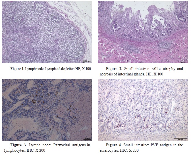
Catarrhal to hemorrhagic enteritis with multifocal crypt necrosis and destruction of crypt intestinal epithelial cells within the small intestine was observed in all animals. Twelve dogs had hemorrhagic enteritis, while eight of them had severe catarrhal enteritis. The intestinal villi were atrophied (Fig. 2). The lumen of the intestines contained large amount of epithelial cell debris as well as inflammatory cells. Lymphocyte infiltration was observed in the mucosa of the small intestine accompanied by the presence of some neutrophils. Some of the samples had hyperplastic gland cells. Lymphoid depletion was evident in the lymph nodes (Fig. 1). The histological findings are presented in Table 1.

Immunohistochemistry
The immunohistochemical staining in this investigation showed that the distribution of the viral antigen was mostly present in the mesenteric lymph nodes of the dogs (Fig. 3). It was also detected in significant amount in the jejunum and the ileum, whereas the duodenum and colon were negative. The antigen distribution within the lymph nodes was mostly visible in the lymphocytes (Fig. 3). In the small intestine, the antigen was located in the epithelial cells of the villi and crypts (Fig. 4). No positive reaction was observed in the other tissues and in control sections. The distribution of PCV2 antigen in the areas with alterations is presented in Table 2.


DISCUSSION

At present the diagnosis of PVE is based on clinical, pathological, virological, and serological examinations (26, 27, 28, 29, 30, 31). The clinical signs in PVE are not pathognomonic and a final diagnosis cannot be determined based on them. Methods for diagnosis of PVE such as serological or fecal examination cannot differentiate an illness as the virus can be present in healthy animals (26, 32). The IHC is a suitable method in cases where pathological signs may not yet be developed in all cases.
This article presented the postmortem histopathological and immunohistochemical findings and made comparison between the two in twenty dogs previously diagnosed or suspected of PVE on the basis of clinical symptoms. The results clearly suggest a correlation between the presence of the CPV antigen and the degree of histopathological lesions in the examined tissues. The accent of the investigation was set on the small intestine, mesenteric lymph nodes, as well as the parenchymal organs such as lungs and spleen. Most significant are the changes affecting the small intestine and the lymphoid tissue.
The changes found in them included villus atrophy in the jejunum and ileum and necrosis of Payer’s patches. These findings are characteristic but not definite findings for PVE as suggested by Evermann at al. (30) and Berkin et al. (32).
The immunohistochemical method provided a direct insight in the presence and the distribution of the CPV antigen in the tissues. According to Dogonyaro (1), Gombac et al. (4), and Decaro et al. (7), the antigen distribution corelates with the organ’s mitotic activity. In this investigation, the detection of PVE was the highest in the jejunum and ileum, which are organs with the highest mitotic activity. The results show that the degree of lymphoid depletion correlates with the amount of CPV antigen in the affected organs. The results provided in this study suggest that the histopathological and the immunohistochemical method can provide a reliable diagnosis for the canine parvovirus infection.

CONCLUSION

The results from the presented study suggest that the observed tissue damage along with the CPV antigen distribution within them is a relevant method for diagnosis of the parvoviral diseases in dogs. In order to improve the diagnostic accuracy, the immunohistochemical method should be included especially when there are inconclusive clinical and pathomorphological findings.

CONFLICT OF INTEREST

The authors declare that they have no potential conflict of interest with respect to the authorship and/or publication of this article.

ACKNOWLEDGMENTS

This work was supported by the project “Aplication of the immunohistochemical method in the diagnostic of virus diseases in domestic animals” funded by the Faculty of Veterinary Medicine - Skopje, Ss. Cyril and Methodius University in Skopje (FVMS-IPR-03).

AUTHORS’ CONTRIBUTIONS

IG and TR wrote the article. SB and EM collected the samples. SB, TN, MFB and TR analized the samples. All authors approved the final version of the study.

References

1. Dogonyaro, B.B. (2010). Molecular characterization of canine parvovirus strains from domestic dogs in South Africa and Nigeria. Doctoral dissertation, University of Pretoria.
2. Appel, M.J., Scott, F.W., Carmichael, L.E. (1979). Isolation and immunisation studies of a canine parco-like virus from dogs with haemorrhagic enteritis. Vet Rec. 105(8): 156-159. https://doi.org/10.1136/vr.105.8.156 PMid:516347
3. Siegl, G., Bates, R.C., Berns, K.I., Carter, B.J., Kelly, D.C., Kurstak, E., Tattersall, P. (1985). Characteristics and taxonomy of Parvoviridae. Intervirology 23(2): 61-73. https://doi.org/10.1159/000149587 PMid:3980186
4. Gombač, M., Švara, T., Tadić, M., Pogačnik, M. (2008). Retrospective study of canine parvovirosis in Slovenia. Slov Vet Res. 45(2): 73-78.
5. Patterson, E.V., Reese, M.J., Tucker, S.J., Dubovi, E.J., Crawford, P.C., Levy, J.K. (2007). Effect of vaccination on parvovirus antigen testing in kittens. J Am Vet Med Assoc. 230(3): 359-363. https://doi.org/10.2460/javma.230.3.359 PMid:17269866
6. Pollock, R.V., Carmichael, L.E. (1982). Maternally derived immunity to canine parvovirus infection: transfer, decline, and interference with vaccination. J Am Vet Med Assoc. 180(1): 37-42.
7. Decaro, N., Elia, G., Martella, V., Campolo, M., Desario, C., Camero, M., Cirone, F., et al. (2006). Characterisation of the canine parvovirus type 2 variants using minor groove binder probe technology. J Virol Methods. 133(1): 92-99. https://doi.org/10.1016/j.jviromet.2005.10.026 PMid:16313976
8. Kahn, C.M., Line, S., Aiello, S.E. (2005). The Merck veterinary manual. Merck & Co. Inc., Whitehouse Station, NJ.
9. Hoskins, D.J. (1998). Canine viral enteritis. In: Greene, C.E. (Ed.), Infectious diseases of the dogs and cats. 2nd ed. (pp. 40-48). Philadelphia: W.B. Saunders Co.
10. Mosallanejad, B., Ghorbanpoor, N.M., Avizeh, R.R.A. (2008). Prevalence of canine parvovirus (CPV) in diarrheic dogs referred to veterinary hospital in Ahvaz. Arch Razi Inst. 63(2): 41-46.
11. Yilmaz, Z., Pratelli, A., Torun, S. (2005). Distribution of antigen types of canine parvovirus type 2 in dogs with hemorrhagic enteritis in Turkey. Turkish J Vet Anim Sci. 29(4): 1073-1076.
12. Ettinger, S.J., Feldman, E.C. (1995). Textbook of veterinary internal medicine. 4th. Ed. Section VIII: The Respiratory System. Philadelphia: W.B. Saunders.
13. Cooper, B.J., Carmichael, L.E., Appel, M.J., Greisen, H. (1979). Canine viral enteritis. II. Morphologic lesions in naturally occurring parvovirus infection. Cornell Vet. 69(3): 134-144.
14. Hayes, M.A., Russel, R.G., Mueller, R.W., Lewis, R.J. (1979). Myocarditis in young dogs associated with a parvovirus-like agent. Can Vet J. 20(5): 126.
15.Robinson, W.F., Huxtable, C.R., Pass, D.A. (1980). Canine parvoviral myocarditis: a morphologic description of the natural disease. Vet Pathol. 17(3): 282-293. https://doi.org/10.1177/030098588001700302 PMid:7368523
16. Bergmann, V., Baumann, G., Fuchs, H.W. (1980). Experiences in the postmortem diagnosis of parvovirus enteritis in the dogs. [Zur postmortalen Diagnostik der Parvovirus-Enteritis des Hundes]. Arch Exp Veterinarmed. 37(1): 127-133. [in German]
17. Breuer, W., Stahr, K., Majzoub, M., Hermanns, W. (1998). Bone-marrow changes in infectious diseases and lymphohaemopoietic neoplasias in dogs and cats-a retrospective study. J Comp Pathol. 119(1): 57-66. https://doi.org/10.1016/S0021-9975(98)80071-6 PMid:9717127
18. Vural, S.A., Alcigir, G. (2011). Histopathological and immunohistological findings in canine parvoviral infection: diagnosis application. Revue Méd Vét. 162(2): 59-64.
19. Maxson, T.R., Meurs, K.M., Lehmkuhl, L.B., Magnon, A.L., Weisbrode, S.E., Atkins, C.E. (2001). Polymerase chain reaction analysis for viruses in paraffin-embedded myocardium from dogs with dilated cardiomyopathy or myocarditis. Am J Vet Res. 62(1): 130-135. https://doi.org/10.2460/ajvr.2001.62.130 PMid:11197551
20. Mulvey, J.J., Bech-Nielsen, S., Haskins, M.E., Taylor, H.W., Eugster, A.K. (1980). Myocarditis induced by parvoviral infection in weanling pups in the United States. J Am Vet Med Assoc. 177(8): 695-698.
21. Desario, C., Decaro, N., Campolo, M., Cavalli, A., Cirone, F., Elia, G., Martella, V., et al. (2005). Canine parvovirus infection: which diagnostic test for virus? J Virol Methods. 126(1-2): 179-185. https://doi.org/10.1016/j.jviromet.2005.02.006 PMid:15847935
22. Waldvogel, A.S., Hassam, S., Stoerckle, N., Weilenmann, R., Tratschin, J.D., Siegl, G., Pospischil, A. (1992). Specific diagnosis of parvovirus enteritis in dogs and cats by in situ hybridization. J Comp Pathol. 107(2): 141-146. https://doi.org/10.1016/0021-9975(92)90031-O PMid:1333495
23. Gjurovski, I., Angelovski, B., Dovenski, T., Mitrov, D., Ristoski, T. (2015). Diagnostic characteristics of circovirus infection in pigs. Mac Vet Rev. 38(1): 73-78. https://doi.org/10.14432/j.macvetrev.2014.11.033 
24. Vural, S.A., Alcigir, G. (2011). Histopathological and immunohistological findings in canine parvoviral infection: diagnosis application. Revue Méd Vét. 162(2): 59-64.
25. Gjurovski, I., Dovenska, M., Kunovska, S.K., Milosevski, J., Trajkov, V.L., Ristoski, T. (2019). Mammary adenocarcinoma with widespread metastasis in a lion. Mac Vet Rev. 42(2): 195-199. https://doi.org/10.2478/macvetrev-2019-0019 
26. Azetaka, M., Hirasawa, T., Konishi, S., Ogata, M. (1981). Studies on canine parvovirus isolation, experimental infection and serologic survey. Nihon Juigaku Zasshi. 43(2): 243-255. https://doi.org/10.1292/jvms1939.43.243 PMid:7289337
27. Danner, K., Oldenburg, U., Weber, S., Arens, M., Krauss, H. (1982). Current epizootiological situation in virus-related canine enteritis. [Derzeitige epizootiologische Situation bei virusbedingten Enteritiden des Hundes]. Praktische Tierarzt. 63, 512 - 518. [in German]
28. Jakov, N., Nenad, M., Andrea, R., Dejan, K., Dragan, M., Aleksandra, K., Marina, R., et al. (2021). Genetic analysis and distribution of porcine parvoviruses detected in the organs of wild boars in Serbia. Acta Veterinaria 71(1): 32-46. https://doi.org/10.2478/acve-2021-0003 
29. Kubašová, I., Štempelová, L., Maďari, A., Bujňáková, D., Micenková, L., Strompfová, V. (2022). Application of canine-derived DSM 32820 in dogs with acute idiopathic diarrhoea. Acta Veterinaria 72(2): 167-183. https://doi.org/10.2478/acve-2022-0014 
30. Evermann, J.F., Abbott, J.R., Han, S. (2005). Canine coronavirus-associated puppy mortality without evidence of concurrent canine parvovirus infection. J Vet Diagn. 17(6): 610-614. https://doi.org/10.1177/104063870501700618 PMid:16475526
31. Url, A., Schmidt, P. (2005). Do canine parvoviruses affect canine neurons? An immunohistochemical study. Res Vet Sci. 79(1): 57-59. https://doi.org/10.1016/j.rvsc.2004.11.003 PMid:15894025
32. Berkin, S., Milli, U., Urman, H.K. (1981). Canine parvoviral enteritis in Turkey. Vet J Ank Uni. 28, 36-49.


Copyright

© 2023 Gjurovski I. This is an Open Access article distributed under the terms of the Creative Commons Attribution-NonCommercial License (http://creativecommons.org/licenses/by-nc/4.0/), which permits unrestricted non-commercial use, distribution, and reproduction in any medium, provided the original work is properly cited.

Conflict of Interest Statement

The authors declared that they have no potential lict of interest with respect to the authorship and/or publication of this article.

Citation Information

Macedonian Veterinary Review. Volume 46, Issue 2, Pages 193-198, e-ISSN 1857-7415, p-ISSN 1409-7621, DOI: 10.2478/macvetrev-2023-0024