Original Scientific Article
The influence of different food types on the morphological characteristics of rat small intestines
Nedžad Hadžiomerović ,
Fuad Babović ,
Anel Vejzović * ,
Aida Bešić ,
Elvir Čičkušić ,
Nejra Dučić ,
Muhamed Katica

Mac Vet Rev 2025; 48 (1): i - ix

10.2478/macvetrev-2025-0010

Received: 16 May 2024

Received in revised form: 28 August 2024

Accepted: 10 October 2024

Available Online First: 23 December 2024

Published on: 20 December 2024

Correspondence: Anel Vejzović, anel.vejzovic@vfs.unsa.ba
PDF

Abstract

Multiple studies have shown the importance of adequate nutrition for animals and humans and its effect on overall health. Therefore, the aim of this study was to investigate the effects of different nutritional regimes on the intestinal health of rats by evaluating different morphological and morphometric characteristics of small intestines, with the emphasis on the villus height:crypt depth ratio (V:C). For the experimental study, 24 clinically healthy adult Wistar rats were used. The rats were randomly divided into 3 groups: the control group (group A) was fed with conventional food, the second group (group B) with bakery products, and the third group (group C) with meat products. Samples of the duodenum and jejunum were collected for detailed morphological and morphometric analysis. A significant increase in the duodenal villi height was reported in group B (661.59 μm) and C (602.83 μm) compared to the control group (475.34 μm). The crypt depth values in the jejunum were significantly higher in group B (191.41μm) and C (246.23 μm) compared with the control (145.14 μm). The jejunal V:C ratio was significantly lower in groups B and C. The study showed significant morphological changes in the intestinal parameters in rats fed predominantly with meat and bakery products. These findings could be applicable in both veterinary and human medicine, underlining the significance of consumed food on gut health.

Keywords: diet, intestines, morphology, rats


INTRODUCTION

Rats belong to the order Rodentia with two incisors in the upper and two in the lower jaw. They are omnivores, with a diet consisting mostly of cereals, plants, different roots, and seeds. Rats’ natural habitats are areas with moderate climate in Asia, mostly around the Caspian Sea. With the human civilization and societies’ development, the population of rats has increased and spread to other areas (1). The rat was the first mammal used for scientific purposes, and together with mice, they are considered as the most commonly used laboratory animals (2). Rats are a suitable model for research of digestive disorders, including chronic inflammatory bowel disease (3), lipid digestion (4), functional dyspepsia (5), intestinal microbiota (6), and many more.
The small intestines are responsible for nutrient transport which occurs through the villi and microvilli (7). The nutrition may affect the size and number of villi, as well as the intestinal physiology. Various physiological and pathological conditions can cause changes in the size and number of the villi. Thus, the functional morphology of the intestinal mucosa is of great importance for the animal’s health (8).
The food consumed has a key role in the health and proper functioning of the intestines and intestinal mucosa. Proteins and carbohydrates are considered to have significant role in the nutrition of humans and animals among other nutritional compounds. Meat represents an important source of proteins and other essential nutrients, acting as a pharmakon with both positive and negative effects on health (9). Meat consumption or highprotein diet helps the regulation of body fat. These and similar high-protein diets have an impact on weight loss and lower the chance of regaining the mass. In addition, proteins provide greater satiety compared to carbohydrates (10, 11). Contrary to it, the overconsumption of meat is associated with obesity in people (12). Some researches show that high intake of red and processed meat can increase the risk of gastrointestinal disease (13, 14). These meat products increase the risk of colorectal cancer. Various supplements are continuously studied for treatment of colorectal cancer. For instance, the honey combination with 5-Fluorouracil has shown positive effects in colon cancer model rats (15). Other researchers have also confirmed the benefits of honey and bee products in treating colorectal cancer (16, 17, 18). The carbohydrates are regarded as the main component of food which provides energy and other nutritive substances. Complex carbohydrates, especially those that contain fibers, are of great importance. In contrast, simple carbohydrates, such as refined sugar and processed cereals, rapidly increase the levels of blood sugar. Constant use of carbohydrates with a high glycemic index can lead to insulin resistency and intestinal inflammation, decreasing mucosal immunity (19). A positive correlation has been established between the villus height-crypt depth ratio (V:C ratio) and gut health (20). This could show the importance of a proper nutritional regime in the diet of animals and humans and its effect on overall health. Thus, the goal of this study was to investigate the effect of different nutritional regimes on V:C ratio. Various morphological and morphometric characteristics of the small intestines were examined with the emphasis on the V:C ratio.

MATERIAL AND METHODS

Animals and ethics
The experimental study included 24 clinically healthy adult Wistar rats. Twelve male and twelve female individuals were used. They were 2 to 3 months of age with an average weight of 200-300 g. The animals were placed in adequate cages with free access to food and water under a controlled temperature (20-23 °C) and humidity (60±10%).
Study permission was obtained from the Ethics Committee of Veterinary Faculty of Sarajevo (Decision no. 07-03-1012-4/21). All procedures performed in studies involving animals were in accordance with the ethical standards of the institution or practice at which the studies were conducted.

Experimental design
The rats were randomly divided into 3 groups. The first group (A) contained eight individuals fed with conventional food for rats, as the control group. The second (B) group was fed with products rich in carbohydrates, and the third group (C) was fed with products rich in meat protein. The animals consumed the food for seven weeks.
The control group (A) consumed a standardized feeding mixture in form of granules that contained balanced nutrient composition in the following proportions: protein 14.0%, raw fibers 14.5%, raw fats 3.0%, raw ash 7.5 %, calcium 1.1%, phosphorus 0.5%, and sodium 0.2%.
After completing the 7-week feeding period, euthanasia was performed by overdose with ketamine hydrochloride (50 mg/kg) injected into the hindlimb musculature. After confirming the absence of vital parameters, the abdominal cavity was opened and all the organs were extirpated. All the thoraco-abdominal organs were weighed separately using a Silver crest laboratory balance (600 g/0.1g) (Owim, Germany). The small intestine and large intestine segments were measured with a ruler. The individual segments of the intestines were subjected to detailed histopathological analysis.

Histopathology and morphometric analysis
For the histopathological examinations, samples of the caudal third of duodenum and middle and caudal third of jejunum were obtained and fixed with 10% buffered formalin. After the conservation process, the tissues were processed in a MICROM Model STP-120 (Thermo Fisher Scientific, USA) rotational tissue processor in standard line with alcohol: 48 hours in 70% alcohol, 24 hours in 96% alcohol, and 24 hours in 100% alcohol. Sample dehydration was performed by immersion in alcohol and toluene solution for 2 hours, and then for 4 additional hours in toluene solution. After dehydration, the samples were molded into paraffin blocks. Using a digital microtome LEICA RM 2145 (Leica, Germany), a 5 μm thick slide was cut from every paraffin block for the standard histopathological staining with hematoxylin-eosin stain (H&E).


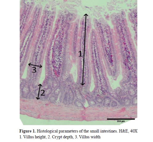
All specimens were observed under an OLYMPUS BX51 light microscope (Olympus, Japan) at magnifications from 40 to 100x. Multiple samples of individual animal duodenum and jejunum were obtained. Villus height, crypt depth, and villus width were measured on multiple sections of duodenal and jejunal samples of each rat. Measurement of the villus height was done from villus tip to villus-crypt junction and the crypt depth from the villus-crypt junction to lower limit of the crypt. Villus width was measured in the lower third of the villus (21). Morphometric analysis and measurement of the mucosa in the small intestines was performed with Cell software (Cell Software Services, UK) and the mucosa parameters were measured as shown in Fig. 1. The villus height:crypt depth (V:C) ratio was estimated by dividing the villus height value by the crypt depth value.

Statistics
The sample size was determined with oneway ANOVA-sample size calculator with 95% confidence interval and 80% power test. The results were recorded in a table as means and standard errors. Using SPSS Software (IBM, USA), Kruskal-Wallis test was performed to test the statistical differences between the parameters while Mann-Whitney U test was used to check the statistical significance among two groups.

RESULTS

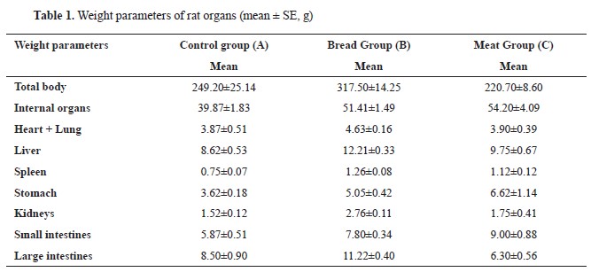
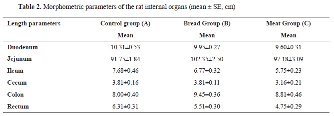
The body weight significantly differed between the groups (Table 1). The highest mean total body weight (317.50±14.25 g) was noted in group B, while the lowest in group C (220.70±8.60 g). The increase in total bodyweight was positively correlated with the organ weight. Duodenum length was nonsignificantly lower and jejunum length was nonsignificantly higher in B and C groups compared to the control (Table 2).
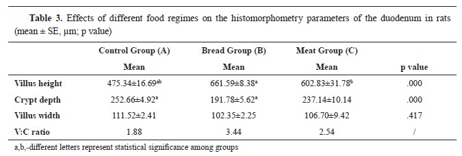
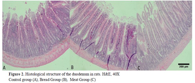
The duodenum differed both in length and in histomorphometry parameters (Fig. 2). The tunica muscularis had similar structure and size in all groups.
The duodenal villi of the control group had similar size with small spaces between them. Group B duodenal villi had larger spaces, especially near the crypts. Several samples in group C had detachments from the villus tips. A significantly higher duodenal villus height was observed in B (661.59±8.38 μm) and C group (602.83±31.78 μm) compared to the control. The villus height was significantly different between the groups, however, the crypt depth was not (Table 3).
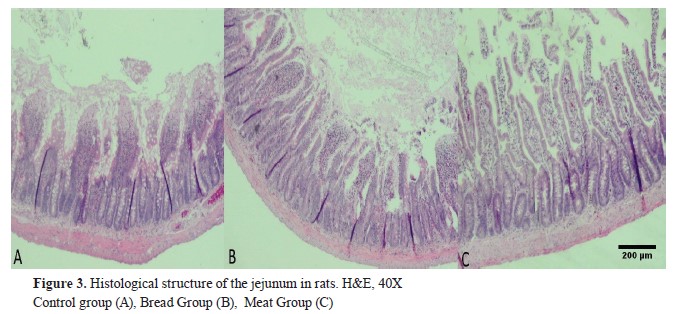
The villi of the jejunal mucosa were evidently lower compared to the duodenal villi (Table 4). Significant amount of desquamated cells were observed on the luminal side of the jejunum compared to the luminal side of the duodenum in all three groups (Fig. 3). The jejunal crypt depth was significantly higher in B (191.41±4.34 μm) and C groups (246.23±9.42 μm) compared to the control (145.14±4.76 μm). The V:C ratio has changed accordingly.


DISCUSSION

The three distinct parts of the small intestines (the duodenum, jejunum and ileum) are responsible for digestion, rendering the chyme compounds to absorbable form, and absorption into the bloodstream. The current study demonstrated that different nutritional regimes caused changes in the weight and length parameters of the alimentary canal. Total body weight was the highest in the group fed with bakery products, and the smallest was noticed in the group fed with meat diet. The weight of the gastrointestinal tract was slightly higher than that reported by Silva-Santana et al. (22). A previous study (23) had reported that small intestine length ranges from 120 to 170 cm, while Silva-Santana et al. (22) recorded a significantly shorter length of 50.6 cm. A recent study has reported small intestines length of 160 cm in Common Brown Rat (Ratus norvegicus) (24). Our study corresponds with the first investigation, and we recorded similar values for all three groups, around 110 cm.
The current study investigated the influence of different food types on intestinal parameters. The hypothesis was that the villus height:crypt depth ratio (V:C) would significantly decrease in groups fed by carbohydrates and meat (Group B and C). The results of the V:C ratio of duodenum did not follow this hypothesis, since the highest V:C ratiowas found in animals fed with bread (group B). This could be explained by Xu et al., which reported that the absorption of glucose mainly occurs in the upper part of small intestine (25). Thus, we underline the findings in jejunal parameters where the main part of nutrients is absorbed. The V:C ratio of the jejunum was significantly lower in rats that consumed bakery products (group B) and meat (group C) compared to the control. This could suggest that non-balanced feeding alters intestinal health, and lowers the intestinal absorption capability. Other studies also mention villi height and crypt depth ratio as a possible indicator of intestinal function (26, 27). In addition, increased villi height and crypt depth are reported to have a positive impact on resistance to intestinal diseases in studies done on weaned piglets (28, 29). 
The increase in villus height has been related to the improved digestion and absorption while it also enhances the digestive enzymes activity (30). Higher crypt depth is correlated with absorptive cells renewal. Researchers reported the increase in crypt depth of turkeys after experimental infection with Escherichia coli and/or Campylobacter jejuni (31, 32). Decrease in crypt depth positively affects digestion since the cell renewal process increases the requirements for energy leading to a lesser absorption and a slower growth rate of animals (33). The lower crypt depth in jejunum of the control group compared to the B and C groups matches the assumption that animals fed with balanced food should show better results of intestinal parameters.
In a recent study, the authors investigated potential gut health biomarkers including the V:C ratio. Broiler chickens were infected with C. jejuni, and treated with CID 2000TM which is used for disinfection of water systems. The results showed changes in V:C ratio in accordance with the treatments. The group which was infected with C. jejuni and not treated with CID showed significant decrease in V:C ratio. Furthermore, the V:C ratio increased in the group which was infected and treated with CID similarly as in the group which was not infected (34).
In a study performed on broiler chickens, a positive correlation was found between the V:C ratio, the number of lactic acid bacteria, and intestinal score (20). A recent study investigated the effect of Pinus pinaster extract supplementation on the intestinal morphology in broiler chickens fed with low protein diets. The jejunal villus height and crypt depth were higher in groups treated with Pinus pinaster extract and encapsulated Pinus pinaster extract, compared to control group (35). This study in broilers shows similarities with the current research where the most notable changes were in histomorphology of jejunum.
Food additives have been frequently investigated in trials with rats, but few have investigated the effect of various food types. Different doses of the Spirulina platensis herb were investigated in rats, where a significant increase in the V:C ratio was recorded in the group with a higher dose (21). It was more than double (5.33±0.46) compared to the control group (2.44±0.10). A study on the influence of methionine on the histomorphological characteristics of the rat intestinal tract, showed significant changes in the size of the villi (36). The study was conducted over a period of 20 days during which two groups received intraperitoneal administration of methionine at doses of 100 mg/kg and 200 mg/kg, respectively. All three parts of the small intestine (duodenum, jejunum, and ileum) showed a significant increase in villus height, particularly the duodenum. The villus height was 389.47 μm in the control, 533.84 μm in the Met100 group, and 525.82 μm in the Met200 group. Additionally, the villus width was higher in the methionine groups (103.22 μm and 106.66 μm, respectively) compared to the control group (69.47 μm).
Predominant feeding with one food type can have adverse effects on V:C ratio as shown in our study. Despite the numerous negative effects of predominant meat protein consumption, it is highly important to be part of a balanced diet. Positive effect of glutamine amino acid as an additive was observed in broiler chickens. Glutamine showed improved trophic effect on intestinal epithelium with increased V:C ratio (37). Conversely, a protein-free diet altered the intestinal architecture and decreased the V:C ratio compared to the group fed with casein (38). Another study showed significantly higher villi height and crypt depth in rats fed with different doses of Soluble Dietary Fiber (Pectin) (39). The villus height was 539.9±25.5 μm in the control and 672.1±63.7 μm in the group that consumed 10% pectin in their meals. Crypt depth had increased from 149.2 μm to 292.5 μm. In a study conducted by Xun et al., a dose-dependent increase of the jejunal V:C ratio was noticed in piglets fed with curcumin-supplemented meals which had higher ratio (2.17) compared to the group that had meals without curcumin (1.48) (40).
Rat nutrition using dominantly bakery products caused significant changes in the blood, including sideropenic hypochromic anemia, with lower erythrocyte and hemoglobin, as well as some atypical forms of red cells, anulocytes, and spherocytes (41).
The above-mentioned studies mostly indicate the positive effects of supplementary products in the rat meals which increase the V:C ratio. This matches with our results where the rats fed with balanced food (group A) had the highest V:C ratio. However, we did not include any supplementary products which could be considered as a limitation of our study.

CONCLUSION

The present study revealed the detailed histological structure of the rat intestines when fed with different foods. The results showed morphological changes in the intestinal parameters in rats fed predominantly with meat and bread diets. A significant increase in the duodenal villi and jejunal crypt depth was recorded in the control which was fed with balanced meals, resulting in highest V:C ratio. The use of confectionery products in laboratory rat nutrition can cause significant changes in their digestive system, and therefore make them not suitable for scientific research.
These findings could be applicable both in veterinary and human medicine, underlining the significance of consumed food on gut health. Further studies are needed to elucidate whether the V:C ratio could be used as reliable gut health indicator.

CONFLICT OF INTEREST

The authors declare that they have no known conflict of interest in the conduction of the current study.

ACKNOWLEDGMENTS

The research was supported by the Veterinary Faculty, University of Sarajevo, and the University of Tuzla.

AUTHORS’ CONTRIBUTION

NH and FB conceived and planned the experiment. MK, AB, NH and FB carried out the experiment. EČ and NH contributed to the interpretation of the results. AV and NH took the lead in writing the manuscript. ND contributed to reviewing and editing. All authors provided critical feedback and helped shape the research, analysis and manuscript.

References

1. Katica, M., Delibegović, S. (2019). Laboratory animals – Basic techniques of the experimental work. Sarajevo: Dobra knjiga [In Bosnian]
2. Hickman, D.L., Johnson, J., Vemulapalli, T.H., Crisler, J.R, Sheperd, R. (2017). Commonly used animal models. In: M.A. Suckow, K. Stewart (Eds.), Principles of animal research for graduate and undergraduate students (pp.117-175). Amsterdam: Elsevier https://doi.org/10.1016/B978-0-12-802151-4.00007-4 
3. Ghattamaneni, N.K.R., Panchal, S.K., Brown, L. (2019). An improved rat model for chronic inflammatory bowel disease. Pharmacol Rep. 71(1): 149-155. https://doi.org/10.1016/j.pharep.2018.10.006 
4. Steingoetter, A., Arnold, M., Scheuble, N., Fedele, S., Bertsch, P., Liu, D., Parker, H.L., Langhans, W., Fischer, P. (2019). A rat model of human lipid emulsion digestion. Front Nutr. 6, 170. https://doi.org/10.3389/fnut.2019.00170 
5. Liang, Q., Yan, Y., Mao, L., Du, X., Liang, J., Liu, J., Wang, L., Li, H. (2018). Evaluation of a modified rat model for functional dyspepsia. Saudi J Gastroenterol. 24(4): 228-235. https://doi.org/10.4103/sjg.SJG_505_17 
6. Tomas, J., Langella, P., Cherbuy, C. (2012). The intestinal microbiota in the rat model: major breakthroughs from new technologies. Anim Health Res Rev. 13(1): 54-63. https://doi.org/10.1017/S1466252312000072 
7. Kiela, P.R., Ghishan, F.K. (2016). Physiology of intestinal absorption and secretion. Best Pract Res Clin Gastroenterol. 30(2): 145-159. https://doi.org/10.1016/j.bpg.2016.02.007 
8. Gulbinowicz, M., Berdel, B., Wójcik, S., Dziewiatkowski, J., Oikarinen, S., Mutanen, M., Kosma, V.M., et al. (2004). Morphometric analysis of the small intestine in wild type mice C57BL/6L -- a developmental study. Folia Morphol. 63(4): 423-430.
9. Leroy, F. (2019). Meat as a pharmakon: an exploration of the biosocial complexities of meat consumption. Adv Food Nutr Res. 87, 409-446. https://doi.org/10.1016/bs.afnr.2018.07.002 
10. Gilbert, J.-A., Bendsen, N.T., Tremblay, A., Astrup, A. (2011). Effect of proteins from different sources on body composition. Nutr Metab Cardiovasc Dis. 21(Suppl 2): B16-31. https://doi.org/10.1016/j.numecd.2010.12.008 
11. Westerterp-Plantenga, M.S., Nieuwenhuizen, A., Tomé, D., Soenen, S., Westerterp, K.R. (2009). Dietary protein, weight loss, and weight maintenance. Annu Rev Nutr. 29, 21-41. https://doi.org/10.1146/annurev-nutr-080508-141056 
12. Wang, Y., Beydoun, M.A. (2009). Meat consumption is associated with obesity and central obesity among US adults. Int J Obes (Lond.). 33(6): 621-628. https://doi.org/10.1038/ijo.2009.45 
13. Pham, N.M., Mizoue, T., Tanaka, K., Tsuji, I., Tamakoshi, A., Matsuo, K., Wakai, K., et al. (2014). Meat consumption and colorectal cancer risk: an evaluation based on a systematic review of epidemiologic evidence among the Japanese population. Jpn J Clin Oncol. 44(7): 641-650. https://doi.org/10.1093/jjco/hyu061 
14. Vieira, A.R., Abar, L., Chan, D.S.M., Vingeliene, S., Polemiti, E., Stevens, C., Greenwood, D., Norat, T. (2017). Foods and beverages and colorectal cancer risk: a systematic review and meta-analysis of cohort studies, an update of the evidence of the WCRFAICR continuous update project. Ann Oncol. 28(8): 1788-1802. https://doi.org/10.1093/annonc/mdx171 
15. Kurtdede, E., Alçığır, M.E., Alperen, A.M., Baran, B., Karaca, O., Gülendağ, E. (2023). Evaluation of the combined effects of Turkish mad honey and 5-fluorouracil in colon cancer model in rats. Ankara Univ Vet Fak Derg. 70(4): 427-435. https://doi.org/10.33988/auvfd.1113279 
16. Erejuwa, O.O., Siti, A.S., Mohd, S.A.W. (2014). Effects of honey and its mechanisms of action on the development and progression of cancer. Molecules. 19(2): 2497-2522. https://doi.org/10.3390/molecules19022497  
17. Subramanian, A., Agnes, J., Vellayappan, M.V., Arunpandian, B., Saravana, K.J., Mahitosh, M., Eko, S. (2016). Honey and its phytochemicals: plausible agents in combating colon cancer through its diversified actions. J Food Biochem. 40(4): 613-629. https://doi.org/10.1111/jfbc.12239 
18. Tawfek, N.S., Al-Azhary, D.B., Hassan, H.F., Esraa, G.M.  (2018). Ameliorative effects of honey and venom of honey bee on induced colon cancer in male albino rats by 1,2 dimethylhydrazine. Cancer Biol. 8(4): 9-20.
19. Arnone, D., Chabot, C., Heba, A.C., Kökten, T., Caron, B., Hansmannel, F., Dreumont, N., et al. (2022). Sugars and gastrointestinal health. Clin Gastroenterol Hepatol. 20(9): 1912-1924.e7. https://doi.org/10.1016/j.cgh.2021.12.011 
20. Nguyen, D.T.N., Le, N.H., Pham, V.V., Parra, E., Forti, A., Hien, T.L. (2021). Relationship between the ratio of villous height: crypt depth and gut bacteria counts as well production parameters in broiler chickens. J Agric Food Dev. 20(3): 1-10. https://doi.org/10.52997/jad.1.03.2021 
21. Asmaz, E.D., Seyidoglu, N. (2022). The prevention role of Spirulina platensis (Arthrospira platensis) on intestinal health. Food Sci Hum Wellness. 11(5): 1342-1346. https://doi.org/10.1016/j.fshw.2022.04.027 
22. Silva-Santana, G., Aguiar-Alves, F., Silva, L.E., Maria, L.B., Jemima, F.R.S., Alexia, G., Mattos-Guaraldi, A.L., Lenzi-Almeida, K.C. (2019). Compared anatomy and histology between Mus musculus mice (Swiss) and Rattus norvegicus rats (Wistar). Preprints. 2019070306. https://doi.org/10.29007/m4db 
23. Hebel, R., Stromberg, M.W. (1976). Digestive system. In: R. Hebel, M.W. Stromberg (Eds.), Anatomy of the laboratory rat (pp. 43-52). Baltimore: Wiliams and Wilkins
24. Katica, M., Bešić, A., Kapo, N., Klaric, S.D., Cickusic, E., Hadžiomerović, N. (2024). Commensal Brown rat (Rattus norvegicus) as a carrier of potential zoonotic parasites in the urban area of Bosnia and Herzegovina. Wien Tierarztl Monat - Vet Med Austria. 111, doc4.
25. Xu, C., Yang, Z., Yang, Z.F., He, X.X., Zhang, C.Y., Yang, H.M., Rose, S.P., Wang, Z.Y. (2023). Effects of different dietary starch sources on growth and glucose metabolism of geese. Poult Sci. 102(2): 102362. https://doi.org/10.1016/j.psj.2022.102362 
26. Awad, W.A., Ghareeb, K., Abdel-Raheem, S., Böhm, J. (2009). Effects of dietary inclusion of probiotic and synbiotic on growth performance, organ weights, and intestinal histomorphology of broiler chickens. Poult Sci. 88(1): 49-56. https://doi.org/10.3382/ps.2008-00244 PMid:19096056
27. Laudadio, V., Passantino, L., Perillo, A., Lopresti, G., Passantino, A., Khan, R.U., Tufarelli, V. (2012). Productive performance and histological features of intestinal mucosa of broiler chickens fed different dietary protein levels. Poult Sci. 91(1): 265-270. https://doi.org/10.3382/ps.2011-01675 PMid:22184453
28. Pu, J., Chen, D., Tian, G., He, J., Zheng, P., Mao, X., Yu, J., et al. (2018). Protective effects of benzoic acid, Bacillus coagulans, and oregano oil on intestinal injury caused by enterotoxigenic Escherichia coli in weaned piglets. Biomed Res Int. 2018, 1829632. https://doi.org/10.1155/2018/1829632 
29. Yao, K., Guan, S., Li, T., Huang, R., Wu, G., Ruan, Z., Yin, Y. (2011). Dietary L-arginine supplementation enhances intestinal development and expression of vascular endothelial growth factor in weanling piglets. Br J Nutr. 105(5): 703-709. https://doi.org/10.1017/S000711451000365X 
30. Prakatur, I., Miskulin, M., Pavic, M., Marjanovic, K., Blazicevic, V., Miskulin, I., Domacinovic, M. (2019). Intestinal morphology in broiler chickens supplemented with propolis and bee pollen. Animals (Basel). 9(6): 301. https://doi.org/10.3390/ani9060301 
31. Kwon, O., Han, T.S., Son, M.Y. (2020). Intestinal morphogenesis in development, regeneration, and disease: the potential utility of intestinal organoids for studying compartmentalization of the cryptvillus structure. Front Cell Devel Biol. 8, 593969. https://doi.org/10.3389/fcell.2020.593969 
32. Rzeznitzeck, J., Breves, G., Rychlik, I., Hoerr, F.J., von Altrock, A., Rath, A., Rautenschlein, S. (2022). The effect of Campylobacter jejuni and Campylobacter coli colonization on the gut morphology, functional integrity, and microbiota composition of female turkeys. Gut Pathog. 14(1): 33. https://doi.org/10.1186/s13099-022-00508-x 
33. Van Nevel, C.J., Decuypere, J.A., Dierick, N.A., Molly, K. (2005). Incorporation of galactomannans in the diet of newly weaned piglets, effect on bacteriological and some morphological characteristics of the small intestine. Arch Anim Nutr. 59(2): 123-138. https://doi.org/10.1080/17450390512331387936 
34. Mantzios, T., Kiousi, D.E., Brellou, G.D., Papadopoulos, G.A., Economou, V., Vasilogianni, M., Kanari, E., et al. (2024). Investigation of potential gut health biomarkers in broiler chicks challenged by Campylobacter jejuni and submitted to a continuous water disinfection program. Pathogens. 13(5): 356. https://doi.org/10.3390/pathogens13050356 
35. Öztap, G., Küçükersan, S. (2023). The effects of Pinus pinaster extract supplementation in low protein broiler diets on performance, some blood and antioxidant parameters, and intestinal histomorphology. Ankara Univ Vet Fak Derg. 70(3): 267-276. https://doi.org/10.33988/auvfd.981159 
36. Seyyedin, S., Nazem, M.N. (2017). Histomorphometric study of the effect of methionine on small intestine parameters in rat: an applied histologic study. Folia Morphol (Warsz). 76(4): 620-629. https://doi.org/10.5603/FM.a2017.0044 
37. Luquetti, B.C., Alarcon, M.F.F., Lunedo, R., Campos, D.M.B., Furlan, R.L., Macar, M. (2016). Effects of glutamine on performance and intestinal mucosa morphometry of broiler chickens vaccinated against coccidiosis. Sci Agric. 73(4): 322-327. https://doi.org/10.1590/0103-9016-2015-0114 
38. Montoya, C.A., Leterme, P., Lalles, J.P. (2006). A protein-free diet alters small intestinal architecture and digestive enzyme activities in rats. Reprod Nutr Dev. 46(1): 49-56. https://doi.org/10.1051/rnd:2005063 
39. Adam, C.L., Williams, P.A., Garden, K.E., Thomson, L.M., Ross, A.W. (2015). Dose-dependent effects of a soluble dietary fiber (pectin) on food intake, adiposity, gut hypertrophy and gut satiety hormone secretion in rats. PLoS One. 10(1): e0115438. https://doi.org/10.1371/journal.pone.0115438 
40. Xun, W., Shi, L., Zhou, H., Hou, G., Cao, T., Zhao, C. (2015). Effects of curcumin on growth performance, jejunal mucosal membrane integrity, morphology and immune status in weaned piglets challenged with enterotoxigenic Escherichia coli. Int Immunopharmacol. 27(1): 46-52. https://doi.org/10.1016/j.intimp.2015.04.038 
41. Katica, M., Gradaščević, N. (2017). Hematologic profile of laboratory rats fed with bakery products. IJRG 5(5): 221-231. https://doi.org/10.29121/granthaalayah.v5.i5.2017.1853 


Copyright

© 2024 Hadžiomerović N. This is an Open Access article distributed under the terms of the Creative Commons Attribution-NonCommercial License (http://creativecommons.org/licenses/by-nc/4.0/), which permits unrestricted non-commercial use, distribution, and reproduction in any medium, provided the original work is properly cited.

Conflict of Interest Statement

The authors declared that they have no potential lict of interest with respect to the authorship and/or publication of this article.

Citation Information

Macedonian Veterinary Review. Volume 48, Issue 1, Pages i-ix, e-ISSN 1857-7415, p-ISSN 1409-7621, DOI: 10.2478/macvetrev-2025-0010