Original Scientific Article
Evaluation of the efficiency of detection of bacterial DNA in milk and tissue samples from cattle, sheep, and goats by conventional and nested PCR targeting Com1, sod and Transposase IS1111 genes of Coxiella burnetii genome
Konstantin Borisov Simeonov * ,
Keytlin Venelinova Todorova ,
Petia Dinkova Genova-Kalou

Mac Vet Rev 2025; 48 (2): i - x

10.2478/macvetrev-2025-0020

Received: 21 June 2024

Received in revised form: 16 February 2025

Accepted: 28 February 2025

Available Online First: 10 April 2025

Published on: 15 October 2025

Correspondence: Konstantin Borisov Simeonov, kbsimeonov@yahoo.com
PDF

Abstract

Q fever is a worldwide zoonosis, caused by Coxiella burnetii, an obligate intracellular bacterium that affects both humans and animals. The serious consequences on human health and the economic losses it causes, require the use of rapid,accurate, and sensitive diagnostic methods for its detection. PCR is the most widely used method for the molecular detection of Coxiella burnetii. Considering available information on the different sensitivity of PCR assays according to the selected genetic targets to be amplified, the present study aimed to compare the effectiveness of conventional and nested PCRs performed with primers Trans1/2, OMP1-4, and CB1/CB2 for the detection of Coxiella burnetii genome in samples, obtained from cattle, sheep and goats. Thirty archival DNAs, extracted from placentae, vaginal swabs, bulk tank milk samples, and cheese were tested. The highest level of detection was found when samples were tested with nested PCR with primers OMP1-4, targeting the Com1 gene (96.3%), and to a lesser extent with conventional PCR (56.7% positivity), performed with primers Trans1/2, encompassing a part of the IS1111 insertion sequence. A correlation was found between the detection efficiency of some primers and the type and origin of the samples. The results show that the sensitivity of the various PCR protocols for the detection of Coxiella burnetii could vary, thus the results obtained with one genetic marker should be interpreted with caution.

Keywords: Coxiella burnetii, ruminants, PCR, primers, detection efficiency

INTRODUCTION

Q fever in humans, known as coxiellosis in animals, is a widespread zoonosis caused by the obligate intracellular Gram-negative proteobacterium Coxiella burnetii. The bacterium is included in category B of particularly dangerous infectious agents, posing a risk to human health (1). It has an extremely wide range of hosts, including almost all domestic animals and wild animal populations (2). The clinical manifestation of the disease varies widely in humans, with subclinical form (60
% of cases), acute form with febrile flu-like symptoms, headache, and pneumonia, and chronic form with severe hepatitis, endocarditis, and other complications which can be fatal if untreated (2, 4). Less common are skin manifestations, osteomyelitis, arthritis, chronic fatigue syndrome, etc. (5). The infection in animals is usually subclinical, however, in ruminants, it can be manifested with abortions, stillbirths, and reproductive problems, leading in some cases to serious economic losses (3, 6). Human infection occurs most often aerogenically, by inhaling aerosolized dust particles contaminated with the pathogen (5). Aborting or parturient infected ruminants are the main sources of infection, shedding a large number of coxiellae with the amniotic fluid and placenta (7). Although to a lesser extent, C. burnetii can be excreted in vaginal secretions, milk, and feces (8). The zoonotic nature of Coxiella burnetii and the economic losses it causes require adequate and accurate diagnostic methods for its detection. At present, the polymerase chain reaction (PCR) is the most widely used method. Several PCR-based diagnostic assays (conventional PCR, nested PCR, real-time PCR, drop digital PCR, and loop-mediated isothermal amplification) have been developed for the detection of C. burnetii DNA in clinical and environmental samples, cell cultures, and foods of animal origin (mainly milk) (9, 10, 11, 12). These methods target one or more specific sequences in the genome, most commonly the plasmid sequences (QpH1 or QpRS) (13) or chromosomal target genes, such as isocitrate dehydrogenase (icd), the superoxide dismutase gene (sod) (14), the outer membrane protein-encoding gene Com1 (15), or the transposase gene in the IS1111 insertion element (16). IS1111 is a preferred target for amplification in PCR assays because of its presence in multiple copies in the genome (from 7 to 110) (17) which increases the sensitivity of the reaction (18, 19). Although PCR or real-time (qPCR) assays have been developed in a multiplex format in which sequences of several targets are simultaneously amplified (20), PCR in routine diagnostics is mainly applied in a separate (singleplex) variant with a primer pair directed against the specific sequence of one target gene. However, several published reports have indicated that the sensitivity and the detection efficiency of the PCR assays could vary according to the genetic markers used as a target for amplification. For example, in samples from Q fever clinical cases in the UK, Marmion et al. (21) identified specific genome sequences of Coxiella burnetii with primers directed against the Com1 gene, and to a lesser extent with others, directed against the 16S rRNA gene, but failed to detect Coxiella burnetii genome with primers, targeting the IS1111 sequence. Interestingly, in another cohort of patients in Australia, the same authors found similar performance of the qPCR assay,establishing positive reactions to all three genetic markers, including IS1111. Kargar et al. (22) and Basanisi et al. (23), also reported differences in the reaction sensitivity when different primers were used in examining milk samples or dairy products. All these facts indicate that if the genetic element corresponding to the primers used is missing or has been altered, amplification may not occur and a false negative reaction may be obtained. In such cases, studying more than one genetic marker in detecting Coxiella burnetii would reduce the chance of not detecting strain variants and would accordingly increase the detection rate. The present study aimed to compare the detection efficiency of conventional and nested PCR assays performed with three different primers in proving the presence of the Coxiella burnetii genome in placental samples, vaginal swabs (VS), bulk tank milk (BTM), and dairy products of various origins (bovine, sheep, and goats). The results were compared with those obtained from testing the same samples using a commercial real-time PCR (qPCR) kit, as well as an in-house developed qPCR procedure.

MATERIAL AND METHODS

Samples
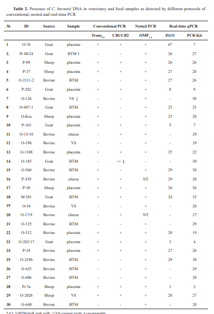
The study included archive genomic DNA extracted from samples scored positive for C. burnetii from previous diagnostic investigations, performed between 2014 and 2024, and retained for further analysis. These included 12 specimens from placentae, 4 vaginal swabs (VS) from cattle, sheep, and goats, 11 bulk tank milk (BTM) samples, and 3 cheese samples. They were analyzed by different conventional PCR protocols according to the primers available. From 2014 to 2020, PCR diagnostics in the laboratory was based on the use of primers CB1/CB2, targeting the superoxide dismutase (sod) gene of the Coxiella burnetii genome and/or Trans1/2 primers, comprising a sequence of the transposase gene of the IS1111 insertion element, while the OMP1-4 -PCR test was introduced later. Sixteen samples were of bovine origin, 8 were obtained from goats, and 6 were from sheep. Upon receiving the diagnostic materials in the laboratory, DNAs were isolated using commercial extraction kits, IndiSpin Pathogen Kit, (INDICAL Bioscience GmbH, Germany) for milk samples and PureLink Genomic DNA Mini Kit (Invitrogen) for tissue samples. After testing for the presence of Coxiella burnetii, extracted DNAs were stored at -20 °C for future reuse. This article does not contain any studies with human participants or animals performed by any of the authors. The research was conducted within the framework of a scientific project (Grant No KP-06-N33/3/2019) funded by the Bulgarian National Science Fund. The authors declare that they have the approval of the institution where they work to use the samples for research activities and to report the scientific results.

Conventional polymerase chain reaction (PCR)
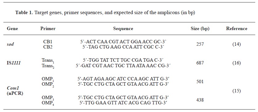
The available DNAs were used as templates in different conventional PCR protocols. PCR reactions were conducted in a total volume of 25 μL. For Trans1/2 and CB1/CB2-PCR, the reaction mixture included 12.5 µL 2x Blank qPCR Master Mix (EURx Ltd, Poland), 2 µL of each primer at a concentration of 10 pmol µL-1, 2 µL target DNA and 6.5 µL nuclease-free H2 O (EURx Ltd, Poland). For the nested PCR with primers OMP1 -OMP2 and OMP3 -OMP4, respectively, the first amplification was performed in a total volume of 25 μL, containing 12.5 μL 2x Blank qPCR Master Mix, 5.5 μL nuclease-free H2 O, 1 μL primer OMP1, 1 μL primer OMP2, and 5 μL target DNA. For the second step, 2 µL amplification products from the first reaction were added to the reaction mixture consisting of 12.5 µL 2x Blank qPCR Master Mix, 8.5 µL nuclease-free H2 O, 1 µL primer OMP1 and 1 µL primer OMP2. The target genes for which the primers were designed, the oligonucleotide sequence, and the expected size of the amplification product are presented in Table 1.

Table 1
All PCR reactions were performed in a DNA thermal cycler BIOER LifeECO at temperature regimes specific to each of the primer pairs used, as follows:
PCR with primers CB1/CB2. A modified procedure of Stein and Raoult (14) was used, with the amplification regimen consisting of an initial denaturation at 94 °C for 5 min, followed by 30 cycles of denaturation at 94 °C for 30 s, annealing at 54 °C for 1 min and elongation at 72 °C for 1 min and a final extension step at 72 °C for 10 min.
PCR with primers Trans1/2. Primers targeted a selected sequence of multicopy insertion element IS1111 in a modified PCR procedure of Berri et al. (16). It included an initial denaturation at 94 °C for 2 min followed by 40 cycles of 94 °C for 30 s, 61 °C for 30 s, and 72 °C for 1 min and final elongation at 72 °C for 5 min.
Nested PCR with primers OMP1 -OMP2 and OMP3 -OMP4 (15). The first amplification consisted of 94 °C for 4 min, 30 cycles of 94 °C for 1 min, 56 °C for 1 min and 72 °C for 1 min. In the second amplification, the temperature regime included denaturation at 95 °C for 4 min and 30 cycles of 94 °C for 1 min, 57 °C for 1 min and 72 °C for 1 min.
Тhe resulting PCR products were analyzed against DNA marker 100-1000 bp Hyper ladder, (Meridian Bioscience Inc.) or Gene Ruler 50 bp DNA Ladder, (Thermo Fisher Scientific Inc.) by electrophoresis in a 1.5 % agarose gel containing ethidium bromide and visualized in a UV transilluminator with a photo documentation system G:BOX (SYNGENE).

Real-time PCR
A bactotype C. burnetii PCR Kit (INDICAL Bioscience GmbH, Germany) was used according to the manufacturer's recommendations. Due to commercial considerations by the manufacturer,  the target genes and oligonucleotide sequences were unknown. Negative and positive controls, as well as an internal (extraction/amplification) control were included in the kit.
In parallel, an in-house procedure of qPCR was developed to detect Coxiella burnetii by targeting a 154-bp long DNA fragment in the transposase of the insertion sequence (IS) element IS1111, using the primers CB_IS1111_0706F 5´-CAAGAAACGTATCGCTGTGGC-3´ and CB_IS1111_0706R 5´-CACAGAGCCACCGTATGAATC-3´ and probe CB_IS1111_0706P 6FAMCCGAGTTCGAAAACAATGAGGGGCTG-TAMRA (24). The in-house procedure consisted of the initial step of denaturation and polymerase activation at 95 °C for 10 min, followed by 40 successive cycles of denaturation at 95 °C for 10 s, hybridization/elongation at 60 °C for 30 s, and cooling at 40 °C for 30 s. DNA extracted from a Bulgarian C. burnetii strain, molecularly characterized by whole genomic sequencing (to be published), and retained in our laboratory, was used as a positive control. PCR-graded H2 O (EURx Ltd, Poland) served as a non-template negative control and was used in each PCR run. The real-time PCR reactions were performed on a CFX Opus 96 Bio-Rad Real-Time PCR system. All samples presenting Ct values ​​lower than the positive control were considered positive. The assay was accepted to be valid when there was no signal in the negative control and the positive control yielded a fluorescence signal with a Ct<35.
Statistical analysis
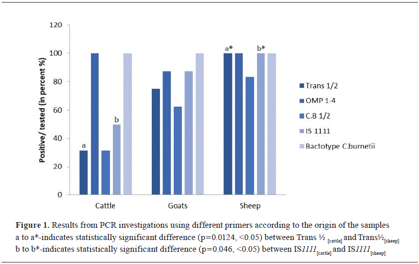
Data analysis was performed using the SPSS Statistics 26.0 (IBM Corp., Armonk, NY, USA) and Excel 2016 (Microsoft, Redmond, WA, USA) software platforms. A p-value of less than 0.05 was considered statistically significant. Chi-square test was used to test the association of tested positive samples with different primers based on 95% statistical significance. Fisher's exact test was used to determine the statistical significance of the differences between the groups.

RESULTS

The presence of C. burnetii DNA in the investigated samples, as detected by conventional PCR using different primers and by real-time PCR tests is summarized in Table 2.
Twelve samples scored positive for three targets (IS1111 plus Com1 plus sod) when tested in conventional PCR and five samples yielded positive results for two C. burnetii targets (Chi-square = 2.24; p<0.05). The rest of the samples were only positive with one of the primers. One sample (O-185) tested with CB1/CB2, was classified as questionable. Due to technical reasons, three of the samples were tested with Trans1/2 and CB1/CB2, but not with OMP1-4 primers. Generally, the sensitivity of each of the conventional PCR protocols defined as the ratio number of positivities to tested in percentages, was as follows: 56.7% (7/30) for PCR performed with the primers Trans1/2 (Chi-square=4.00; p<0.05); 96.3% (26/27) for OMP1-4-PCR (Chi-square=4.00; p<0.01) and 50.0% (15/30) for the primers CB1/CB2 (Chi-square=3.85; p<0.05). A marginally significant association between the detection efficiency of some of the primers and the species origin of the samples was found (Fig. 1).
In addition, differences in detection efficiency were found depending on the sample type; the largest number of samples testing positive for the three markers was obtained from placentae (10/12) and the lowest number of positives was observed in the BTM samples (1/11). All samples included in the study, regardless of whether they scored positive for one, two, or all three C. burnetii targets in conventional PCR, gave a positive result when tested with a commercial real-time PCR kit. The sensitivity of IS1111 real-time PCR was 70%. The Ct values ​​varied significantly among the samples, and differences were found in the average values ​​of the different types of samples, as follows: Ctplacentae-17.08/15.77; CtBTM-27.9/26.7; Ctcheese-28/29; CtVS-28.5/29.0. The reactions were validated when all positive controls from the test runs were positive and all non-template/negative controls gave no fluorescence signal.


DISCUSSION

Multiple reports have shown that conventional PCR protocols targeting different genetic markers also differ in their effectiveness in detecting the Coxiella burnetii genome. The results of our study were consistent with this conclusion. In fact, only 12 out of 30 tested samples of bovine, ovine, and goat origin scored positive in PCR with all three primers, and the rest of the reactions were positive with only one or two of the primers.
The highest percentage of samples - 96.3 % (26/27) tested positive in PCR, performed with OMP1-4 primers amplifying the Com1 gene. Originally designed to detect Coxiella burnetii DNA in human blood sera (15), this genetic target has been used in PCR to detect the DNA of the pathogen and also in other types of samples, including dairy (22) and ruminant abortion material (25). Using this format, we found positives among all types of specimens, including placentas, VS, BTM, and cheese. As a rule, nested PCR increases PCR sensitivity and leads to at least a 10,000-fold enhancement in PCR product over non-specific products that could be co-amplified when using only the outer primers (26, 27). In contrast to OMP1-4, the other two primer pairs were used in a one-step PCR. Another factor that probably matters in this case is that Com1 is highly conserved and therefore, reduced primer binding due to sequence variability is less likely to occur (15). It is also unlikely that this high detection rate is due to a false-positive PCR since OMP1-4 PCR has been shown to specifically detect only Coxiella burnetii but not other related bacteria (15, 26). The reaction showed efficiency, especially in BTM samples where the risk of contamination is minimal (28). The specificity of the reaction in the present study was evidenced by repeated negative results for the negative controls and by the fact that all samples that tested positive with these primers were also positive with the commercial real-time PCR kit.
Several comparative studies point out PCR with primers directed against IS1111 as the most sensitive one due to the presence of many copies of the target gene in the Coxiella burnetii genome, generating more amplicons, respectively. Substantial superiority of this PCR protocol has been described over PCR targeting Com1 and 16S rRNA genes (22), icd (29, 20), etc. In marked contrast, the percentage of samples in our study that reacted positively with these primers was 56.7 %, significantly lower than the level of detection obtained with OMP1-4. Although slightly exceeding the sensitivity of conventional PCR, qPCR performed with primers spanning the sequence of the same gene was not positive in the samples that were positive with Com1, as well as with bactotype C. burnetii kit. These contradictions result from several factors. There is a debate about the existence of Coxiella burnetii strains in which IS1111 is completely absent (21, 30). A recent publication reports this strain in marine mammals as well (31). On the other hand, it has been proven that there are significant differences between the isolates, including missing elements and sequence alterations within and near the IS1111 element coding regions (19). In strains that differ significantly in terms of IS1111, there is a high probability that amplification will not occur when using primers designed against sequences of this genetic marker. The presumption that these differences are genetically determined, is partially confirmed by the fact that the in-house IS1111 realtime PCR was positive in samples from sheep and goats, but mostly negative in samples from cattle. Thus, it can be speculated that the Coxiella burnetii genotype circulating in the bovine population in Bulgaria differs from the strains found in small ruminants and was not detected with the primers used in our study. However, confirmation of this assumption requires sequence analysis.
The analysis of the data obtained from conventional and real-time PCR targeting IS1111 showed a strong association between the percentage of samples testing positive and their type (placentas, BTM, VS, and cheese). In fact, 100 % (12/12) of the placentas reacted positively in the reactions performed against this genetic marker; however, in the BTM samples, this percentage was only 27.3% (3/11). All placental samples were from clinical cases, most often abortions. In contrast, most of the BTM samples (7/11) were collected from cattle herds with a positive immune status for Coxiella burnetii as detected after serological screening but showing no clinical manifestations. Accordingly, it can be assumed that the bacterial load in the clinical samples significantly exceeded that in the milk samples. All this makes it much more likely that the low detectability with Trans1/2 primers is due to the presence of a very low copy number of the Coxiella burnetii genome, which is below the detection limit. Indirectly, this was confirmed by the results from real-time PCR, in which the fluorescence signal increased significantly earlier when placentae were tested than in the other samples. To increase detection with primers directed against IS1111, nested PCR has been proposed (27).
In the study, the lowest level of detection-50.0 % (15/30) was observed when the reactions were performed with the primers CB1/CB2. This is in agreement with others, reporting a lower sensitivity of this test, as compared to PCR directed against other genetic targets (23). Moreover, one of the samples (O-185), which scored "positive" in a previous study, was reassessed as "negative" for CB1/CB2, because the size of the electrophoretic band was different from the specific band of Coxiella burnetii. A similar problem was also reported by other authors (26). Interestingly, in bactotype C. burnetii qPCR, this sample showed an increase in fluorescence signal with a Ct value within the range of the positive control and was also counted as positive.
Finally, an association was found between the detection efficiency achieved with the different primers and the origin of the samples. The possible presence of PCR inhibitors in the sample is also important since it has been reported that PCRs for different target sequences are not equally susceptible to inhibition by coextracted substances (32). The obtained results indicated that many factors could influence the output of PCR for the detection of Coxiella burnetii; therefore, the choice of suitable primers should be made according to the type, origin and quality of extracted DNA of the examined sample.

CONCLUSION

The data presented here show that the test results from conventional PCR should be interpreted with caution when the test is performed in a singleplex format. Even performed with the most sensitive genetic target such as IS1111, false negative results may be obtained if the initial bacterial load in samples is below the level of detection, or primer binding is reduced due to variability in the Coxiella burnetii DNA. Thus, performing a conventional PCR test with more than one genetic marker, or combining it with real-time PCR, would reduce the chance of missing the presence of this infection.

CONFLICT OF INTEREST

The authors declare that they have no financial or non-financial conflict of interest regarding authorship and publication of this article.

ACKNOWLEDGMENTS

This study was supported by the Bulgarian National Science Fund under Grant No KP-06-N33/3/2019. entitled: "Molecular genetic identification and creation of an archival genomic bank of the circulating human and animal C. burnetii genotypes and determination of their role as particularly dangerous infectious agents causing epizootic and epidemiological outbreaks on the territory of the Republic of Bulgaria"

AUTHORS' CONTRIBUTION

KS conceptualized the study, supervised the sample analysis and wrote the manuscript. KT and PG were included in the hypothesis formulation. KT carried out the sample analysis, prepared graphs and tables and participated in the manuscript writing. PG participated in editing the manuscript and contributed to the practical performance of this research. All authors have revised and approved the final version of the manuscript.


References

  1. Madariaga, MG, Rezai, K., Trenholme, GM, Weinstein, RA (2003). Q fever: a biological weapon in your backyard. Lancet Infect Dis. 3(11): 709-721. https://doi.org/10.1016/S1473-3099(03)00804-1 PMid:14592601
  2. Angelakis, E., Raoult, D. (2010). Q fever - review. Vet Microbiol. 140(3-4): 297-309. https://doi.org/10.1016/j.vetmic.2009.07.016   PMid:19875249
  3. Maurin, M., Raoult, D. (1999). Q fever. Clin Microbiol Rev. 12(4): 518-553. https://doi.org/10.1128/CMR.12.4.518 PMid:10515901 PMCid:PMC88923
  4. Melenotte, C., Protopopescu, C., Million, M., Edouard, S., Carrieri, MP, Eldin, C., Angelakis, E., et al. (2018). Clinical features and complications of Coxiella burnetii infections from the French National Reference Center for Q fever. JAMA Network Open. 1(4): e181580. https://doi.org/10.1001/jamanetworkopen.2018.1580 PMid:30646123 PMCid:PMC6324270
  5. Eldin, C., Mélenotte, C., Mediannikov, O., Ghigo, E., Million, M., Edouard, S., Mege, JL, et al. (2017). From Q Fever to Coxiella burnetii infection: a paradigm change. Clin Microbiol Rev. 30(1): 115-190. https://doi.org/10.1128/CMR.00045-16 PMid:27856520 PMCid:PMC5217791
  6. Agerholm, JS (2013). Coxiella burnetii associated reproductive disorders in domestic animals - a critical review. Acta Vet Scand. 55(1): 13. https://doi.org/10.1186/1751-0147-55-13 PMid:23419216 PMCid:PMC3577508
  7. Astobiza, I., Barandika, JF, Ruiz-Fons, F., Hurtado, A., Povedano, I., Juste, RA, García-Pérez, AL (2011). Coxiella burnetii shedding and environmental contamination at lambing in two highly naturally infected dairy sheep flocks after vaccination. Res Vet Sci. 91(3): e58-63. https://doi.org/10.1016/j.rvsc.2010.11.014 PMid:21168178
  8. Arricau-Bouvery, N., Souriau, A., Lechopier, P., Rodolakis, A. (2003). Experimental Coxiella burnetii infection in pregnant goats: Excretion routes. Vet Res. 34(4): 423-433. https://doi.org/10.1051/vetres:2003017 PMid:12911859
  9. Berri, M., Laroucau, K., Rodolakis, A. (2000). The detection of Coxiella burnetii from ovine genital swabs, milk and fecal samples by the use of a single touchdown polymerase chain reaction. Vet Microbiol. 72(3-4): 285-293. https://doi.org/10.1016/S0378-1135(99)00178-9 PMid:10727838
  10. Fournier, PE, Raoult, D. (2003). Comparison of PCR and serology assays for early diagnosis of acute Q fever. J Clin Microbiol. 41(11): 5094-5098. https://doi.org/10.1128/JCM.41.11.5094-5098.2003 PMid:14605144 PMCid:PMC262519
  11. Van den Brom, R., van Engelen, E., Roest, HI, van der Hoek, W., Vellema, P. (2015). Coxiella burnetii infections in sheep or goats: an opinionated review. Vet Microbiol. 181(1-2): 119-129. https://doi.org/10.1016/j.vetmic.2015.07.011 PMid:26315774
  12. Sahu, R., Rawool, DB, Vinod, VK, Malik, SVS, Barbuddhe, SB (2020). Current approaches for the detection of Coxiella burnetii infection in humans and animals. J Microbiol Methods. 179, 106087. https://doi.org/10.1016/j.mimet.2020.106087 PMid:33086105
  13. Zhang, GQ, Hotta, A., Mizutani, M., Ho, T., Yamaguchi, T., Fukushi, H., Hirai, K. (1998). Direct identification of Coxiella burnetii plasmids in human sera by nested PCR. J Clin Microbiol. 36(8): 2210-2213. https://doi.org/10.1128/JCM.36.8.2210-2213.1998 PMid:9665993 PMCid:PMC105014
  14. Stein, A., Raoult, D. (1992). Detection of Coxiella burnetti by DNA amplification using polymerase chain reaction. J Clin Microbiol. 30(9): 2462-2466. https://doi.org/10.1128/jcm.30.9.2462-2466.1992 PMid:1401016 PMCid:PMC265524
  15. Zhang, GQ, Nguyen, SV, To, H., Ogawa, M., Hotta, A., Yamaguchi, T., Kim, HJ, et al. (1998). Clinical evaluation of a new PCR assay for detection of Coxiella burnetii in human serum samples. J Clin Microbiol. 36(1): 77-80. https://doi.org/10.1128/JCM.36.1.77-80.1998 PMid:9431924 PMCid:PMC124811
  16. Berri, M., Arricau-Bouvery, N., Rodolakis, A. (2003). PCR-based detection of Coxiella burnetii from clinical samples. Methods Mol Biol. 216, 153-161. https://doi.org/10.1385/1-59259-344-5:153 PMID: 12512362
  17. Hoover, TA, Vodkin, MH, Williams, JC (1992). A Coxiella burnetti repeated DNA element resembling a bacterial insertion sequence. J Bacteriol. 174(17): 5540-5548. https://doi.org/10.1128/jb.174.17.5540-5548.1992 PMid:1324903 PMCid:PMC206497
  18. Klee, SR, Ellerbrok, H., Tyczka, J., Franz, T., Appel, B. (2006). Evaluation of a real-time PCR assay to detect Coxiella burnetii. Ann NY Acad Sci. 1078, 563-565. https://doi.org/10.1196/annals.1374.111 PMid:17114778
  19. Denison, AM, Thompson, HA, Massung, RF (2007). IS1111 insertion sequences of Coxiella burnetii: characterization and use for repetitive element PCR-based differentiation of Coxiella burnetii isolates. BMC Microbiol. 7, 91. https://doi.org/10.1186/1471-2180-7-91 PMid:17949485 PMCid:PMC2104537
  20. de Bruin, A., de Groot, A., de Heer, L., Bok, J., Wielinga, PR, Hamans, M., van Rotterdam, BJ, Janse, I. (2011). Detection of Coxiella burnetii in complex matrices by using multiplex quantitative PCR during a major Q fever outbreak in The Netherlands. Appl Environ Microbiol. 77(18): 6516-6523. https://doi.org/10.1128/AEM.05097-11 PMid:21784920 PMCid:PMC3187144
  21. Marmion, BP, Storm, PA, Ayres, JG, Semendric, L., Mathews, L., Winslow, W., Turra, M., Harris, RJ (2005). Long- term persistence of Coxiella burnetii after acute primary Q fever. QJM 98(1): 7-20. https://doi.org/10.1093/qjmed/hci009 PMid:15625349
  22. Kargar, M., Rashidi, A., Doosti, A., Najafi, A., Ghorbani-Dalini, S. (2015). The sensitivity of the PCR method for detection of Coxiella burnetii in the milk samples. ZJRMS 17(6): e988. https://doi.org/10.17795/zjrms988
  23. Basanisi, MG, La Bella, G., Nobili, G., Raele, DA, Cafiero, MA, Coppola, R., Damato, AM, et al. (2022). Detection of Coxiella burnetii DNA in sheep and goat milk and dairy products by droplet digital PCR in southern Italy. Int J Food Microbiol. 366, 109583. https://doi.org/10.1016/j.ijfoodmicro.2022.109583 PMid:35182931
  24. Edouard, S., Raoult, D. (2016). Lyophilization to improve the sensitivity of qPCR for bacterial DNA detection in serum: the Q fever paradigm. J Med Microbiol. 65(6): 462-467. https://doi.org/10.1099/jmm.0.000253 PMid:27008653
  25. Jones, RM, Twomey, DF, Hannon, S., Errington, J., Pritchard, GC, Sawyer, J. (2010). Detection of Coxiella burnetii in placenta and abortion samples from British ruminants using real-time PCR. Vet Rec. 167(25): 965-967. https://doi.org/10.1136/vr.c4040 PMid:21262712
  26. Ogawa, M., Setiyono, A., Sato, K., Cai, Y., Shiga, S., Kishimoto, T. (2004). Evaluation of PCR and nested PCR assays currently used for detection of Coxiella burnetii in Japan. Southeast Asian J Trop Med Public Health. 35(4): 852-855.
  27. Mares-Guia, MAMM, Guterres, A., Rozental, T., Ferreira, MDS, Lemos, ERS (2018). Clinical and epidemiological use of nested PCR targeting the repetitive element IS1111 associated with the transposase gene from Coxiella burnetii. Braz J Microbiol. 49(1): 138-143. https://doi.org/10.1016/j.bjm.2017.04.009 PMid:28899604 PMCid:PMC5790644
  28. Abiri, Z., Khalili, M., Kostoulas, P., Sharifi, H., Rad, M., Babaei, H. (2019). Bayesian estimation of sensitivity and specificity of a PCR method to detect Coxiella burnetii in milk and vaginal secretions in sheep and goat samples. J Dairy Sci. 102(6): 4954-4959. https://doi.org/10.3168/jds.2018-15233 PMid:31005328
  29. Klee, SR, Tyczka, J., Ellerbrok, H., Franz, T., Linke, S., Baljer, G., Appel, B. (2006). Highly sensitive real-time PCR for specific detection and quantification of Coxiella burnetii. BMC Microbiol. 6, 2. https://doi.org/10.1186/1471-2180-6-2 PMid:16423303 PMCid:PMC1360083
  30. Rolain, JM, Raoult, D. (2005). Molecular detection of Coxiella burnetii in blood and serum during Q fever. QJM 98(8): 615-617. https://doi.org/10.1093/qjmed/hci099 PMid:16027172
  31. Gardner, B., Bachmann, N., Polkinghorne, A., Hufschmid, J., Tadepalli, M., Marenda, M., Graves, S., et al. (2023). Novel marine mammal Coxiella burnetii-genome sequencing identifies a new genotype with potential virulence. Pathogens. 12(7): 893. https://doi.org/10.3390/pathogens12070893 PMid:37513739 PMCid:PMC10386718
  32. Huggett, JF, Novak, T., Garson, JA, Green, C., Morris-Jones, SD, Miller, RF, Zumla, A. (2008). Differential susceptibility of PCR reactions to inhibitors: an important and unrecognized phenomenon. BMC Res Notes. 1, 70. https://doi.org/10.1186/1756-0500-1-70 PMid:18755023 PMCid:PMC2564953


Copyright

Copyright: © 2025 Simeonov KB This is an open-access article published under the terms of the Creative Commons Attribution License which permits unrestricted use, distribution, and reproduction in any medium, provided the original author and source are credited.

Competing Interests

The authors have declared that no competing interests exist.

Citation Information

Macedonian Veterinary Review. Volume 48, Issue 2, Pages i-x, e-ISSN 1857-7415, p-ISSN 1409-7621, DOI: https://doi.org/10.2478/macvetrev-2025-0020