Abstract
Second-degree burns typically require a longer healing period and can develop hypertrophic scars. Collagen plays a crucial role in treating and healing skin wounds. This study aimed to investigate the effect of topically applied nano-collagen extracted from catfish skin on rabbit burn wounds by assessing the production of collagen and the development of fibroblasts in the healing wounds. The nano-collagen material was produced in two stages by using NaOH and alcohol solutions in the first, and acetic acid solution in the second phase. The viscous solution obtained in this process was adjusted to a specific pH, was centrifuged, and was mixed with Vaseline before being lyophilized. Thirty skin samples from burn wounds were obtained from blemish-free rabbits. Each rabbit was treated by inflicting a 50 mm2 burn wound on the back. They were divided into three subgroups: a control group (G1, n=10) without wound treatment, a group topically treated with nano-collagen cream on a daily basis for one week (G2, n=10) and a group treated by sewing catfish skin onto their wounds (G3, n=10). Measurements were taken daily during the first week to monitor any possible post-burn contractions. G2 had significantly the lowest wound diameter after 7 days of treatment (35.00±1.21 mm). It had significantly the lowest wound contraction (8.200±0.042 kg/cm 2 ) on 21 days of the treatment and had higher observable vascularization and development of hair follicles in the wounds compared to the other groups. In conclusion, the topical application of catfish skin nano-collagen cream significantly reduces second-degree burn wounds in rabbits and improves the healing process.
Keywords: nano-collagen, catfish skin, burn wound, skin, rabbits
INTRODUCTION
Burns are a common injury which can range from mild first-degree burns to third-degree burns. The second stage of inflammation involves the outer (epidermis) and deeper skin layers (dermis). These burns can be painful and may take a long time to heal, leading to discomfort and possible complications (1).
Collagen is a useful biomaterial commonly used in industrial applications. Various studies have investigated alternative sources of this protein, such as aquatic animals (2) using various methods. The collagen production, purity, and molecular integrity during extraction relies on the biomolecule's temperature stability which can vary in different fish species from tropical and temperate climates (3).
Collagen is abundant in the human body and plays an important role in wound healing (4). Nano-collagen is composed of small collagen molecules, allowing it to be better absorbed and penetrate the skin. Along with its immunity-boosting effects, collagen has also been shown to have antibacterial and anti-inflammatory properties that may help reduce the risk of infection and inflammation in burns. It supports faster healing by stimulating collagen production in the skin, increasing skin elasticity, and reducing scars. The small size of nano-collagen allows it to penetrate the skin deeply, increasing its effectiveness in promoting wound healing (5).
Many of the bioactive compounds found in marine species have potential applications in the pharmaceutical and cosmetic industries. Numerous studies have investigated the use of marine collagens, being easily accessible, naturally compatible, and water-soluble biomaterials (6).
Catfish skin is a byproduct of the food industry and is otherwise discarded as waste. This byproduct of the industry can be used as a resource for the production of wound-healing medications (7).
The use of nano-collagen obtained from catfish skin yielded efficient results in the treatment of various skin diseases, including burns (8). Given its small and elastic nature, collagen easily penetrates the skin, allowing faster healing, reduces the risk of complications, and minimizes inflammation (4). These characteristics render it an effective and sustainable natural material for the treatment of second-degree burns and skin inflammations. Nano-collagen effect on skin burns could include: improved healing by inducing collagen synthesis necessary for skin regeneration; minimized scarring by enhancing the skin's structural integrity during the healing process; improved skin elasticity and texture; and potentially reduce the risk of infection by exhibiting antimicrobial properties (9, 10).
Natural collagens obtained from catfish skin in the fish industry, can reduce the need for producing synthetic collagens or harvesting it from live animals (11). The environmentally friendly harvesting of natural collagens makes this process efficient and cheap (3).
The study aimed to test an extraction method of nano-collagen from catfish skin and to assess its therapeutic effect on second-degree skin burns in rabbits.
MATERIAL AND METHODS Nano-collagen extraction from catfish skin
Five catfish were purchased at a nearby market. After the skins were removed, they were chopped into small pieces, triple-washed in distilled water, and then frozen at -40 ºC.
In order to extract nano-collagen from catfish skin, two major phases were followed. In the first phase, a 0.05 M NaOH solution was employed for 12 h, with a 1:20 (w/v) solution exchanged every 6 h. Then, the samples were cleaned with ice-cold distilled water to make a pH-neutral solution. As the samples were exposed to a 1:2 (v/v) rated 10% butyl alcohol solution for 24 h, with the solution being changed on 12 h, they were washed and distilled at 4 °C to extract collagen.
In the second phase, a 0.5 M concentrated 1:40 (w/v) rated acetic acid solution was used for 72 h, with 24-hour-long changes. After the viscous solution was distilled, it was mixed with 0.9 M sodium chloride and 0.9 M carbonate-bicarbonate buffer solution until the pH was 8.87. A 40-minute centrifugation was then carried out at 4 °C and 11,000 g. The post-configuration material was subjected to three 12-hour dialyses, with the initial two dialyses in ice-cold distilled water, and the final two dialyses in a 0.1 M acetic acid solution. When used to treat burns, the dried post-dialysis ASC (
12) material was combined with Vaseline at 1:10 (w/w) after being frozen and lyophilized. The electro-spinning technology involved two main procedures, electro-spinning melting and electro- spinning solutions. Electro-spinning generally applies strong electrostatic forces to produce fibers. However, to make ultra-fine fibers, this approach involved an electrical charging of the polymer solution or melting at a high voltage (about 10–20 KV) (
13). Hence, the melted polymer or solution in the syringe pump comes first. Then, to create a pendant drop, the solution was pressed to the tip. The application of the voltage potential with the submerged electrode in the syringe helped generate free charges inside the polymer. The presence of the electric field caused the pendant to elongate. Finally, the surface tension was overcome when the potential applied reached a critical value.
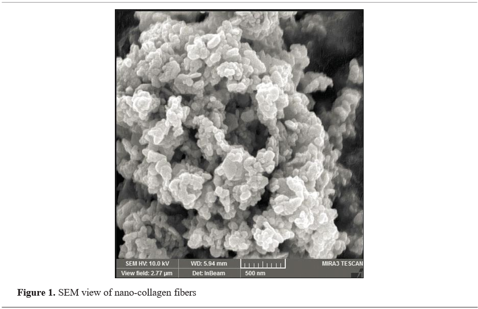
Nano-collagen samples were examined with a scanning electron microscope to view nano- collagen fibers, which would scaffold as little as 75 µm, as shown in
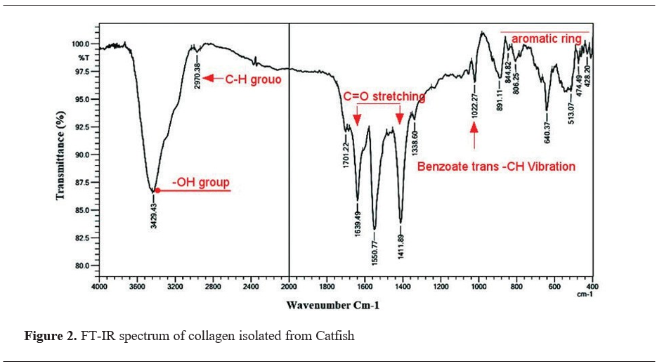
Fig. 1, while Fourier transform infrared spectroscopy (FTIR), shown in
Fig. 2, was used to analyze the chemical structure.
Electron microscopy analysis (SEM) was used to identify the morphological and quantitative properties of nanoparticles: shape, size, level of separation, and general appearance. A scanning electron microscope was used to analyze the microstructure of fish collagen and fish-scale collagen nanoparticles. Following lyophilization, the collagen sample was perforated and fastened to an adhesive carbon stub for imaging purposes. Using a scanning electron microscope, the nano- collagen sample was analyzed to view the nano- collagen fibers, which might scaffold as little as 75 µm (
14).
A Fourier Transform Infrared Spectroscopy analysis was conducted using an FTIR spectrometer (8400S, Shimadzu, Japan). FT-IR analysis was used to detect the functional groups on the nano- collagen. Various absorption peaks were detected at 3,429.43, 2,970.38, 1,701.22, 1,639.49, 1,550.70, 1,411.80, 1,335.60, 1,022.20, 891.10, and 806.30 cm, which correspond with the different functional groups: OH- group, C-H group, benzoate -trans-CH-vibration, and aromatic ring structure (
Fig. 2).
Even though infrared spectroscopy is sensitive to the chemical structure of molecules, it is, however, suitable for the determination of proteins and polypeptides in different states, concentrations, and environments, and useful for determining the secondary structure of proteins and polypeptides (
15, 16).
Thirty New Zealand white rabbits, kept in the animal enclosure of the College of Veterinary Medicine at the University of Al-Qadisiyah, were used in this study. These rabbits, which weighed 2.5±0.5 kg, and showed no skin lesions, were kept in regular conditions. To apply treatment, the rabbits were put into three 10-rabbit subgroups. The first group (G1) received no therapy and thus was considered as control group. The second group (G2) received a week-long coating of nano-collagen every day. The third group (G3) had square-shaped pieces of catfish skin (CFS) sewn onto their burns. All rabbits underwent conventional surgery. The burn device was composed of thermal sensor and a hot metal stylus. One of the two parts that made up the metal stylus was the aluminum probe which heated and cooled rapidly. The temperature sensor was situated adjacent to the probe’s tip. Digital displays made it easy to customize the temperature and timer to suit individual preferences. The device was adjusted for the purpose of inflicting burn wounds to the rabbit model. Each rabbit was inflicted with a burn wound on the back skin in an area of 50 mm2 with 100 °C for 15 sec. The wound depth was similar in all animals by adjusting the exposure time and temperature. Diameters were measured daily in the first week. Every single stage of the investigation was conducted in accordance with the recommendations for the use of experimental animals by the College of Veterinary Medicine at the University of Al-Qadisiyah. The study was approved by the College of Veterinary Medicine at the University of Al-Qadisiyah (approval no. 5420/9/7).
The morphological changes of the wounds were recorded and monitored throughout the treatment. The wound contraction/tension force was monitored daily for ten days using a modified Tensiometer (kg/cm2). The wound diameter was measured in millimeters. The histopathological skin samples were obtained from anesthetized rabbits. A tiny piece of the burnt tissue was removed using a sterile punch biopsy tool. The biopsy was typically small, with a diameter of 4-6 mm, in order to reduce stress, minimize surrounding tissue damage, and obtain sufficient tissue volume for the analysis. The samples were collected after seven, fourteen, and twenty-one days after the infliction of the burn wounds in all groups. The specimens were stored in a 10% buffered formalin solution and were processed for histological examination. This involved preserving, dehydrating, embedding, sectioning, and staining. The samples were stained with hematoxylin and eosin, and were sectioned in 6 µm slices (17).
Statistical analysis
To compute the data findings, ANOVA software version 32 was used to statistically evaluate these findings, with a significance level of p<0.05.
RESULTS Burn wound contraction
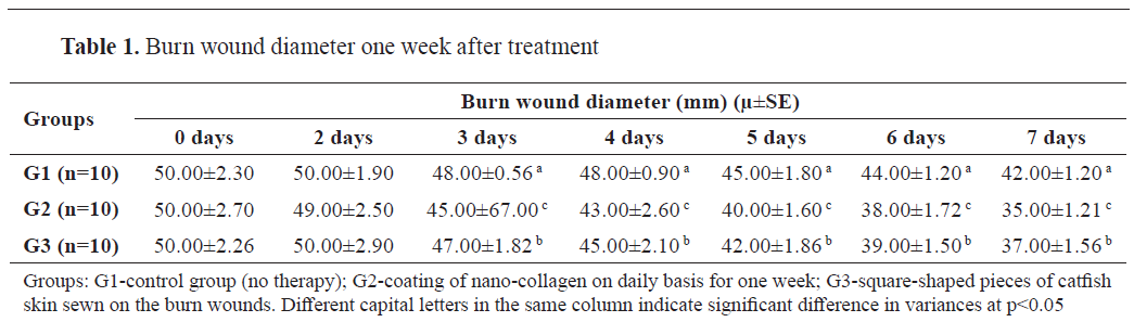
The burn wound diameter had a significant decrease in G1, G2, and G3, from 50 mm to 42 mm, 35 mm, and 37 mm, respectively, after seven days of the treatment. The lowest wound diameter was recorded in the second group during the seventh day (35±1.21 mm) (Table 1).
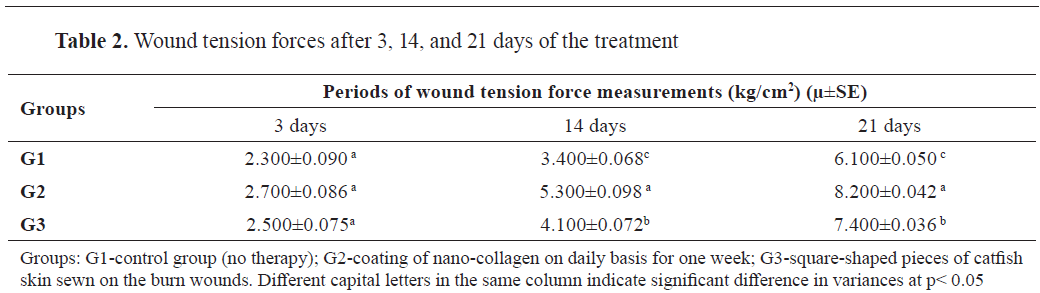
Burn wound tension force
As shown in Table 2, the wound tension forces varied across the three groups after 3, 14, and 21 days. G1 significantly increased from 2.3 kg/cm2 at day 2 to 6.1 kg/cm2 at day 21. G2 had the highest but non-significant increase from 2.7 kg/cm2 at day 3 to 8.2 kg/cm2 at day 21. G3 had significantly increased from 2.5 kg/cm2 at day 3 to 7.4 kg/cm2 at day 21.
Histopathological evaluation
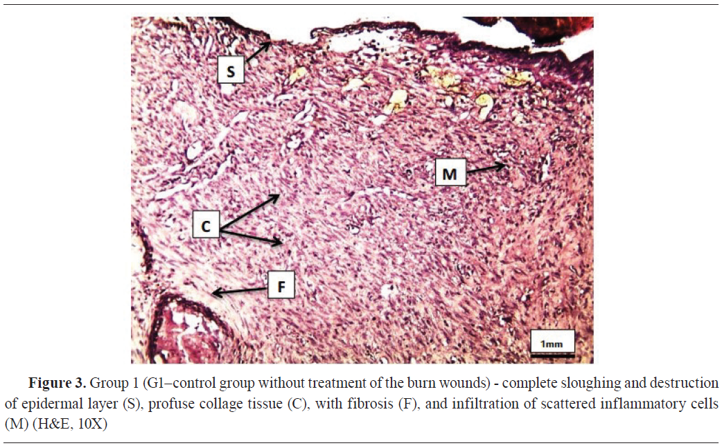
After one-week-treatment, the three groups had observable histopathological changes. G1 developed total epidermal sloughing and disintegration, extensive fibrotic collage tissue, and dispersed inflammatory cell infiltration (Fig. 3).
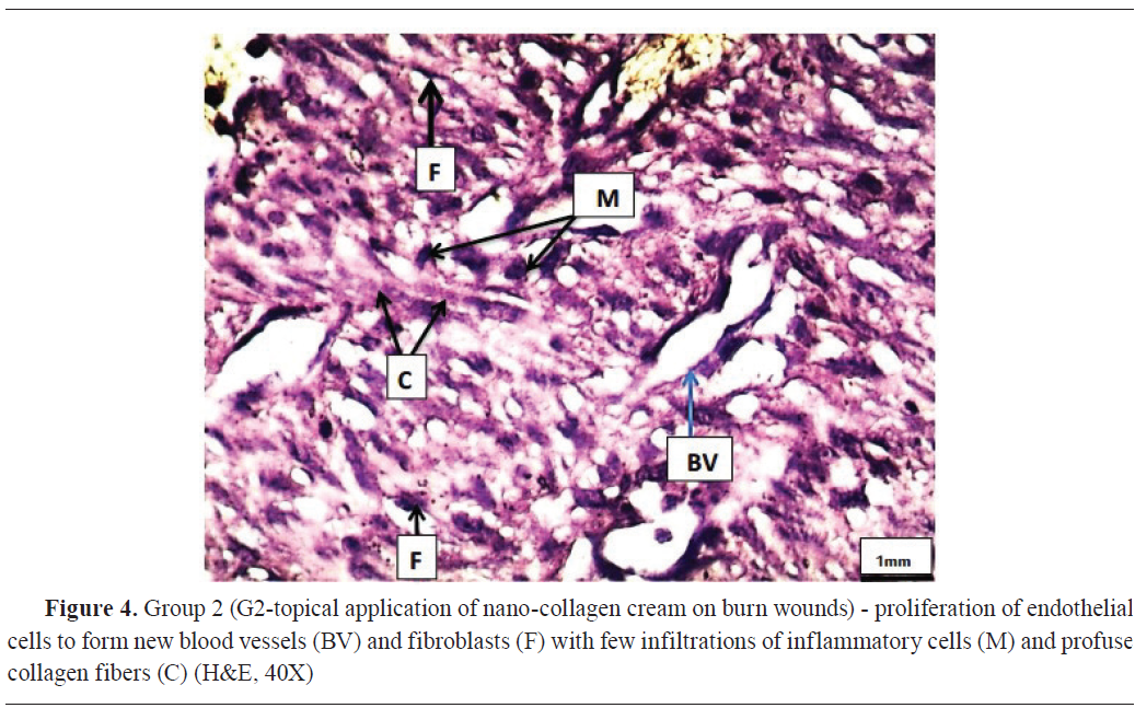
G2 had fibroblasts and endothelial cells proliferation, inflammatory cells infiltration, and abundant collagen fibers in the dermis (Fig. 4).
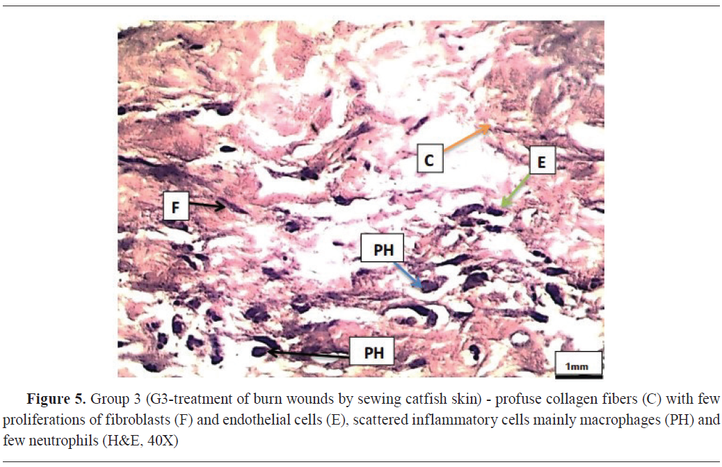
G3 had abundant collagen fibers, proliferated fibroblasts and endothelial cells, and dispersed inflammatory cells, mostly macrophages and neutrophils (Fig. 5).
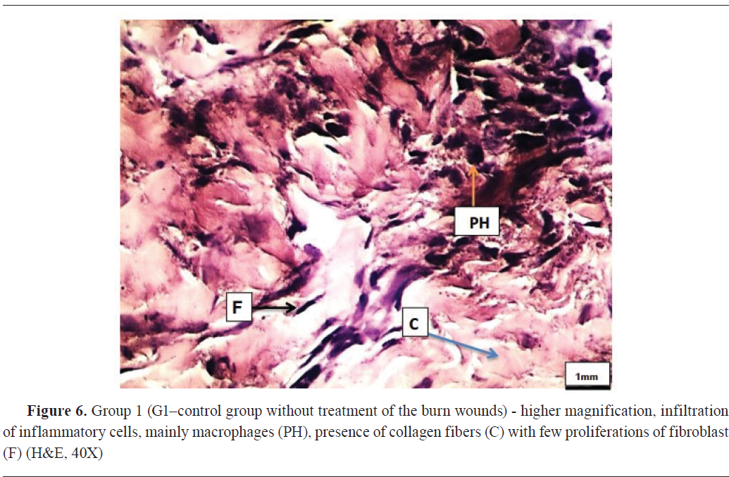
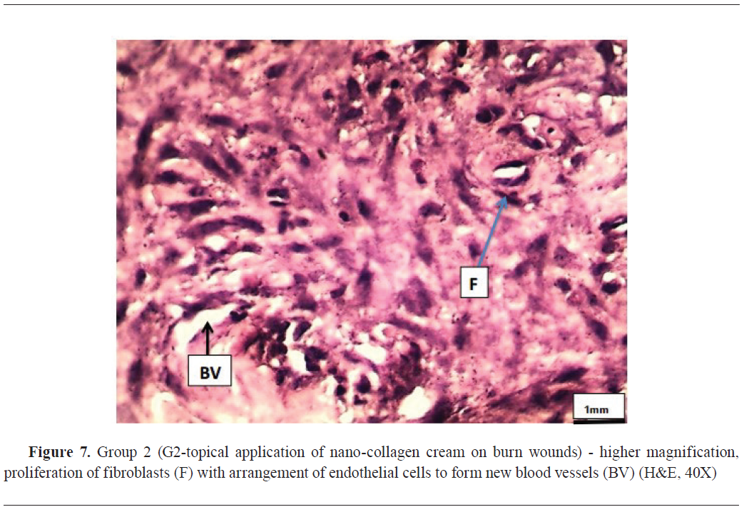
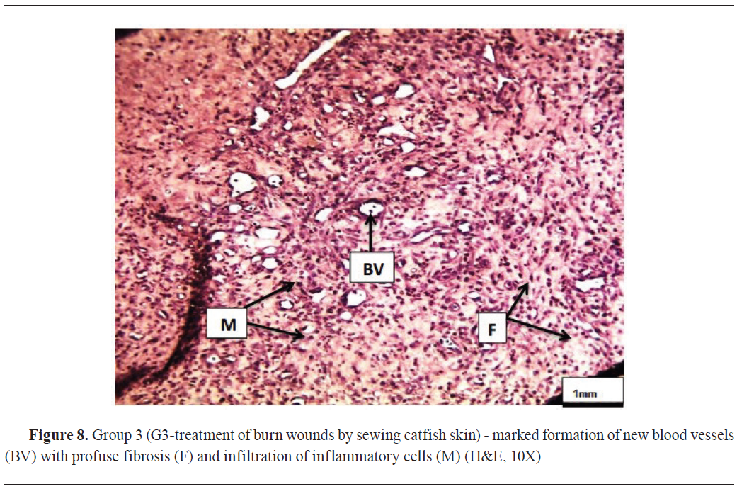
In the second week of the treatment, G1 had infiltrating inflammatory cells, primarily macrophages, collagen fibers, and minimal fibroblast growth (Fig. 6). G2 showed fibroblasts proliferation combined with endothelial cell organization which indicated formation of new blood vessels (Fig. 7). Likewise, G3 had a significant development of new blood vessels with extensive fibrosis and an infiltration of inflammatory cells (Fig. 8).
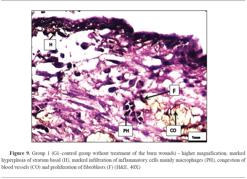
In the third week after the treatment, G1 showed significant hyperplasia of the basal layer, marked with an infiltration of inflammatory cells, primarily macrophages, constriction of blood vessels, and fibroblast proliferation (
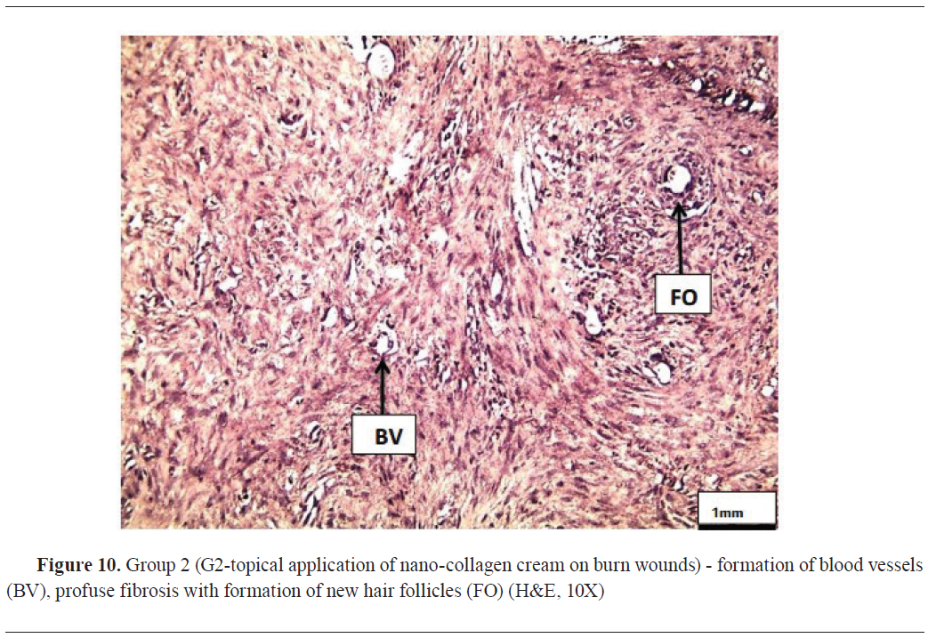
Fig. 9). Similarly, G2 showed blood vessel development, extensive fibrosis, and the growth of new hair follicles (
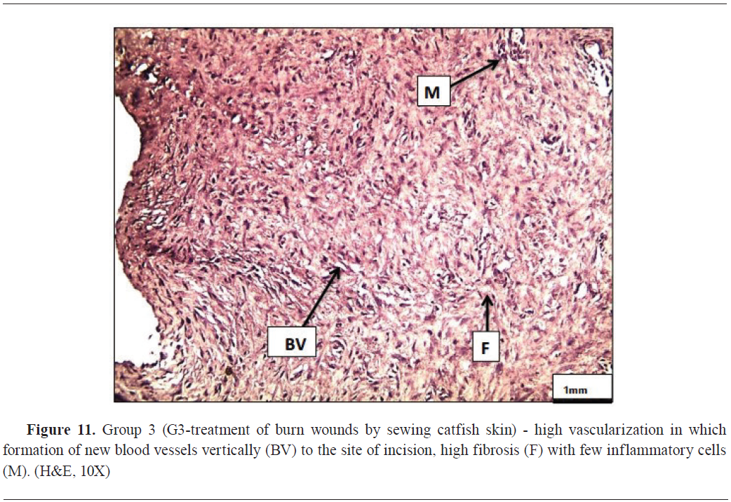
Fig. 10). G3 had vascularization perpendicularly to the incisions, high level of fibrosis, and minimal inflammatory cells infiltration (
Fig. 11).
DISCUSSION
Burn wounds remain sterile for the first 24–48 h after which they get infected with skin flora. Gram-negative organisms predominate after 5–7 days (
18). Therefore, burn dressings minimize bacterial counts to reduce possible overt infection and provide a moist environment for optimal healing (
19).
The collagen FTIR spectra obtained from the results are consistent with Zhang et al. (
20), where the development of amide A bands in the region of 3292–3315 cm
-1 demonstrated the involvement of peptides’ N–H groups in hydrogen bonding. At 1656 cm
-1, the Amide I band was detected. Collagen has an amide II band at 1538–1548 cm
-1 and an amide III band between 1,232–1,238 cm
-1. The characteristic amino-acids and entire collagen molecules are generally represented by FTIR (
21).
A healing reaction, also known as wound contraction, minimizes the amount of the tissue defect and reduces the wound area. Myofibroblasts from the surrounding tissues are mainly responsible for this effect by drawing newly synthesized collagen fibers in the injured tissue towards its center (
2). The actin and myosin proteins in myofibroblast exert the contracting force. In the extracellular matrix, the actin, myosin, and newly synthesized collagen fibers co-form a web-like sticky mesh which facilitates the wound contraction (
22).
Group 2 had a significant contraction (35 mm) at seven days after the treatment which corresponded with the increased vascularization, accelerated fibroblast proliferation, and the stimulated cell expansion, and was in line with the report from Dhivya et al. (
23).
The first-week histopathological changes in G1 revealed complete epidermal sloughing and destruction, fibrosis-driven profuse collage tissue, and infiltration of scattered inflammatory cells, which was in agreement with Hussien et al. (
24), who reported similar changes. In the same context, G2 had dermic granulation tissue. The dermic growth is characterized by proliferation of endothelial cells and fibroblasts, infiltration of inflammatory cells, and profuse collagen fibers (
25). Similarly, G3 revealed the same dermic changes observed in G2 above. These changes were reported by Andini et al. (
26), stating that nano-collagen can cause effects similar to those caused by skin burns.
In the second week, G1 developed an infiltration of inflammatory cells, mainly macrophages and some collagen fibers, as well as a minimal proliferation of fibroblasts. These changes are similar to those reported by Vujičić et al. ( 27 ). G2 had a proliferation of fibroblasts, which, along with endothelial cells, co-form new blood vessels. These results are in line with Mbese et al. ( 28 ), who indicated that a two-week application of nano-collagen can progressively stimulate healing. Similar changes were observed in G3 by formation of new blood vessels, profuse fibrosis, and the infiltration of inflammatory cells, similarly reported by Munahi et al. ( 29 ).
Following three weeks after the treatment, histopathological changes were characterized by variable findings in the three treatment groups (hyperplasia of the basal layer, infiltration of inflammatory cells, particularly macrophages, congestion of blood vessels, and proliferation of fibroblasts in G1), similarly reported by Hussein et al. ( 24 ). The changes in G2 (blood vessels, profuse fibrosis, and new hair follicles) were in line with the findings of Shpichka et al. ( 30 ). G3 changes were characterized with high vascularization (new blood vessels were formed perpendicular to the incision), high fibrosis, and a few inflammatory cells which were in agreement with Mulder et al. ( 31 ).
CONCLUSION
Topical application of nano-collagen significantly enhanced the healing of burn wounds by increasing fibroblast count, collagen, and overall re-epithelization in rabbits. No immunological reactions were observed during the treatment.
CONFLICT OF INTEREST
The authors declare that they have no financial or non-financial conflict of interest regarding authorship and publication of this article.
ACKNOWLEDGMENTS
Authors would like to acknowledge the Department of Surgery and Obstetrics at College of Veterinary Medicine, Al-Qadisiyah University, Al-Qadisiyah, Iraq.
AUTHORS' CONTRIBUTION
AKM made the critical review, supervision, data collection and final approval of the manuscript. AAH conducted the conceptualization and data analysis. RHF was involved in writing and AIT in the conceptualization of the manuscript.

 10.2478/macvetrev-2025-0021