Porcine Circovirus 2 (PCV2), a member of the family Circoviridae and the genus Circovirus, is associated with several syndromes that are collectively referred to as porcine circovirus associated disease (PCVAD). These include PCV2 systemic disease (PCV2-SD), formerly known as post-weaning multisystemic wasting syndrome (PMWS), PCV2-reproductive disease (PCV2-RD), PCV2-lung disease (PCV2-LD), PCV2-enteric disease (PCV2-ED), porcine dermatitis and nephropathy syndrome (PDNS), PCV2-subclinical infection (PCV2-SI), and acute pulmonary edema (
1). Comprehensive knowledge of PCV2 biology and PCVAD is essential for effective disease control (
2) as diseases associated with PCV2 have a substantial impact on pig-producing countries and represent one of the most economically significant health problems in the global swine industry.
To date, four species within the genus
Circovirus have been described: PCV type 1 (PCV1), PCV type 2 (PCV2), PCV type 3 (PCV3), and PCV type 4 (PCV4), with PCV2 recognized as the predominant genotype worldwide (
1, 3). Porcine circoviruses are widely distributed in both domestic pigs and wild boars (
4), although they have occasionally been detected in non-porcine species, including ruminants (cattle, goats, and deer), rodents (mice and rats), carnivores (dogs, minks, and foxes), insects (flies and mosquitoes), and shellfish (
5). PCV2, which is recognized as the primary etiological agent of PCV2-SD, was first identified in association with PMWS in weaning piglets in Canada in 1991 and represents the smallest known autonomously replicating virus in vertebrates (
6, 7). After the syndrome was defined by Harding and Clark in 1997 (
6), PCVAD was recognized as the major cause of production losses in pig herds across the principal swine-producing regions of the world.
Among the nine currently recognized genotypes of PCV2 – designated PCV2a to PCV2i – three major genotypes, namely PCV2a, PCV2b, and PCV2d, have been reported worldwide, with PCV2b emerging as the predominant genotype in many regions (
8, 9). Although all nine identified genotypes of PCV2 have been detected in domestic pigs (
10), only seven of these genotypes (a, b, d, e, f, g, and h) have so far been reported in wild boar populations (
11).
Clinical manifestations of PCVAD in pigs most commonly include wasting (98.1%), inappetence (90.4%), diarrhea (77.2%), dyspnea (75.1%), lymphadenopathy (44.8%), central nervous system signs (39.6%), and death (96.8%) (
2). At the microscopic level, lesions in lymphoid tissues may present as lymphoid depletion, histiocytic infiltration, the presence of inclusion bodies, and multinucleated giant cells (
12).
The diagnosis of PCVAD is based on the evaluation of clinical signs, the identification of characteristic microscopic lesions in affected organs – such as lymphoid tissues, lungs, liver, heart, kidneys, and intestines – and the detection of PCV2 antigen or viral DNA within these lesions (
13). Several laboratory techniques are commonly used for PCV2 detection. Immunohistochemistry (IHC),
in situ hybridization (ISH), and polymerase chain reaction (PCR) are among the most frequently applied methods. IHC and ISH enable localization of the virus within infected tissues or cells while preserving histological architecture and providing detailed cellular information, whereas PCR represents a rapid and highly sensitive technique that can also be applied to samples obtained from live animals (
14). In addition, enzyme-linked immunosorbent assay (ELISA), particularly the INGEZIM Circo IgG test, has been used to detect PCV2 antibodies in wild boar populations (
15).
Previous studies have indicated that PCV2 transmission occurs from infected to susceptible pigs through both direct contact and indirect airborne routes (
16). Moreover, numerous investigations conducted in different countries have demonstrated that PCV2 genotypes detected in wild boars are genetically very similar to those circulating in domestic pigs (
17). This genetic similarity suggests a potential ecological interaction between wild boars and domestic pigs that may facilitate the transmission and maintenance of PCV2. In Europe, PCV2 has been detected in wild boar populations in most countries, including Greece, Poland, Germany, and Italy (
4, 18, 19, 20). Given the high mobility of wild boar populations and the documented genetic similarity of PCV2 strains circulating across Europe, investigating the circulation of PCV2 in Estonia may provide valuable insight into the broader epidemiological situation of the virus in the region.
Laboratory detection of PCV2 in wild boar populations have primarily relied on real-time PCR for the detection of viral DNA in tissue samples, particularly tonsils and lymph nodes (
15, 21). These results have demonstrated that the prevalence of PCV2 varies considerably between geographical regions, with reported prevalence rates of 13.52% in Transylvania (
21), 75.6% in Poland (
19), and 50.7% in southwestern Germany (
15).
PCV2 has the potential to circulate between wild boars and domestic pigs, thus monitoring the presence of the virus in wild populations is of considerable importance (
18). The variability in PCV2 prevalence among wild boar populations across different geographical regions highlights the need for further studies aimed at improving the understanding of the epidemiology and genetic characteristics of the virus, as well as its potential implications for animal and public health.
The high prevalence of PCV2 in wild boar populations highlights the need to develop preventive measures to reduce the risk of viral transmission between wild and domestic pigs (
22). Furthermore, the detection of PCV2 in wild boars has important implications for understanding the epidemiology and genetic diversity of the virus in wildlife reservoirs (
9, 20, 23, 24, 25). High levels of PCV2 antibodies detected in wild boars indicate that the virus is widely distributed within these populations.
According to the available literature, wild boars may act as potential reservoirs and spreaders of PCV2 to domestic pigs. Although studies conducted in several European countries have demonstrated the widespread presence of PCV2 in wild boar populations, no studies have yet investigated the distribution of PCV2 in wild boars in Estonia. Therefore, the aim of the present study was to assess the seroprevalence of PCV2 in wild boars in Estonia and to evaluate its distribution among female and male animals across three different age groups.
We hypothesized that PCV2 circulates widely within wild boar populations in Estonia and that the seroprevalence of the virus does not differ significantly between age groups or sexes. Since no data on PCV2 prevalence in Estonian wild boars are currently available, the aim of this study was to assess the seroprevalence of PCV2 in wild boar populations in Estonia according to age and sex, and to evaluate tissue-level histopathological changes and antigen localization in order to support the serological findings.
MATERIAL AND METHODSAnimals and sampling procedureBlood and mesenteric lymph node samples from 433 wild boars were investigated. The samples were collected from all counties of Estonia and included wild boars found dead and hunted animals. Wild boars found dead, including animals killed in road traffic accidents or shot due to illness, were sampled across the whole country irrespective of the health status of the area (passive surveillance). Blood samples from wild boars were collected by hunters immediately after hunting, whereas for non-hunted wild boars (including animals found dead due to natural causes or traffic accidents), blood was collected post-mortem directly from the heart or large veins. Lymph nodes were collected by official veterinarians within 24 hours of death or hunting. Further details about the sampling procedures and the results of other laboratory tests have been described elsewhere (
26, 27).
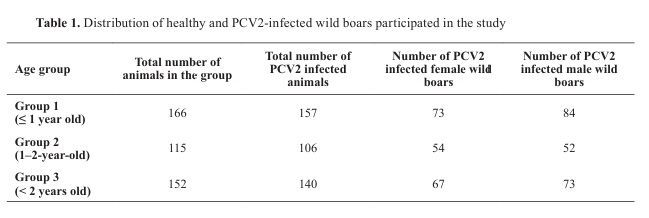
The animals were divided into three age groups. The first group included wild boars up to one year of age, the second group consisted of animals aged 1-2 years, and the third group included wild boars older than 2 years (
Table 1). All groups were further subdivided according to sex in order to evaluate the distribution of PCV2 in female and male cohorts.
Blood samples were analyzed using the ELISA method with the INGEZIM Circo IgG 11.PCV.K.1® assay (Ingenasa, Spain). Optical density (OD) was measured at 450 nm using a Power Wave XS reader (BioTek®), with a cutoff value of 0.3 according to the manufacturer’s instructions.
Histopathological and immunohistochemical analysisTissue sections from mesenteric lymph nodes of a subset of wild boars, predominantly seropositive for PCV2, were used for histopathological and immunohistochemical (IHC) analyses. Formalin fixed, paraffin-embedded tissue sections (4–5 µm) were deparaffinized and rehydrated in alcohol, followed by routine hematoxylin-eosin staining (
28) and immunohistochemical staining using the EnVision+™ System (DakoCytomation, Denmark).
For immunohistochemistry, tissue sections underwent antigen retrieval in citrate buffer (pH 6.0) for 20 min, followed by blocking of endogenous peroxidase activity with 0.6% hydrogen peroxide. The sections were incubated at 37 °C for 30 min with a monoclonal antibody against PCV2 (Danish Institute for Food and Veterinary Research; dilution 1:300) diluted in antibody diluent (Dako, S0809). Detection was performed using the EnVision+™ polymer–HRP system (DakoCytomation, Denmark) with 3,3′-diaminobenzidine (DAB) as the chromogen. Nuclei were counterstained with Harris hematoxylin. Negative control sections were processed using antibody diluent (Dako, S0809) instead of the primary antibody.
The histopathological changes and immunolocalization of PCV2 were evaluated visually by three independent scientists in a blinded analysis. The samples were photographed using an AxioCam HRc camera connected to a Zeiss Axioplan 2 Imaging microscope (Carl Zeiss, Germany).
Statistical analysisStatistical analysis was performed using Python software (version 3.13.0, 2024). Differences in PCV2 seroprevalence between age groups and between sexes were evaluated using appropriate statistical tests. A p-value of <0.05 was considered statistically significant.
Ethical statementThe samples used in this study were collected in 2016 within the framework of the Estonian African swine fever (ASF) disease control and surveillance program in accordance with the European Commission Implementing Decision 2014/709/EU approved by the Estonian University of Life Sciences. The material included samples from wild boars found dead as well as from hunted animals submitted for routine surveillance. No animals were killed specifically for the purposes of this study. The collected samples were obtained as part of official disease monitoring activities and were approved for use in scientific research and publication.
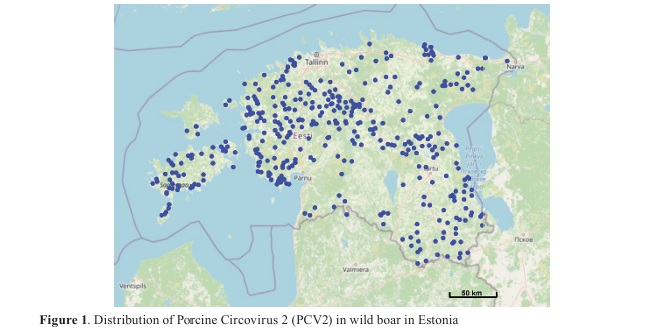
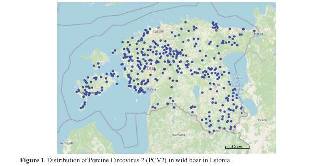
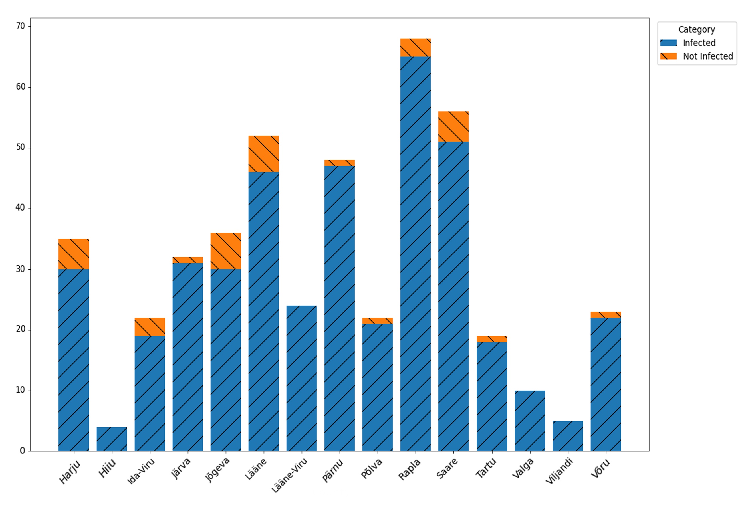
RESULTSELISAThe studies by using ELISA method showed widespread distribution of PCV2 in wild boars in Estonia as the ELISA test detected PCV2 antibodies in approximately 92% of samples (n=433) (
Fig. 1).
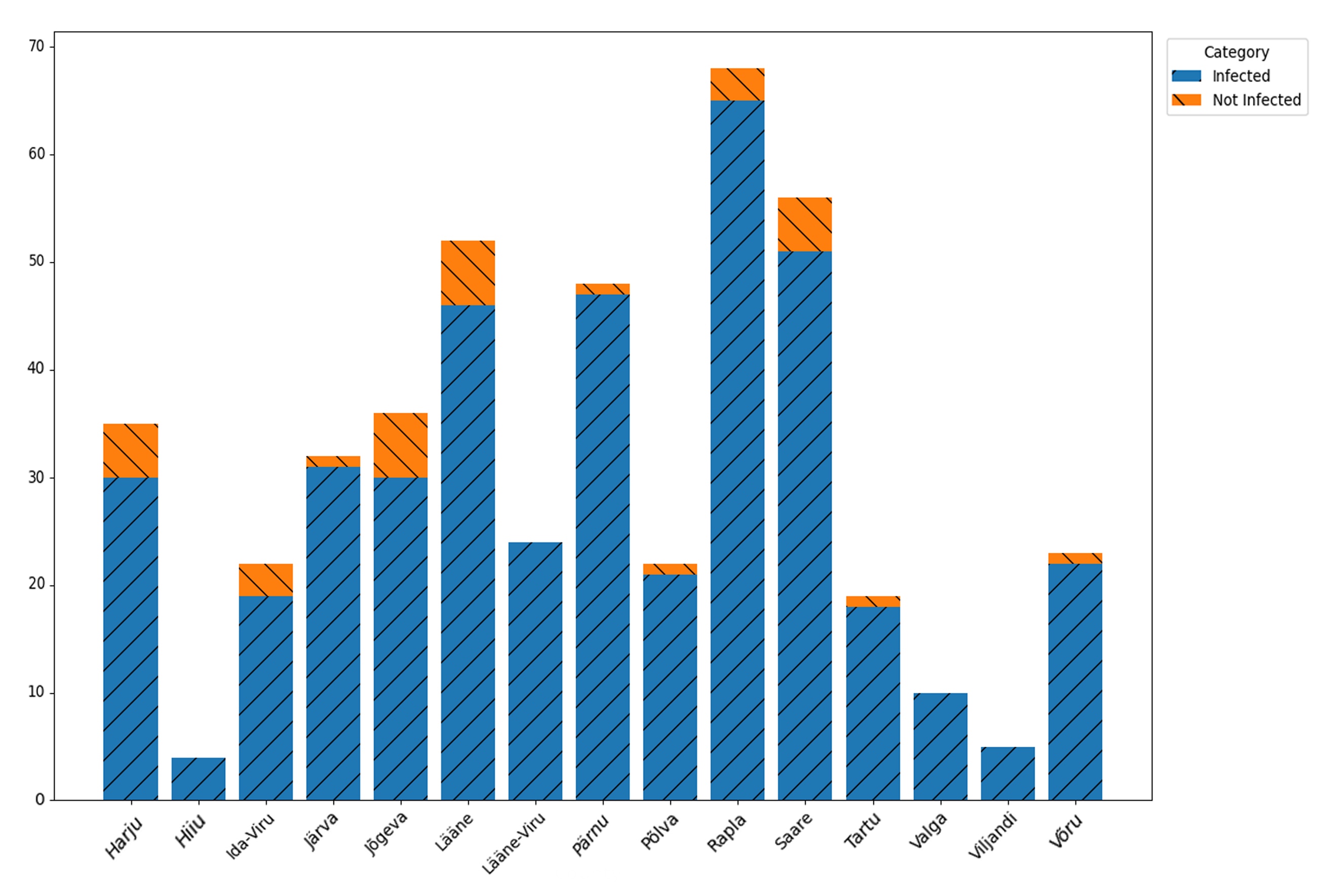
The PCV2-infected wild boars were found in all Estonian counties. In the western and southern counties of Estonia (Hiiu, Lääne-Viru, Valga, and Viljandi), the virus was widespread, with no seronegative animals detected among the sampled wild boars. (
Fig. 2).
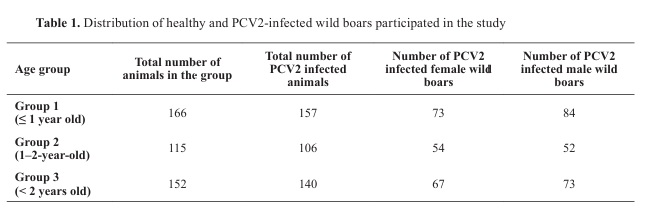
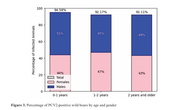
In the first age group (0-1 year), there were 94.58% positive cases, of which 44% females and 51% males; in the age group (1-2 years), there were 92.17% positive cases, of which 47% females and 45% males; in the age group (over 2 years), there were 92.11% positive cases, of which 43% females and 49% males. The studies revealed the highest persentage of PCV2 positive cases among the wild boars up to one year of age (94.58%). In 1 - 2 and older animals there were fewer positive cases (92.17% and 92.11%, respectively) (
Table 1).
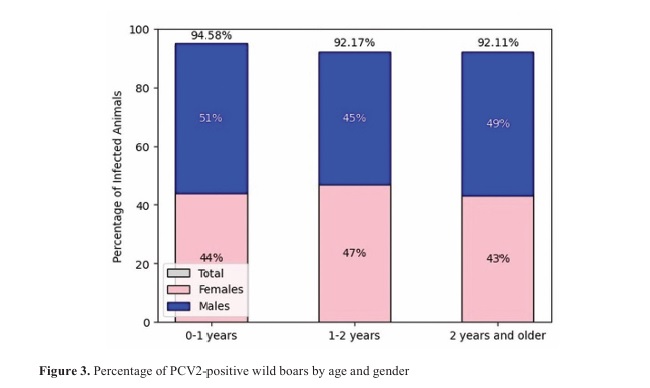
Comparing the distribution of PCV2 between female and male wild boars, the study revealed that in one to two years old animals the distribution of PCV2 between female and male wild boars was comparatively similar, accounting for 47% (54/115) and 45% (52/115), respectively. However, in the 0-1 years old wild boars’ group and in wild boars over 2 years there were slightly more positive cases in male wild boars (
Fig. 3).
Statistical analysis revealed no significant differences in PCV2 positivity among age groups (p=0.199) or between genders (p=0.453).
Age (p=0.199) and gender groups (p=0.453) did not show significant differences.
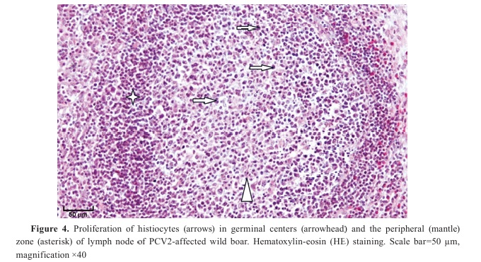
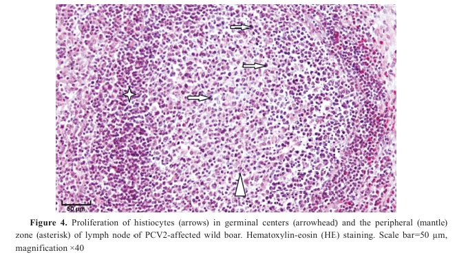
Histopathology and immunohistochemistryThe routine histology staining with hematoxylin and eosin showed depletion of lymphocytes and proliferation of histiocytes in PCV2-affected animals which were noted to be the highest in germinal centres and peripheral zone (mantle zone) of the lymphatic nodules of the mesenteric lymph nodes (
Fig 4).
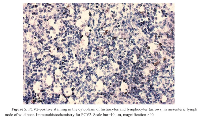
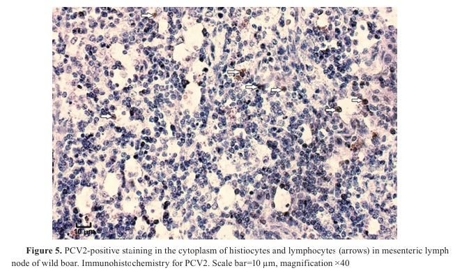
IHC investigation revealed PCV2-positive staining in the cytoplasm of lymphocytes and histiocytes (
Fig. 5).
The negative controls of PCV2 infected lymph nodes containing antibody diluent instead of primary antibodies, showed no specific staining. In our study, all seropositive wild boars selected for histological examination displayed PCV2 positive immunostaining in lymphocytes and histiocytes, whereas no PCV2 antigen was detected in seronegative animals.
 DISCUSSION
Porcine Circovirus 2 (PCV2), a member of the family Circoviridae, is a small non-enveloped virus that primarily infects domestic pigs and wild boars and is associated with a wide spectrum of clinical manifestations, ranging from wasting, inappetence, and respiratory disorders to severe and frequently fatal disease conditions (
13). PCV2’s ability to persist in the environment, its high mutation rate, and its interaction with host immune systems make it particularly difficult to control, further exacerbating its impact on the swine industry globally. Its role in the etiology of multiple disease syndromes makes its detection and monitoring essential for effective disease management.
Among the diagnostic approaches used for the detection of PCV2, the enzyme-linked immunosorbent assay (ELISA) represents one of the most widely applied serological methods. In ELISA testing, the presence of PCV2 specific antibodies in a sample is detected through a colorimetric reaction that is typically quantified by measuring absorbance at defined wavelengths (
29). One of the major advantages of ELISA is its capacity to process a large number of samples within a relatively short period of time, making it particularly suitable for large-scale epidemiological investigations such as surveillance studies of wildlife populations (
30). Furthermore, ELISA allows the detection of antibodies in serum or blood samples, enabling relatively non-invasive sample collection (
31). This feature is especially advantageous in wildlife research, including studies involving wild boar populations, where obtaining samples by more invasive methods can be difficult.
Despite these advantages, several limitations of ELISA should be considered when applying the method to PCV2 diagnostics. Most importantly, ELISA detects the presence of antibodies, which may indicate previous exposure to the virus rather than an active infection (
14). Consequently, the method may be less suitable for identifying animals with acute PCV2 infection, since antibody production typically occurs some time after initial viral exposure. In addition, false-positive results may occur due to potential cross-reactivity with antibodies directed against other pathogens, and the assay does not allow differentiation between individual PCV2 genotypes (
5). Nevertheless, ELISA remains an important and widely utilized diagnostic tool, particularly for population-level surveillance and for identifying areas with a high prevalence of PCV2 exposure (
32). In several European countries, including Poland, Germany, and Hungary, ELISA has been used in wildlife monitoring programs to detect PCV2 antibodies in wild boars, providing valuable information about the distribution of the virus and its potential involvement in cross-species transmission (
15, 19, 21). In addition to wildlife surveillance, ELISA is routinely used in domestic pig populations to evaluate herd immunity and to monitor the effectiveness of vaccination programs (
5).
The correlation between serological status and histopathological findings was also evaluated in this study. Wild boars that tested positive for PCV2 antibodies by ELISA exhibited pronounced histopathological changes in the mesenteric lymph nodes, including lymphocyte depletion and proliferation of histiocytes, particularly in germinal centers and the peripheral (mantle) zone (
Fig. 4). Immunohistochemistry confirmed the presence of PCV2 antigen in the cytoplasm of lymphocytes and histiocytes in these animals (
Fig. 5). Although ELISA cannot distinguish between active and past infections, the consistent presence of histopathological lesions and antigen detection in seropositive animals supports a link between serological exposure and tissue-level infection. This observation further suggests a correlation between ELISA seropositivity and tissue-level detection of PCV2. In contrast, ELISA-negative animals (controls) showed minimal to no histopathological changes, and no PCV2 antigen was detected by IHC, highlighting the specificity of the observed lesions for PCV2 infection. This correlation between serological positivity and tissue pathology strengthens the evidence that PCV2 is widely circulating in Estonian wild boar populations and underlines the relevance of combining serological and histopathological approaches for a comprehensive understanding of PCV2 infection dynamics.
In addition to the limitations of ELISA, genotyping of PCV2 plays a crucial role in understanding the epidemiology and evolutionary dynamics of the virus. Different PCV2 genotypes (PCV2a–i) have been associated with varying pathogenicity, geographic distribution, and temporal emergence, which can influence both clinical outcomes and the effectiveness of vaccination strategies (
8, 9, 11). While serological methods such as ELISA provide valuable information on exposure and population level prevalence, they do not differentiate between genotypes. This represents a limitation in assessing the full epidemiological picture, particularly when evaluating the potential for genotype shifts or the introduction of new variants into domestic or wild populations. Incorporating genotyping data would allow for a more precise understanding of transmission pathways between wild boars and domestic pigs, help identify dominant circulating strains, and inform targeted disease control measures. Future studies combining serology with molecular characterization are therefore recommended to enhance the assessment of PCV2 circulation, genotype diversity, and associated risks to swine populations.
The present study provides new information on the distribution of PCV2 in wild boar populations in Estonia, using ELISA as the primary diagnostic method and supporting the serological findings with PCR and immunohistochemistry (IHC). The results of our study demonstrate a high prevalence of PCV2 exposure among wild boars, with 92% of the analyzed ELISA samples testing positive for PCV2 antibodies. These findings are consistent with observations reported from other European countries, where similarly high seroprevalence rates have been documented in wild boar populations. For example, Malmsten et al. (
33) reported a seroprevalence of 99% among wild boars in Sweden, indicating widespread exposure to the virus within the population. Comparable results have also been reported in studies conducted in Poland and Romania (
19, 21), further supporting the conclusion that PCV2 infection is widely distributed across Europe. Considering that porcine circoviruses are ubiquitous in both domestic and wild pig populations, the detection of PCV2 in a large proportion of the analyzed wild boar samples was not unexpected (
20). The presence of PCV2 in wild boars highlights their potential role as reservoirs and vectors of the virus, thereby facilitating its transmission between wild and domestic pig populations, a phenomenon that has been previously reported in several regions (
17).
An important observation of the present study is the variation in PCV2 prevalence among different age groups of wild boars. The highest prevalence was detected in younger animals, particularly in individuals aged between 1 and 2 years, suggesting that this age group may be more susceptible to infection or may experience higher levels of exposure. This age-related pattern is consistent with previous studies indicating that younger animals are more likely to become infected with PCV2, potentially due to increased contact rates with other infected animals or environmental sources of infection (
1). In contrast, the lower prevalence observed among older wild boars (over 2 years of age) may reflect the development of immunity following earlier exposure to the virus. Such immunity may result either from previous infections or from passive transfer of maternal antibodies during early life.
When the distribution of PCV2 antibodies was analyzed according to sex, differences between male and female wild boars were observed, although these differences were less pronounced than those related to age. These observations raise questions regarding possible sex-related differences in susceptibility to infection or exposure risk. Behavioral factors, including territorial behavior and the social structure of wild boar populations, may influence contact patterns and consequently affect transmission dynamics. However, additional studies would be required to determine whether the observed differences are indeed associated with sex-specific biological or behavioral factors.
Currently, in addition to serological testing methods such as ELISA, immunohistochemistry and in situ hybridization are considered to represent the gold standard techniques for the detection of PCV2 in the context of diagnosing porcine circovirus associated disease (PCVAD) (
34). During PCV2 infection, characteristic histopathological changes occur in lymphoid tissues, particularly in lymph nodes. Histiocyte proliferation is mainly observed within germinal centers, which are sites where B lymphocytes undergo proliferation, differentiation, and maturation. In response to viral infection, increased infiltration of histiocytes, including macrophages and dendritic cells, occurs as part of the host immune response (
35). Although germinal centers represent the primary location of histiocyte proliferation, the peripheral zones of lymphoid tissues may also demonstrate increased histiocytic infiltration due to virus-induced alterations in immune regulation. In the present study, immunohistochemistry was used to confirm the presence of PCV2 in tissue samples. The results of our histopathological and immunohistochemical analyses correspond well with previously published studies, demonstrating that histiocytes, multinucleated giant cells, and macrophages within lymphoid tissues represent the principal target cells of PCV2 infection (
36). Typical microscopic lesions include lymphocyte depletion in lymphoid tissues accompanied by diffuse histiocytic infiltration (
37).
The detection of PCV2 in wild boars also has important implications for the epidemiology of porcine circovirus-associated disease in both wildlife and domestic pig populations. The identification of PCV2 in wild boars inhabiting areas near swine farms suggests that these animals may contribute to the epidemiological maintenance and spread of the virus. In regions where wild boar populations overlap with domestic pig production systems, the risk of viral transmission between wild and domestic animals is increased, thereby complicating efforts to control PCV2 infections in commercial pig herds (
16). Consequently, the findings of the present study emphasize the importance of including wildlife surveillance within broader biosecurity strategies aimed at preventing the introduction and spread of PCV2 in domestic pig populations.
Additionally, the findings underscore the need for more targeted preventive measures to mitigate the risks of virus transmission between wild and domestic pigs. The detection of PCV2 in wild boars in Estonia, together with the high prevalence observed in certain regions, underscores the necessity for further research focusing on the ecology and population dynamics of wild boars and their interactions with domestic pigs. Such studies could help identify critical contact points between wild and domestic animals that facilitate PCV2 transmission and thereby support the development of more effective disease control strategies.
CONCLUSIONThis study presents the first nationwide assessment of Porcine Circovirus 2 (PCV2) in wild boar populations in Estonia. The high seroprevalence of PCV2 antibodies (92%) detected across all counties and in all age and sex groups indicates widespread exposure to the virus. Histopathological and immunohistochemical findings confirmed PCV2-associated lesions and antigen localization in lymphoid tissues, supporting the serological results. The absence of significant differences between age and sex groups suggests that viral circulation occurs broadly within the wild boar population. These results set an important epidemiological baseline for Estonia and highlight the need for continued surveillance to monitor potential interactions between wild and domestic pig populations.
CONFLICT OF INTERESTThe authors declare that they have no financial or non-financial conflict of interest regarding authorship and publication of this article.
ACKNOWLEDGMENTSThe authors wish to thank Mrs. Mare Tamm for her laboratory assistance.
AUTHORS’ CONTRIBUTIONTJ made the conceptualization, methodology, investigation, data curation, formal analysis, wrote the original draft, the review and editing of the manuscript. TS and TS was involved in conceptualization, methodology, investigation and formal analysis. PH performed the conceptualization, formal analysis, was included in writing of the original draft, the review and editing. All authors contributed to the revision of the manuscript, addressed the reviewers’ comments, and approved the final version.


 10.2478/macvetrev-2026-0019