Anatomical variations of the celiac trunk (CT), superior mesenteric artery (SMA) and inferior mesenteric artery (IMA) have significant clinical relevance, particularly in abdominal surgery and interventional radiology. In this study, a cross sectional analysis was performed on 60 contrast-enhanced CT angiography scans of the abdominal aorta in adult patients. The evaluation included the vertebral level of origin, branching patterns, and arterial diameters of the CT, SMA, and IMA, with variations classified according to established anatomical classifications and analyzed using descriptive statistical methods. The results demonstrated that typical branching anatomy was present in 95% of cases, while anatomical variations were identified in 5% of patients. The celiac trunk most commonly originated at the level of the T12–L1 intervertebral disc (91.70%). The classical trifurcation (Uflacker Type I) was observed in 95% of cases. The SMA showed an independent origin in 98.33% of patients, whereas the IMA originated independently in all examined cases. The mean arterial diameters were 6.94±1.18 mm for the CT, 6.56±1.11 mm for the SMA, and 2.73±0.52 mm for the IMA. Although classical vascular anatomy predominates, the presence of clinically relevant variations highlights the importance of their preoperative identification. High-resolution 128-slice MDCT angiography proves to be a reliable modality for accurate vascular mapping and effective surgical planning.
The abdominal aorta represents the continuation of the thoracic aorta after passing through the aortic hiatus of the diaphragm at the level of the twelfth thoracic vertebra (T12). It extends along the anterior surfaces of the vertebral bodies from T12 to L4, as well as anterior to the intervertebral discs and the anterior longitudinal ligament. It gives rise to ventral, lateral, dorsal, and terminal branches (
1).
The paired lateral branches include the inferior phrenic arteries, middle suprarenal arteries, renal arteries, and gonadal arteries. The dorsal branches consist of four pairs of lumbar arteries. The terminal branches are the right and left common iliac arteries and a smaller median sacral artery (
2).
The abdominal aorta and its visceral branches, such as the celiac trunk and the superior and inferior mesenteric arteries, constitute the primary source of blood supply to the organs of the digestive system. Variations in their anatomy occur relatively frequently and are of significant clinical importance, as they may influence the course and safety of various medical and surgical interventions.
The celiac trunk typically arises from the abdominal aorta at the level between T12 and L1 and divides into three terminal branches: the left gastric artery, the common hepatic artery, and the splenic artery. However, anatomical variations may occur, including differences in the branching pattern and number of branches arising from the celiac trunk, as well as complete absence of the celiac trunk (
3, 4).
The superior mesenteric artery (SMA), which arises from the anterior surface of the abdominal aorta at the level of L1, may demonstrate early branching or an unusual origin, findings that are highly relevant when selecting and performing surgical approaches (
4, 5).
Accurate identification of these variations is crucial in the planning of numerous surgical procedures. The development of modern high resolution imaging technologies, particularly 128-slice computed tomography (CT), has significantly enhanced the ability to study vascular anatomy noninvasively. This method provides detailed axial and three-dimensional visualization, enabling clinicians to accurately depict arterial branching patterns and identify anatomical variations (
6, 7).
Despite the increasing use of this modality in clinical practice, anatomical studies utilizing this method remain limited in our region. To address this gap, the expected results of this study will provide valuable reference material for clinicians and contribute to greater safety and effectiveness in performing surgical procedures involving these blood vessels.
Comparative studies in various animals, such as pigs, monkeys, dogs, cats, rabbits, and goats, have shown that the branching patterns of the abdominal aorta and its visceral arteries can differ among species (
8, 9, 10). In mammals, such as pigs, primates, and other species, the branching pattern shows similarities to the human model, but it also demonstrates species-specific differences in vessel length, branching angles and the origin of arterial branches. In pigs, a common origin with the cranial mesenteric artery is frequently reported, whereas in primates the pattern more closely resembles that of humans (
11). These comparative findings are relevant for experimental surgery, transplantation medicine, and the selection of appropriate animal models in biomedical research. Understanding these interspecies variations provides context for human anatomical research and helps highlight the clinical importance of recognizing variations in vascular anatomy. In experimental veterinary medicine, particularly in pigs as a model organism, precise vascular anatomy is crucial for transplantation research, the development of new surgical techniques, and the testing of interventional procedures (
12). Therefore, knowledge of these anatomical variations has a direct impact on the safety, success, and precision of veterinary treatment.
The aim of this study was to analyze the anatomical variations of the abdominal aorta and its visceral branches in humans using 128-slice CT angiography (CTA) and to assess their clinical significance. According to the literature, the highest number of variations in branching is expected to be observed in the celiac trunk, while the branching pattern shows a high degree of similarity between humans and animals.
MATERIAL AND METHODSAll procedures performed in studies involving human participants were in accordance with the ethical standards of the institutional and/or national research committee and with the 1964 Helsinki declaration and its later amendments or comparable ethical standards. The study was approved by the Ethics Committee of the Faculty of Medicine, Ss. Cyril and Methodius University in Skopje, North Macedonia (Approval No. 03-5505/4). All patient data were handled with strict confidentiality and used solely for research. All data were anonymized, and patient confidentiality was protected throughout the research process. The study was conducted for academic purposes to advance medical knowledge and improve clinical and anatomical understanding.
In this cross-sectional study, а total of 60 CTA scans of the abdominal aorta (36 males and 24 females) were analyzed in patients older than 18 years (male and female) who, for justified clinical reasons and with an appropriate referral diagnosis, were referred to the University Institute of Radiology.
Patients with a history of previous surgical or endovascular interventions involving the abdominal aorta and its branches were excluded from the study. Patients with contrast allergy, claustrophobia, pregnancy, vascular abnormalities (aneurysms, stenoses, occlusions), chronic renal insufficiency, and CTA scans with significant artifacts that interfered with assessment or rendered the scans technically non-interpretable were also excluded.
The examination was performed using a multidetector CT scanner (MDCT, Siemens Definition AS, 128 slices, SYNGO software; gantry rotation speed 0.5 s; tube voltage 120 kV; tube current 80 mAs; collimation 0.75 mm; pitch 0.6; interval 128×0.625; delayed scanning interval 2 s).
Following acquisition, the images were analyzed using Vitrea® Advanced Visualization Software. The image processing and reconstruction were performed at a dedicated workstation using four visualization techniques: maximum intensity projection (MIP), multiplanar reconstruction (MPR), shaded surface display (SSD), and four dimensional CTA with volume rendering (VR).
CTA images were evaluated for the origin, level of emergence, and diameters of the celiac trunk, superior mesenteric artery, and inferior mesenteric artery. Anatomical variations were independently identified and classified by two experienced radiologists. Rare or unclear variants were described in detail and included in the analysis if clearly visualized; if reliable classification was not possible, they were excluded from the study.
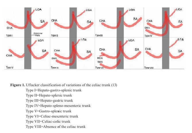
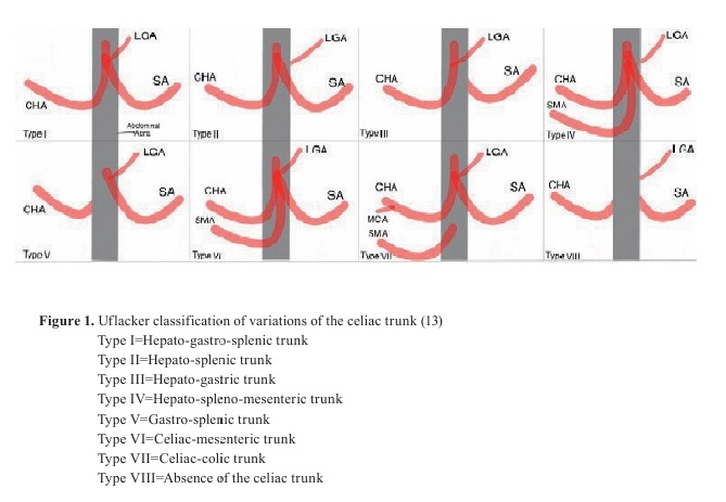
Variations in the branching pattern of the celiac trunk (the most common variation) have been described according to the Uflacker classification (
Fig. 1), which defines eight types of anatomical variations (
13). The nomenclature of these variations is based on the arteries forming the respective configurations. Typically, the celiac trunk represents a trifurcation into the gastro hepatic-splenic trunk. A combination of two arteries instead of three results in a bifurcation.
Statistical analysisDescriptive statistics were used for data analysis, and the results were presented as absolute numbers and percentages.
RESULTS
Sixty CTA scans were evaluated. Typical branching of the anterior branches of the abdominal aorta was identified in 57 cases (95%), while variations in branching pattern and number of branches were observed in 3 cases (5%).
Among the 60 analyzed CTA scans, the celiac trunk most commonly originated at the level of the T12/L1 intervertebral disc (91.70%). Other levels of origin included the upper border of L1 (3.33%), the body of L1 (3.33%), and the body of T12 (1.67%).
The SMA most frequently originated from the body of L1 (91.70%), followed by the upper border of L1 (4.97%) and the L1/L2 intervertebral disc (3.33%). The IMA most commonly originated at the level of the L3 vertebral body (93.30%), followed by the L3/L4 intervertebral disc (3.33%), the L2/L3 level (1.67%), and the upper border of L4 (1.67%).
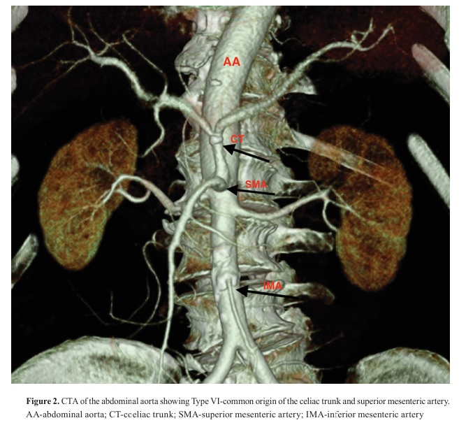
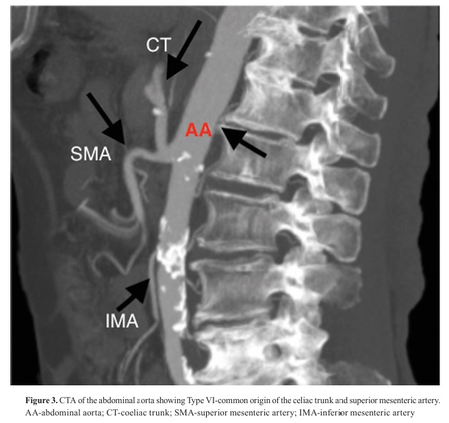
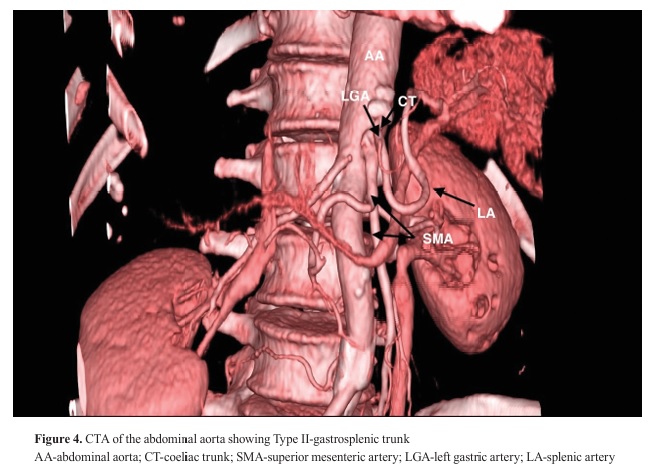
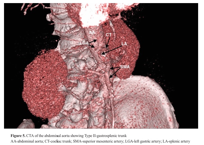
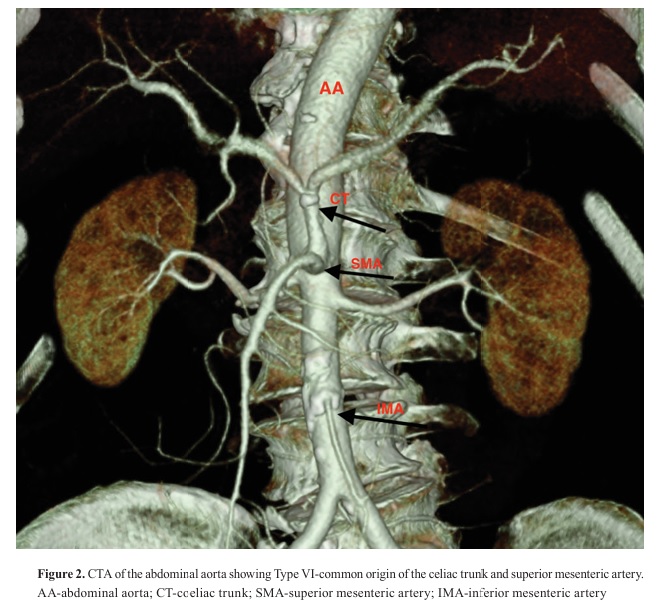
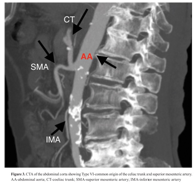
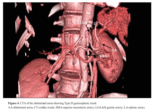
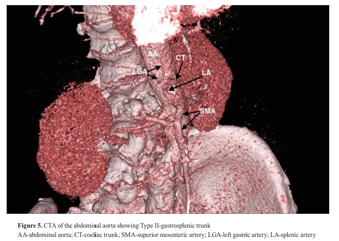
Three variations of the celiac trunk were observed. According to Uflacker’s classification, the most common pattern was Type I – classical trifurcation – in which the celiac trunk divides into the common hepatic, left gastric, and splenic arteries. Type I was observed in 57 (95%) of cases. Other variants included Type VI - common origin of the celiac trunk and SMA (
Fig. 2 and
Fig. 3) in 1.67%, and Type V - gastrosplenic trunk (
Fig. 4 and
Fig. 5) in 3.34%. No cases of Types II, III, IV, VII, or VIII were observed.
The SMA demonstrated independent origin from the abdominal aorta (Type I) in 98.33% of cases, while common origin with the celiac trunk (Type II) was observed in 1.67%. The IMA showed independent origin in 100% of cases. The mean diameter of the celiac trunk was 6.94±1.18 mm. The mean diameter of the SMA was 6.56±1.11 mm, while the mean diameter of the IMA was 2.73±0.52 mm.
 DISCUSSION
During human embryonic development, four omphalomesenteric arterial roots form from the abdominal aorta, interconnected by a single ventral longitudinal anastomosis. Among these four roots, the two middle regress during development, while the first and fourth remain connected through the longitudinal anastomosis. From this common anastomosis, the splenic, left gastric, and common hepatic arteries later develop, while the SMA forms from the fourth omphalomesenteric root. If any of these arteries persist or, conversely, regress more than usual, various vascular variations of the celiac trunk or the SMA may occur (
14). Understanding this embryological basis is essential for accurate radiological interpretation and preoperative planning.
In the past, preoperative evaluation of patients who were candidates for hepatic resection included a combination of non-invasive and invasive diagnostic methods, including computed tomography (CT), magnetic resonance imaging (MRI), CT arterial portography, and conventional angiography. In modern clinical practice, however, the integration of CT with three-dimensional CTA is preferred to provide better visualization of vascular anatomy.
Multidetector computed tomography (MDCT) with contrast application (CTA) allows examinations with isotropic resolution. Hence, within a short period of just a few seconds, multiplanar and multivolumetric imaging can be performed with a resolution of less than 1 mm, covering large anatomical regions (
9).
CTA additionally enables precise visualization of the arterial phase, vascular wall, lumen, and surrounding tissues from multiple angles. Although CTA offers many advantages, this method also has its limitations. The greatest disadvantage is exposure to ionizing radiation. There is also a possibility of an allergic reaction to iodine or other components of the contrast agent (however, alternative gadolinium-based contrast agents can be used in patients with iodine allergy) (
9, 10). Moreover, the potential nephrotoxicity of contrast agents should be considered.
Detailed preoperative recognition of anatomical variations of the blood vessels reduces the need for extensive surgical exploration, significantly decreases the risk of intraoperative vascular injuries, andimproves surgical outcomes (
15, 16).
The abdominal aorta vascularizes the gastrointestinal organs through three unpaired anterior branches: the celiac trunk (
truncus coeliacus), the superior mesenteric artery (
a. mesenterica superior), and the inferior mesenteric artery (
a. mesenterica inferior). These branches typically arise from the abdominal aorta skeletotopically at the level of T12 to L3/4 vertebral bodies (
1, 17). Anatomically, the celiac trunk usually divides into three branches: the left gastric artery (LGA), the common hepatic artery (CHA), and the splenic artery. In this study, the analyzed CTA images showed that the celiac trunk most frequently originated at the level of the T12-L1 intervertebral disc in 91.7% of cases, consistent with the descriptions of Ahluwalia et al. (
7).
The results of our study showed a lower degree of variability in the level of origin of the celiac trunk. This contrasts with the findings of Khoa et al., who identified a wider range and greater diversity of anatomical variations (
18). The mean diameter of the celiac trunk was 6.94±1.18 mm, which aligns with previously published data (
18, 19, 20), although slightly smaller than those described in Western populations, such as the study by Dyches (
21). Anatomical classification according to Uflacker (
13) showed the classical trifurcation model (Type I) in 95% of cases analyzed in our study, which largely corresponds with the results reported by Song et al. (89.1%) (
22) and Osman et al. (90.5%) (
23).
Although the classical trifurcation model of the celiac trunk predominated in our sample, present in 95% of cases, indicating a relatively low frequency of anatomical variations, other studies report a significantly broader spectrum of variations. The literature describes hepatosplenic trunks, common origin of the celiac trunk with the superior mesenteric artery, as well as the presence of accessory arteries, including inferior phrenic and pancreatic arteries, as shown in the study by Randrianalison et al. (
6). Despite the lower incidence in our population, recognizing such variants remains critically important, as failure to identify them may lead to intraoperative bleeding, vascular injuries, or inadequate perfusion, especially during hepatobiliary and pancreatic surgical interventions (
5, 10).
Regarding the SMA, its most common origin from the body of the L1 vertebra (91.7%) aligns with classical anatomical descriptions (
3, 4). The mean diameter of 6.56±1.11 mm corresponds with values reported in the study by Hao et al. (
24). According to Kornafel’s classification, in 98.33% of our cases the SMA had an independent origin (Type I), while only 1.67% were classified as Type II, indicating a common origin with the celiac trunk. This result is consistent with the findings of Khoa et al. (
18), who also reported a dominant prevalence of independent origin of the SMA, with minimal frequency of common origin with the celiac trunk. The similarity between these results further confirms that the classical anatomical model of SMA origin is predominant in most populations. Although these variations are rare, their recognition is important as they may influence surgical outcomes, organ transplantation, and the treatment of mesenteric ischemia (
14, 25). There is a significant need for anatomical references adapted to specific populations, rather than relying solely on universal anatomical norms. Surgeons and interventional radiologists should be prepared for possible unexpected anatomical variations, especially during high-risk interventions involving gastrointestinal organs such as the liver, pancreas, or colon.
The use of 128-slice CT in this medical field is particularly useful. The high resolution of CT combined with multiplanar, and 3D reconstruction processing allows precise visualization of the arterial origin level, their branching, and possible anatomical variations (
18). This significantly enhances safety in surgical planning and improves procedural outcomes. Moreover, preoperative identification of anatomical variations, such as replaced hepatic arteries, accessory branches, or common trunks, can help prevent intraoperative complications, including bleeding, ischemia, and procedural failure (
26).
In addition to the findings reported in humans, comparable anatomical variations have also been described in animal species. Comparative studies in animals provide valuable insight into the morphological variability of the celiac and mesenteric arteries, which can inform both experimental research and the understanding of evolutionary and interspecies differences. In animals, the celiac artery has been described as presenting possible variations at different levels (
27). In the study by Abidu-Figueiredo et al. (
28), the artery emerged in a single morphological form without the presence of a celiac–mesenteric trunk, which is consistent with the findings reported by Bednarova et al. (
29). In carnivores, it is generally described at the level of the first lumbar vertebra. This observation aligns with the findings of Abidu Figueiredo et al. (
28), where this level was identified in 76.7% of the examined cases. According to the same authors, the classical trifurcation into the hepatic, splenic, and left gastric arteries was observed in only 33.3% of the animals, whereas the most frequent arrangement was the presence of a gastrolienal trunk (56.7%), indicating interspecies variation in branching morphology.
The cranial mesenteric artery most commonly presents as a single vessel arising independently from the abdominal aorta. This pattern has been observed in cats by Estruc et al. (
30), in rock cavies by Queiroz et al. (
31), in crab-eating raccoons by Morais Lima et al. (
32), and in nutrias by Machado et al. (
33, 34). However, the presence of a common celiac–mesenteric trunk has also been described in goats by Ferreira et al. (
35), in dogs by Schmidt and Schoenau (
36), and in cats by Roza et al. (
37) and Estruc et al. (
30). The caudal mesenteric artery most commonly originates as a single branch from the abdominal aorta. This pattern has been reported in New Zealand rabbits by Machado et al. (
34), in nutrias by Machado et al. (
33), in rock cavies by Queiroz et al. (
31), and in cats by Estruc et al. (
30). Nevertheless, the variations have been described,including absence of the caudal mesenteric artery with vascular supply provided by the left colic artery arising from the cranial mesenteric artery. This variation was described in nutrias by Culau, Azambuja, and Campos (
38) and mentioned in cats by Estruc et al. (
30). The vertebral level of origin also varies among species, occurring around the fifth lumbar vertebra in dogs, between the fifth lumbar and the first sacral vertebra in cats, and between the fifth and seventh lumbar vertebrae in rabbits (
39). Such comparative anatomical insights support more accurate interpretation of vascular patterns across species and clinical contexts, underscoring their relevance for experimental research and clinical decision-making in both human and veterinary medicine.
CONCLUSIONIn this study, the typical branching pattern of the abdominal aorta was observed in the majority of cases (95%), whereas anatomical variations were identified in a smaller proportion (5%), including celiac trunk bifurcation and a common celiac mesenteric trunk. Although the classical vascular configuration predominated, the detected variations emphasize the importance of careful preoperative imaging, as unrecognized vascular anomalies may significantly affect surgical and endovascular procedures. In veterinary medicine, knowledge of celiac trunk variations is also important for safe surgical and diagnostic interventions, as it reduces the risk of vascular complications. High-resolution 128-slice computed tomography therefore represents a valuable tool for accurate preoperative assessment and improved procedural safety in both human and veterinary medicine.
CONFLICT OF INTERESTThe authors declare that they have no financial or non-financial conflict of interest regarding authorship and publication of this article.
ACKNOWLEDGMENTSThe authors would like to thank the University Institute of Radiology in Skopje for providing access to the CT angiographic data used in this study.
AUTHORS’ CONTRIBUTIONJZ supervised the research, manuscript preparation and participated in the analysis of the CTA images. AP designed and conducted the study and drafted the manuscript. BZ and AD participated in the analysis of the CTA images. SN and VA facilitated the imaging analyses at the University Institute of Radiology and acted as CTA observers. JK assisted with the technical preparation of the manuscript. LP contributed comparative anatomical analysis, providing veterinary anatomical insight for the comparison between human and animal anatomy.


 10.2478/macvetrev-2026-0020