Original Scientific Article
Computer tomography (CT) scans as a diagnostic tool for interpretation of S10 plastinated slides from dog cadaver
Lazo Pendovski
*
,
Dimitar Bozhinovski
,
Ksenija Ilievska
,
Plamen Trojachanec
,
Vlatko Ilieski
Received: 09 February 2022
Received in revised form: 08 April 2022
Accepted: 11 April 2022
Available Online First: 18 April 2022
Published on: 15 October 2022
Correspondence: Lazo Pendovski, lpendovski@fvm.ukim.edu.mk
Abstract
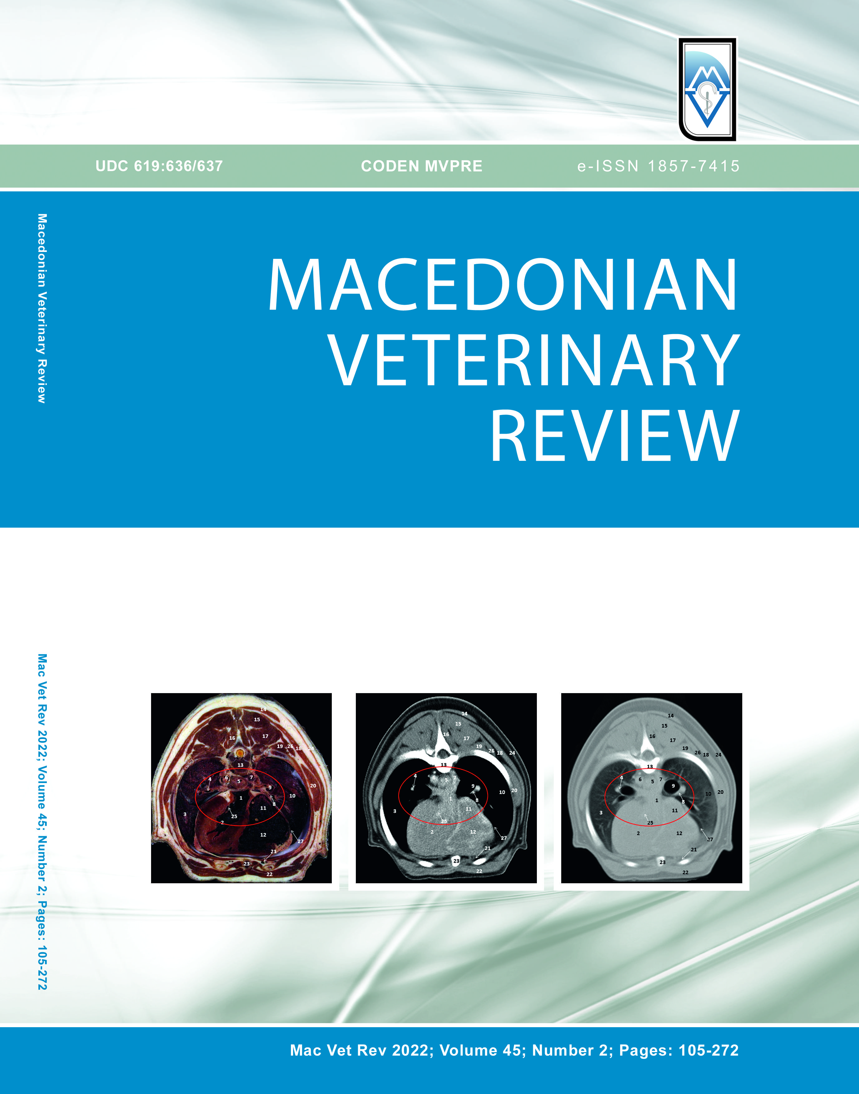
Computed tomography (CT) is a routine method for the diagnosis of pathological structures in the body and has been widely used in veterinary medicine as an advanced diagnostic imagining tool in veterinary clinics. However, interpretation of CT scans requires detailed knowledge of topographical animal anatomy and usually has limited scan resolution due to the ambiguous relationship between signal intensity and tissue composition. The aim of the study was to assess the morphometric similarities between S10 plastinated slides and computer tomography (CT) scans and their usability as compatible paired diagnostic methods. A 3-year-old euthanized dog cadaver was scanned on SHIMADZU SCT/6800TXL scanner immediately post-mortem, then frozen at -80 °C to preserve the correct anatomical position, and plastinated with a standardized procedure. Semi-transparent transversal slices (5 mm) were obtained from the head, thoracic, and lumbar sections of the body. The S10 plastinated slides and CT scans contained fine and small anatomical structures with high similarity. The spatial relationships of all anatomical structures on the serial S10 platinates were in the correct anatomical position. In conclusion, S10 transversal slices showed high similarity with the CT scans and allowed identification of the corresponding morphological structures. The S10 thin plastinated transversal slices could be used for additional interpretation of CT transversal scans at veterinary clinics and as a didactical tool for veterinary students.
Keywords: S10 plastination, computer tomography (CT), topographic anatomy, dog
References
- Brenner, D.J., Hall, E.J. (2007). Computed tomography - an increasing source of radiation exposure. NEJM 357(22): 2277-2284. https://doi.org/10.1056/NEJMra072149 PMid:18046031
- Randall, E.K. (2016). PET-computed tomography in veterinary medicine. Vet Clin North Am Small Anim. 46(3): 515-533. https://doi.org/10.1016/j.cvsm.2015.12.008 PMid:27068445
- Samii, V.F., Biller, D.S., Koblik, P.D. (1998). Normal cross-sectional anatomy of the feline thorax ana abdomen: comparasion of computed tomography and cadaver anatomy. Vet Radiol Ultrasound. 39(6): 504-511. https://doi.org/10.1111/j.1740-8261.1998.tb01640.x PMid:9845186
- Pendovski, L., Ilieski, V., Ursic, M., Petkov, V., Popovska-Percinic, F., Travnikar, B., Fazarinc, G. (2011). Anatomical correlation between dog transversal S10 plastinated thin sections and computer tomography (CT) images. Proceedings of the Days of Veterinary Medicine (pp. 124-125), Ohrid, R. Macedonia
- Bozinovski, D., Popovska-Percinic, F., Petkov, V., Adamov, N., Nikolovski, M., Ilieski, V., Pendovski, L. (2018). Tools for interpretation of computer tomography (CT) split images at dog: study with S10 thin plastinated specimens (p. 200), Ohrid, R. Macedonia
- Latorre, R., Arencibla, A., Gil, F., Rivero, M., Ramirez, G., Vaquezauton, J.M., Henry, R.W. (2003). P-40 and S10 plastinated slices: an aid to interpreting MR images of the equine tarsus. J Int Soc Plast. 18, 14-22.
- Jones, D.G. (2002). Re-inventing anatomy: the impact of plastination on how we see the human body. 15(6): 436-440. https://doi.org/10.1002/ca.10040 PMid:12373732
- Fasel, J.H.D. (1988). Use of plastinated specimens in surgical education and clinical practice. 1(3): 197-203. https://doi.org/10.1002/ca.980010306
- Pendovski, L., Urscic, J., Brankovic, V., Ilieski, V., Popovska-Percinic, F., Petkov, V., Travnikar, B., Fazarinc, G. (2013). Use of S10-thin plastinated slices for interpretation computer tomography (CT) scans: applied study for dog anatomy. Proceedings of the 7th Meeting of the Young Generation of Veterinary Anatomists, July, 17-20 (pp. 31), Leipzig, Germany
- Pendovski, L., Petkov, V., Popovska-Percinic, F., Ilieski, V. (2008). Silicone plastination procedure for production of thin, semitransparent tissue slices: a study using the pig kidneys. J Int Soc Plast. 23, 10-16.
- Șora, M.C. (2016). The general protocol for the S10 technique. Res Clin Med. 1(1): 14-18.
- Sora, M.C., Latorre, R., Baptista, C., López, A.O. (2019). Plastination-a scientific method for teaching and research. Anat Histol Embryol. 48(6): 526-531. https://doi.org/10.1111/ahe.12493 PMid:31573113
- Pauline, S., Rabi, R.S., Sridhar, G., Suganthy, R. (2015). Comparison of CT numbers of organs before and after plastination using standard S-10. Clin Anat. 28(4): 431-435. https://doi.org/10.1002/ca.22514 PMid:25708008
- Stefanie, O., Gernot, Sc. (2007). Computed tomography in small animals - basic principles and state of the art applications. Vet J. 173(2): 254-271. https://doi.org/10.1016/j.tvjl.2005.12.014 PMid:16516508
- Rodríguez, M.J., Latorre, R., López-Albors, O., Soler, M., Aguirre, C., Vazquez, J.M., Querol, M., Agut, A. (2008). Computed tomographic anatomy of the temporomandibular joint in the young horse. Equine Vet J. 40(6): 566-571. https://doi.org/10.2746/042516408x322166
- Arredondo, J., López, A.O., Agut, A., Gil, F., Soler, M., Rodriguez, M.J., Latorre, R. (2008). Epoxy plastinated slices of the temporomandibular joint of the cat are used to assess high resolution computed tomography. J Int Soc Plast. 23, 61-62.
- Sora, M.C., Brugger, P.C., Strobl, B. (2002). Shrinkage during E12 plastination. J Int Soc Plast. 17, 23-27.
- Donoso, E.S., Sora, M.C., Probst, A., Budrass, K.D., Konig, H.E. (2009). Mesoscopic structures of the equine toe demonstrated by using thin slice plastination (E12). Wiener Tierärztliche Monatsschrift. 96(11/12): 286-291.
Copyright
© 2022 Pendovski L. This is an open-access article published under the terms of the Creative Commons Attribution License which permits unrestricted use, distribution, and reproduction in any medium, provided the original author and source are credited.
Conflict of Interest Statement
The authors have declared that no competing interests exist.
Citation Information
Macedonian Veterinary Review. Volume 45, Issue 2, Pages 127-135, e-ISSN 1857-7415, p-ISSN 1409-7621, DOI: 10.2478/macvetrev-2022-0018, 2022

 10.2478/macvetrev-2022-0020